Crimson Publishers High Impact Journals

Showing posts with label journal of Surgical Medicine. Show all posts

Thursday, August 18, 2022

Intestinal Intussusception in an Adult Affected by Vanek Tumor: A Case Report_Crimson Publishers

Intestinal Intussusception in an Adult Affected by Vanek Tumor: A Case Report by Morazán AD in Surgical Medicine Open Access Journal_journal of Surgical Medicine


Abstract

Inflammatory fibroid polyp (IFP) is a rare benign lesion, originating from the submucosa in the gastrointestinal tract. It generally appears as an isolated benign lesion, rarely located at the level of the ileum. Its origin is controversial. Clinical presentation varies depending on its location; invagination and obstruction are the most common indicative symptoms when the polyp is located at the level of the small intestine. We report the case of a 60-year old patient with abdominal pain, nausea and vomiting and a personal history of intermittent constipation. Radiological imaging objectified ileo-ileal invagination completely obstructing the ileum light. Segmental resection of the obstructed ileal segment and terminalterminal anastomosis were performed. The final diagnosis of IFP was established using histological examination.

Keywords: Inflammatory fibroid polyp; Ileum; Invagination

Introduction

Adult intussusception is relatively rare, constituting only 1% of patients with bowel obstructions. Adult intussusception is mostly caused by tumors and 80% of the tumors associated with small bowel intussusception are benign. Vanek tumor (inflammatory fibroid polyp) is a rare pseudo-tumorous lesion of the gastrointestinal tract first described by Vanek in 1949 [1]. It occurs most frequently in the gastric antrum of adults, inflammatory fibroid polyp (IFP) rarely cause ileal intussusception [2]. The IFP is a benign lesion that arises from the submucosa of the gastrointestinal (GI) tract, most commonly in the antrum (70%) and ileum (20%) and, only occasionally, in the duodenum and jejunum. Its frequency from 1% to 4% of diagnoses among benign lesions, and usually occurs between the fifth and the seventh decade of life [3]. The underlying cause of IFPs is still unknown. Many have suggested etiologies possibly related to chemical, physical, or metabolic triggers. Genetic study of IFP showed mutations in platelet derived growth factor alpha (PDGFRA) in some cases. The frequency of mutations among case series ranges from 21.7% to 69.6%. Activating PDGFRA mutations occur in exons 12, 14 and 18. It also shows for the presence of androgen receptor-positive cells whose location corresponds with the distribution of Ki67- positive cells. When localized in the small bowel, the presenting symptoms are colicky abdominal pain and obstruction. Intussusception due to IFPs is uncommon; moreover, jejunojejunal and ileoileal intussusception has only rarely been reported [4-14]. We present the case of a 60-year-old male presenting with symptoms preceding an intestinal obstruction caused by an ileoileal intussusception with an inflammatory fibroid polyp acting as the lead point.

Clinical Case

A 60-year-old male patient, married, farmer, from Tegucigalpa, with a history of a month and a half of evolution, insidiously presenting moderately intense abdominal cramps, diffuse location with predominance in the right iliac fossa and mesogastrium, short duration, accompanied by nausea, vomiting twice a week, which is attenuated after the intake of analgesics and antispasmodics. Evaluated by general practitioner and managed with antispasmodics, changes in lifestyle and diet. The patient continued with insidious, sporadic, and self-limited episodes of pain until one day prior to admission presented an exacerbation with increased intensity without attenuating the intake of analgesics and antispasmodics, so he went to the emergency surgery assessment general. Pathological personal history of systemic arterial hypertension managed with irbersartan 300mg orally every day, benign prostatic hyperplasia treated with alfuzosin once daily, grade I obesity. On physical examination, the patient presented complaining with pain facies, hemodynamically stable with vital signs of BP 130/90mmhg, FC 96x, FR 16x, temperature of 36.5 ℃ without data of respiratory distress, oxygen saturation of 98% with ambient air , chest with well-ventilated lung fields with vesicular murmur, globular abdomen at the expense of adipose tissue, hypoactive intestinal noises 2 per minute, without distension, percussion without alterations, soft, depressible, moderate pain on deep palpation in the iliac fossa and right flank, no involuntary muscular resistance, no data of peritoneal irritation, no palpable tumors or visceromegalies. Genitals and inguinal region without alterations. No stool to the rectal touch. Laboratory tests were requested with the following results: Hb 12.1g/dL, H to 38.5mL/dL, VCM 91.9μm3, WBC. 10,800/mm3, neutrophils 72.2%, platelets 474,000/mm3; Biochemical: Glucose 100mg/dL, Na 140mEq/L, K 4.4mEq/L, creatinine 1.0mg/dL, BUN 13.8mg/dL, urea 32.5mg/dL, Alb 5.1g/dL, TGO 23U/L, TGP 36U/L.

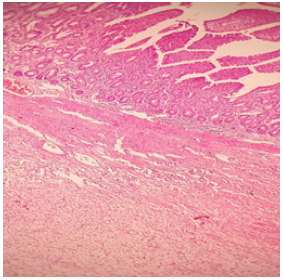
Subsequently, his study was complemented by requesting abdominal tomography. In which a proximal ileal loop was observed with the presence of distal cystic tumor that measures approximately 3.5cm with proximal dilation of this intestinal loop with a hydro-air level, related to intestinal invagination. No data of intestinal obstruction, free fluid, or pneumoperitoneum were observed (Figure 1). He was admitted for surgical treatment with a diagnosis of intestinal invagination. A diagnostic laparoscopy was performed and turned in open laparotomy with the following findings, a firm consistency tumor in the small intestine at the level of the ileum 7cm long and 3.5cm in diameter, occupying the entire lumen of the intestine, with an ileo-ileal invagination segment of approximately 15cm (Figure 2). The tumor was evaluated small intestine segment which measures 7cm long by 3.5cm in diameter with a light brown, smooth, shiny serosa, wall 4cm thick, trabeculated mucosa preserved in the intestinal wall, a nodular mass measuring 4 by 3.5cm, gray, cream attached directly to the wall, clean surgical edges of soft consistency and gelatinous appearance, well defined, not encapsulated with moderate amount of non-foul, non-hot inflammatory fluid (Figure 3). It was decided to perform resection and ileo-ileum anastomosis, the patient evolved satisfactorily and began diet 24 hours post-operative and discharged 4 days later without complications. Histological reported myxoid-like lesion made up of elongated fibroblasts, also with an inflammatory infiltrate polyp accompanied by eosinophils. It grows in the submucosa, pushes and ulcerates the submucosa, with an acute inflammatory reaction. well-defined tumor without cell atypia or mitosis, normal surgical edges (Figure 4).

Figure 1: Proximal ileal loop with the presence of distal cystic tumor that measures approximately 3.5cm with proximal dilation of this intestinal loop with a hydro-air level, related to intestinal invagination.
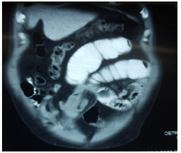

Figure 2: Firm consistency tumor in the ileum 7cm long and 3.5cm in diameter, occupying the entire lumen of the intestine, with an ileo-ileal invagination.
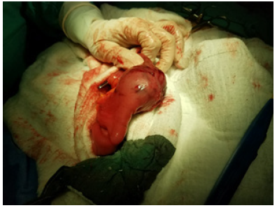

Figure 3: The tumor with a light brown, smooth, shiny serosa, trabeculated mucosa preserved in the intestinal wall, a nodular mass, cream attached directly to the wall, clean surgical edges of soft consistency and gelatinous appearance, well defined, not encapsulated.
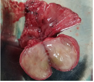

Figure 4: Histological reported myxoid-like lesion made up of elongated fibroblasts, also with an inflammatory infiltrate polyp accompanied by eosinophils.

References

  1. Daum O, Hes O, Vanecek T, Benes Z, Sima R, et al. (2005) Vanek’s Tumor (Inflammatory Fibroid Polyp). Report of 18 cases and comparison with three cases of original Vanek’s series. Annals of Diagnostic Pathology 7(6): 337-347.
  2. Akbulut S, Mahsuni M, Cakabay Bahri, Sule Bakir, Ayhan Senol (2009) Giant inflammatory fibroid polyp of ileum causing intussusception: a case report. Cases J 2: 8616.
  3. Fazzio C, Madeira E (2015) Inflammatory fibroid polyp (Vanek’s polyp): a case report and literature review. J Bras Patol Med Lab 51(2): 117-120.
  4. Bays D, Anagnostopoulos G, Katsaounos E, Filis P, Missas (2004) Inflammatory fibroid polyp of the small intestine causing intussusception: A report of two cases. Dig Dis Sci 49(10): 1677-1680.
  5. Jukic Z, Ferencic Z, Radulovic P, Mijic A (2014) Estrogen and androgen receptors in inflammatory fibroid polyp (Vanek’s Tumor): Case report. Anticancer Res 34(12): 7203-7206.
  6. Lee C, Yuen MK (2014) Inflammatory fibroid polyps causing intussusception in adult patients: Two case reports and review of literature focusing on radiological features. Hong Kong J Radiol 17: 271-276.
  7. Rossi P, Montuori M, Balassone V, Ricciardi E, Anemona L, et al. (2012) Inflammatory fibroid polyp. A case report and review of the literature. Ann Ital Chir 83(4): 347-351.
  8. Ahtil R, Bensghir M, Meziane M, Houba A, Jaafari A, et al. (2017) Rare etiology of subocclusive syndrome: inflammatory fibroid polyp of the ileum, about a clinical case. Pan Afr Med J 26: 146.
  9. Madriz DeHaan P, Martínez Y, Gutiérrez F (2014) Intestinal intussusception due to an inflammatory fibroid polyp (Vanek's tumor). Legal Medicine of Costa Rica-Virtual Edition 31(1): 1409-1415.
  10. Paikos D, Moschos J, Tzilves D, Koulaouzidis A, Kouklakis G, et al. (2007) Inflammatory fibroid polyp or Vanek’s tumour. Dig Surg 24: 231-233.
  11. Yakan S, Calıskan C, Makay O, Denecli AG, Korkut MA (2009) Intussusception in adults: Clinical characteristics, diagnosis and operative strategies. World J Gastroenterol 15(16): 1985-1989.
  12. Gara N, Falzarano J, Limm W, Namiki T (2009) Ileal inflammatory fibroid polyp causing chronic ileocolic intussusception and mimicking cecal carcinoma. World J Gastrointest Oncol 1(1): 89-92.
  13. Bassam Abboud (2015) Vanek’s tumor of the small bowel in adults. World J Gastroenterol 21(16): 4802-4808.
  14. Zakaria A, Daradkeh S (2012) Jejunojejunal intussusception induced by a gastrointestinal stromal tumor. Case Rep Surg 2012: 173-680.
For more articles in journal of Surgical Medicine
Please click on below link:  https://crimsonpublishers.com/smoaj/

Monday, May 30, 2022

Candida Tropicalis Infection of the Knee Joint in an Immunocompromised Pediatric Patient: A Case Report_Crimson Publishers

Candida Tropicalis Infection of the Knee Joint in an Immunocompromised Pediatric Patient: A Case Report by Benjamin Sookhoo in Surgical Medicine Open Access Journal_journal of Surgical Medicine


Abstract

Background: Candida septic arthritis is a debilitating condition affecting joint function. Candida tropicalis, an organism found in normal human flora is noted to be the third most common pathogenic yeast in the elderly and immunosuppressed population. Infections are rare in the US, typically limited to the neonate and elderly populations. Most infections occur in the south America and southeast Asian regions. In the last 2 decades, Tropialis infection rates have risen impart due to antifungal resistance. We present a case of Candida tropicalis infection in the knee of a 13-year-old female with a past history of relapsed Acute Myeloid Leukemia following bone marrow transplant, pancytopenia and graft vs. host disease.

Methods: Patient case is thoroughly discussed, and a review of the current literature performed.

Result: Patient underwent multiple open irrigation and sharp debridement as well as arthroscopic complete synovectomy in addition to medical management. Patient remained pancytopenic throughout her course and eventually underwent granulocyte transfusion. During her course, she developed a relapse of her AML and subsequently expired. Limited case reports exist in the literature, with no recent reports of Candida arthritis in an adolescent in the United States.

Discussion: Candida septic arthritis remains a rare but potentially devastating condition, particularly in the immunocompromised patient population. Candida arthritis should be high on the treating physician’s suspicion in immunocompromised patients presenting with new onset joint pain. Aggressive medical and surgical management is recommended, and current literature has shown a high cure rate with early treatment.

Introduction

Septic arthritis due to Candida species is a relatively rare infection typically seen in patients with immunocompromising conditions. While C. albicans is the most commonly encountered fungal species affecting humans, C. tropicalis has demonstrated increasing resistance to anti-fungal medications leading to persistent and difficult to control infections [1-15]. C. tropicalis is part of the normal human microbiota and is typically found on the skin and mucosal surfaces of the body. It is the third most common Non-Candida albicans Candida species (NCAC) isolated in clinical practice and primarily found in tropical regions, particularly Asia and South America [1,8]. Few reports of Tropicalis septic arthritis in North America have been published. We present a case report of a confirmed monoarticular C. tropicalis septic arthritis in a pediatric patient with chemotherapy induced granulocytopenia the setting of relapsed acute myeloid leukemia that lead to systemic candidiasis and patient mortality.

Case Report

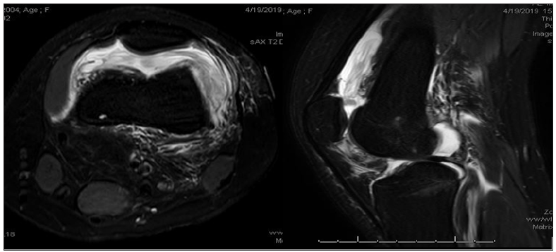
Our patient is a 13-year-old Caucasian female who presented to our institution with relapsed AML following bone marrow transplant with clinical signs and symptoms concerning for graft vs. host disease. She had undergone previous chemotherapy treatment a little over a month prior to her admission and had an indwelling catheter. The patient was scheduled for intrathecal chemotherapy administration and was undergoing preoperative evaluation when it was noticed she had a diffuse rash and endorsing febrile episodes as well as diffuse joint pain. Pre-operative labs showed of white count of 0.1 cells/mm3 as well as elevated inflammatory markers. She was admitted to our Children’s hospital for further work up and evaluation. 2 days into her hospital stay, patient awoke in the middle of the night new onset severe left knee pain and swelling with inability to bear weight to the extremity. An MRI of the knee (Figure 1), showed a moderate effusion to the joint. An arthrocentesis was performed and synovial fluid culture demonstrated fungal growth that was identified as C. tropicalis.

Figure 1: MRI of the knee showing moderate effusion.


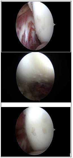
Figure 2: TArthroscopic views of the knee showing plaque formation on the femoral condyles.


Patient was started on IV Micafungin and was taken to the operating room where a formal irrigation and debridement was performed. A large amount of dark, viscous and serosanguinous fluid was encountered and evacuated from the joint along with a large of necrotic looking synovial tissue in the lateral gutter of the knee joint. Following initial I&D, patient continued to have febrile episodes and rapidly progressive skin rash. Initial medical work up including echocardiogram and CT scans of the abdomen and pelvis showed no evidence of vegetations or hepato-splenic candidiasis. Over the next 2 weeks, our patient subsequently underwent 3 additional open knee irrigations with sharp debridement of the synovium due to failure of response, despite adjustments in anti-fungal medications, including addition of Amphotericin B. Patient subsequently underwent a complete an arthroscopic synovectomy to better reduce her fungal load. An abundance of white plaques was noted on her femoral condyles that were determined to be yeast colonies on culture, (Figure 2).

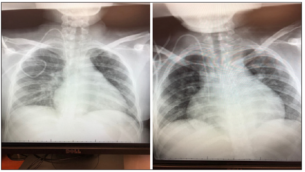
In the two weeks following her final surgery, patient continue to have significant pain and swelling to the left knee, however, she remained afebrile. Her overall clinical picture soon began to decline, and repeat echocardiogram demonstrated a pericardial effusion with loculations that was not present on initial examination. Chest radiographs taken 2 weeks apart, (Figure 3), show enlarging of the cardiac silhouette. Her renal function subsequently began to decline as well, secondary to the Amphotericin B. Amphotericin B was discontinued and she was started on Voriconazole. Her oncology and pediatric medical teams decided to initiate a granulocyte transfusion to due to her continued pancytopenia and poor response to medical and surgical treatment of her infection. Her granulocyte transfusion was not well tolerated, and the patient developed respiratory distress in addition to worsening kidney function. CBC following her granulocyte transfusion showed peripheral blasts cells that were confirmed by flow cytometry to be relapsed AML. After a family meeting with the medical teams, a decision was made to place the patient on hospice and comfort measures. She was discharged from the hospital and expired at home the following day.

Figure 3: Chest radiographs obtained 2 weeks apart demonstrating enlarged cardiac silhouette.


Discussion

Septic arthrosis with Candida tropicalis infection is a rare infection in the United States. Patients at risk for developing this infection include those with immunocompromising conditions including malignancy, HIV/AIDS, neonates, patients with prolonged ICU stays with or without mechanical ventilation, those with indwelling central venous catheters, prolonged broad-spectrum antibiotic use, direct inoculation or previous surgery [1-2]. Systemic infections with tropicalis are common in Asian and South American countries, where various studies have shown to be the third most commonly isolated species in patients with systemic candidemia. C. tropicalis produces more persistent infections than C. albicans leading to longer hospital stays [3-5,9]. Recent studies have also shown that C. tropicalis is becoming increasingly resistant to antifungals as a result of C. tropicalis’ various virulent factors, including biofilm formation, adhesion molecules, cell wall hydrophobicity as well as extracellular proteases and phospholipases [6]. It has been shown to have a higher mortality rate when compared to all other non-Candida Albicans candidemia (NCAC) species with mortality rates surpassing C. albicans [1,8].

Septic joint arthrosis and osteomyelitis caused by Candida species are typically seeded by hematogenous dissemination, direct inoculation or direct extension from nearby focus of infection. Knee joints are the most common site of infection, owning to the highly vascular synovium, which lacks a limiting basement membrane allowing for easy passage for joint inoculation [10]. In pediatric patients, open epiphyseal plates allow for hematogenously disseminated organisms to extend into the joint as well as the metaphysis, setting the stage for osteomyelitis [1,7]. Candida arthritis presents with a clinical picture similar to bacterial septic arthritis, however, fever may not be a presenting symptom and inflammatory markers may only be moderately elevated, particularly in immunocompromised hosts. The most common presenting symptoms include joint pain and effusion with limitations to range of motion and weightbearing secondary to pain. Surgical findings include thickened fibrotic synovium, cartilage damage and purulence [9,10].

The current literature regarding septic arthritis with C. tropicalis in pediatric populations is sparse with the vast majority of case reports emerge from outside of the United States [11-14]. Gamaletsou et al. [1] performed a systematic review of the literature of 112 cases of adult and pediatric with confirmed diagnosis of Candida arthritis. They found that C. tropicalis infection occurred in 14% of the cases identified and was the second most common recovered species behind C. albicans. 36% of the patients identified in their study pediatric, with the vast majority being neonates. 78% of the patients were cleared of the infection with either medical therapy alone or in conjunction with surgical intervention. However, while there was no significant difference between patients treated with medical therapy alone or combine with surgery, there were far fewer deaths with the latter. McCullers and Flynn reported on a case of tropicalis arthritis and osteomyelitis in a 5-year-old male following chemotherapy treatment for acute lymphocytic leukemia. This infection was successfully treated with a prolonged course of amphotericin B and rifampin. Their review of the literature found 11 additional cases involving adult and pediatric patients, with 3 originating from their institution. They reported a 11.2% risk of invasive disease among immunocompromised patients with culture positive C. tropicalis compared to 2% risk in those colonized with C. albicans.

The Infectious Disease Society of America set forth clinical practice guidelines for the treatment of Candida septic arthritis. Management of native joint septic candidiasis that received strong recommendation included fluconazole for a minimum of 6 weeks or an echinocandin such as caspofungin, micafungin or anidulafungin for 2 weeks followed by a minimum of 4 weeks of fluconazole. Surgical drainage was indicated for all cases of septic arthritis. The use of intra-articular amphotericin B has been reported, however, this modality remains controversial.

Conclusion

We present a case of monoarticular C. tropicalis arthritis in an adolescent female with relapsed acute myeloid leukemia that was unsuccessfully treated despite aggressive medical and surgical intervention. There are few reports in the literature of septic joint candidiasis caused by C. tropicalis in the pediatric population, and to our knowledge, none have been reported in North America involving the adolescent age group that resulted in patient death. Most cases reported in the literature have shown C. tropicalis septic arthritis and osteomyelitis to be successfully treated with prolonged administration of anti-fungal agents and surgical management. While this condition presents with a clinical picture similar to bacterial arthritis, the clinician should have a high suspicion for Candida arthritis in patients with immunocompromising conditions and with indwelling catheters with new onset joint pain and effusion to reduce patient morbidity and mortality.

References

  1. Gamaletsou MN, Rammaert B, Bueno MA, Sipsas NV, Moriyama B (2015) Candida arthritis: Analysis of 112 pediatric and adult cases. Open Forum of Infectious Disease 3(1): ofv207.
  2. Zuza Alves DL, Silva Rocha WP, Chaves GM (2017) An update on Candida tropicalis based on basic and clinical approaches. Front Microbiol 13(8): 1927.
  3. Kontoyiannis DP, Vaziri I, Hanna HA, Boktour M, Thornby J (2001) Risk factors for Candida tropicalis fungemia in patients with cancer. Clin Infect Dis 33(10): 1676-1681.
  4. Pappas PG, Kauffman CA, David RA, Cornelius JC, Kieren AM (2016) Clinical practice guideline for the management of candidiasis: 2016 update by the infectious disease’s society of America. Clinical Infectious Diseases (62)4: e1-e50.
  5. Sónia S, Negri M, Henriques M, Oliveira R, Williams DW (2012) Candida glabrata, Candida parapsilosis and Candida tropicalis: Biology, epidemiology, pathogenicity and antifungal resistance. FEMS Microbiology Reviews 36(2): 288-305.
  6. JA McCullers, Flynn PM (1998) Candida tropicalis osteomyelitis: Case report and review. Clinical Infectious Diseases 26(4): 1000-1001.
  7. Wang HP, Yen YF, Chen WS, Chou YL, Tsai CY (2007) An unusual case of Candida tropicalis and Candida krusei arthritis in a patient with acute myelogenous leukemia before chemotherapy. Clin Rheumatol 26(7): 1195-1197.
  8. Fanning S, Mitchell AP (2012) Fungal biofilms. PLoS Pathog 8(4): e1002585.
  9. McCarty TP, Pappas PG (2016) Invasive candidiasis. Infect Dis Clin North Am 30(1): 103-124.
  10. Krcmery V Jr, Mrazova M, Kunova A, Grey E, Mardiak J (1999) Nosocomial candidaemias due to species other than Candida albicans in cancer patients. Aetiology, risk factors, and outcome of 45 episodes within 10 years in a single cancer institution. Support Care Cancer 7(6): 428-431.
  11. Bariteau JT, Warvasz GR, McDonnell M, Fischer SA, Hayda RA (2014) Fungal osteomylietis and septic arthritis. J Am Acad Ortho Surg 22(6): 390-401.
  12. Hu XR, He JS, Ye XJ, Zheng WY, Wu WJ (2008) Candida tropicalis arthritis in a patient with acute leukemia. Zhongguo Shi Yan Xue Ye Xue Za Zhi 16(5): 1215-1218.
  13. Sim JP, Kho BC, Liu HS, Yung R, Chan JC (2005) Candida tropicalis arthritis of the knee in a patient with acute lymphoblastic leukemia: Successfully treatment with caspofungin. Hong Kong Med J 11(2): 120-123.
  14. Vicari P, Feitosa PR, Chauffaille ML, Yamamoto M, Figueiredo MS (2003) Septic arthritis as the first sign of candida tropicalis fungaemia in an acute lymphoid leukemia patient. Braz J Infect Dis 7(6): 426-428.
  15. Weisse ME, Person DA, Berkenbaugh JT (1993) Treatment of candida arthritis with flucytosine and amphotericin B. J Perinatol 13(5): 402-404.

Publishers: https://crimsonpublishers.com/

For more articles in journal of Surgical Medicine
Please click on below link: https://crimsonpublishers.com/smoaj/

Monday, February 1, 2021

Same Story with Different Endings in HER2- Positive Breast Cancer: Why the Benefit of Pertuzumab is Robust in the Metastatic Scenario and Modest in the Adjuvant Setting_ Crimson Publishers

 Same Story with Different Endings in HER2- Positive Breast Cancer: Why the Benefit of Pertuzumab is Robust in the Metastatic Scenario and Modest in the Adjuvant Setting? by Jayesh Sagar* in Surgical Medicine Open Access Journal_ Journal of Surgical Medicine


Abstract
The addition of pertuzumab to chemotherapy and trastuzumab yielded an impressive improvement in the outcomes of metastatic HER2-positive breast cancer patients [1]. Intriguingly, the same magnitude of benefit could not be reproduced with pertuzumab in the adjuvant setting, being the reasons for this discrepancy unknown [2,3]. In this manuscript, we discuss clinical and biological differences between metastatic and early-stage HER2-positive breast cancer, and conclude by proposing potential explanations for the distinct magnitudes of benefit of pertuzumab in different disease settings.

Magnitude of Risk Reduction
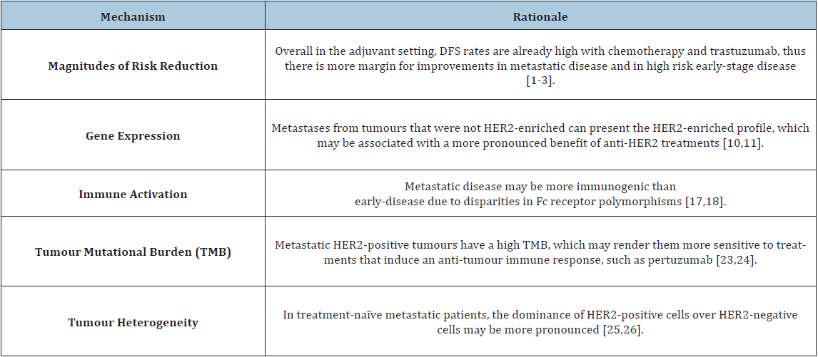
When evaluating a new treatment in the context of a clinical trial, events occurring in experimental and control arms are compared [4]. Early-stage HER2-positive breast cancer patients treated with adjuvant chemotherapy and trastuzumab had a 87.8% recurrencefree survival rate at 6 years as per the recently updated results of the APHINITY trial [3]. In the metastatic setting, however, the perspective is different: only 20% of patients receiving chemotherapy and trastuzumab remain alive and progression-free at 3 years [1]. Therefore, events are more frequent in the metastatic setting than in early-disease. In other words, there is more room for improvement in metastatic disease, whereas in the adjuvant setting chemotherapy and trastuzumab already yield high Disease-Free Survival (DFS) rates.

Illustrating this hypothesis, the addition of pertuzumab to trastuzumab and chemotherapy in the metastatic setting yields a 32% relative reduction in the risk of progression, which translates into an 8.2% absolute increase in Progression-Free Survival (PFS) at 3 years, whereas in the adjuvant setting pertuzumab yields a 24% relative reduction in the risk of recurrence at 6 years, translating into a modest 2.8% absolute improvement in invasive DFS (iDFS) [1-3]. When considering only node-positive patients (who present a higher risk of recurrence), the benefit of adjuvant pertuzumab becomes more pronounced (28% relative reduction in recurrence risk yielding a 4.5% absolute 6-year iDFS improvement).

https://crimsonpublishers.com/smoaj/fulltext/SMOAJ.000560.php

Crimson Publishers: https://crimsonpublishers.com/

For more articles in Journal of Surgical Medicine,
Please click on below link: https://crimsonpublishers.com/smoaj/

Thursday, April 4, 2019

Perforated Appendicitis Preoperatively complicated by Multiple Intra-Abdominal Abscesses | Crimson Publishers

Perforated Appendicitis Preoperatively complicated by Multiple Intra-Abdominal Abscesses by Tarik Zahouani in Surgical Medicine Open Access Journal

Acute appendicitis is the most common surgical emergency in the pediatric population [1]. Complications are seen in 30 to 40% of cases, and include perforated, gangrenous, intra-abdominal abscess and peritonitis [1]. The rate of perforated appendicitis is higher in children compared to adults and varies from 30% to 74% [2]. We report a case of perforated appendicitis preoperatively complicated by multiple intra-abdominal abscesses.


For more articles in Surgical Medicine Open Access Journal, follow the URL: https://crimsonpublishers.com/smoaj/index.php
For more articles in Crimson Publishers, follow the URL: https://crimsonpublishers.com/

Visual-cognitive Skills and Physical Qualities in Elite Soccer: Practical Considerations for Training and Return-to-Play Protocols: Crimson Publishers

Visual-cognitive Skills and Physical Qualities in Elite Soccer: Practical Considerations for Training and Return-to-Play Protocols by Lukasz...