Crimson Publishers High Impact Journals

Thursday, March 31, 2022

Biomedical Waste Management in A Multispecialty Hospital_Crimson Publishers

 Biomedical Waste Management in A Multispecialty Hospital by Vivek Kumar Garg in COJ Reviews & Research_Journal of Pharmaceutical Sciences Review and Research


Abstract

Any waste which is generated during the diagnosis, treatment, or immunization of human beings or animals or in research activities pertaining thereto or in the production or testing of biologicals, is called biomedical waste. The current study is based on BMW in multispecialty hospital at civil hospital, Barnala, India. The material used in the papers regarding BMW management is the Questionnaire, prepared with different topics. Our objective in the study was to evaluate the good points as well as the lacunas in the protocols adopted in the hospital waste management and its disposal, in the multispecialty civil hospital. In the present study, a list of departments of civil hospital, Barnala was made. There are following departments like General Medicine, General Surgery, Gynecology, Pediatrics, Ophthalmology, ENT, Dental, Skin, Orthopedics, Blood bank, OT, Wards, Indoor etc. The results obtained showed lack of awareness of bio medical waste management and its handling even in qualified medical staff. The reason being its complex is system and rules that has not become part of daily curriculum till now. We have studied one month regular protocol followed for bio medical waste collection by the hospital staff. During the study, we have observed that the hospital is properly managing their biomedical waste. Regularly, the hospital segregates the waste according to the specified categories and color coding. Regarding the capabilities and risks of biomedical waste treatment alternatives, it must be emphasized that the only treatment technologies that are usually used to treat pathological waste are the incineration and mechanical/ chemical disinfection systems.

Keywords: Biomedical waste; Healthcare; Multispecialty; Hospitals

Introduction

Any waste which is generated during the diagnosis, treatment, or immunization of human beings or animals or in research activities pertaining thereto or in the production or testing of biologicals, is called biomedical waste [1]. Medical care is indispensable for our life. But the waste generated during medical care needs attention. The nosocomial (hospital acquired) infections are the result of hospital waste. The hazardous and toxic parts of waste from healthcare establishments comprising infectious, biomedical and radioactive material as well as sharps needles, constitute a grave risk. The rag pickers and waste workers are often worst affected. Diseases like cholera, typhoid, plague, tuberculosis, hepatitis, AIDS, Diphtheria, Malaria, etc. pose grave public health risks [2-4]. With a judicious planning and management, the risk of spread of disease can be considerably reduced. The rules framed by the Ministry of Environment and Forests (MoEF), Govt. of India, known as Bio Medical Waste Management and Handling Rules, 1998, provides uniform guidelines and code of practice for the whole nation [5,6]. In Schedule I of the Bio Medical Waste Management rules 1998 (Annexure II), waste has been categorized into 10 points [2,4,7]. There are total 6 schedules in BMW management.

Schedule I: (Table 1)

Table 1: Schedule I.
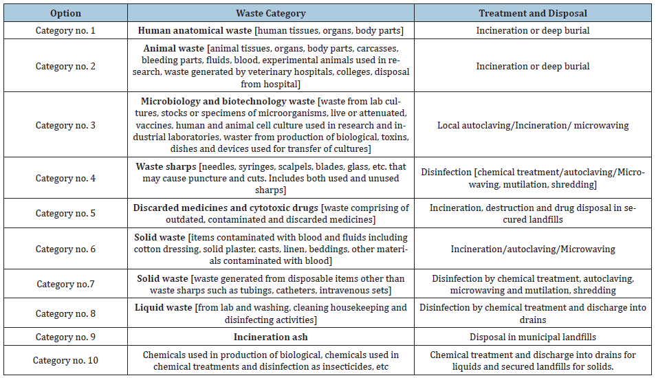

Schedule II: (Table 1)

Table 2: Schedule II: Colour coding charts and type of container for disposal of BMW is as follows.


Schedule III: Labels for BMW containers/Bags

Schedule IV: Label for transport of BMW containers/Bags

Schedule V: Standards for treatment and disposal of BMW standards for incinerators.

Schedule VI: Schedule for waste treatment facilities like Incinerator/Autoclave/Microwave system.

Materials and Methods

The current study is based on BMW in multispecialty hospital at civil hospital, Barnala, India. The material used in the papers regarding BMW management is the Questionnaire, prepared with different topics.

Methods- The questionnaire was prepared by keeping following points in mind.

  1. Awareness of the importance of BMW management
  2. What is BMW
  3. Types of BMW
  4. BMW management at different levels- means at small clinics to big hospitals to medical and dental colleges.
  5. Classification of BMW
  6. Isolation of BMW
  7. Packaging of BMW and its disposal practices

Selection criteria to distribute questionnaire- The first step is preparation of questionnaire, study sample selected, organized and finalized. The second stage included distribution of questionnaire, analyzing the collected data and determining the response. Selection criteria for the persons are determined- The person must be part of healthcare, graduate or undergraduate, knowledge of English language, medical or paramedical staff, male or female, small clinics, primary health centers, polyclinics, multispecialty hospitals, government civil hospitals.

Aims and Objectives

To evaluate the steps taken in the management of hospital generated bio medical waste. Our objective in the study was to evaluate the good points as well as the lacunas in the protocols adopted in the hospital waste management and its disposal, in the multispecialty civil hospital. In the present study, a list of departments of civil hospital, Barnala was made. There are following departments like General Medicine, General Surgery, Gynecology, Pediatrics, Ophthalmology, ENT, Dental, Skin, Orthopedics, Blood bank, OT, Wards, Indoor etc. It took one month to record bio medical waste generation of all departments separately for Red, Yellow, Blue and White containers/bags. Then we added the waste for Red of all departments into one head, Yellow of all departments into one head, Blue of all departments into one head and White of all departments into one head. This all was infected waste. The non-infected waste e.g. expired medicines were put in Black colored container. But we did not include non-infected waste in our study. Since, our topic is focused on the protocols, that means we have to keep eye on the proper segregation of the waste before putting into their respective color-coded containers in their respective departments.

Result

Red Bag- Solid waste, POP casts, infected cotton & dressing, infected bandages, items containing blood & body fluid like extracted teeth, cysts, granulomas

Yellow Bag- Anatomical waste Microbiology & Biotechnology waste. Wastes from lab cultures or specimen of microbes live or attenuated vaccines, human and animal cell cultures, waste from production of biological, toxins, dishes & devices.

Blue Bag- Disposable plastic glasses, Plastic Tubings, IV sets, syringes, Gloves, Solid waste other than sharp needles/blades.

White Container- Sharp needles/objects that may cause cuts, Needles, Blades, expired injections, cut glasses.

Black Container- All above mentioned color bags contain Infected Waste. But Black color bag contains Non-Infected waste. e.g. cytotoxic drugs, chemical waste, expired drugs tablets & capsule form.

We studied one-month 1st November 2017 to 30th November 2017, BMW collection and disposal and its Protocols followed in the Civil Hospital, Barnala (Table 3) (Figure 1-5).

Table 3: BMW November 2017, civil hospitaal, Barnala.
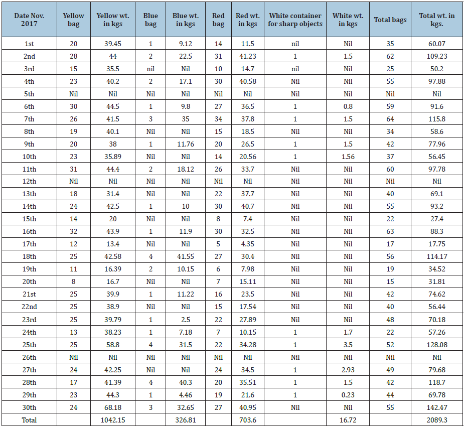

Figure 1: Comparison of average yellow bag waste week wise for the month of November 2017.
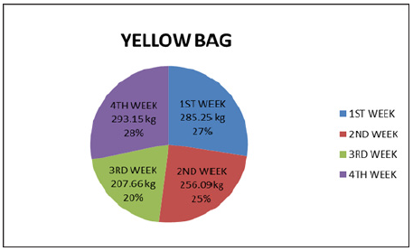

Figure 2: Comparison of average red bag waste week wise for the month of November 2017.
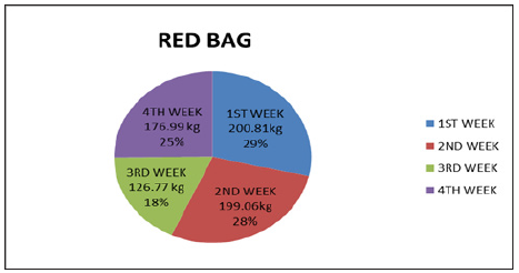

Figure 3: Comparison of average blue bag waste week wise for the month of November 2017.


Figure 4: Comparison of average white bag waste week wise for the month of November 2017.
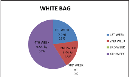

Figure 5: BMW report of November 2017 civil hospital, Barnala.
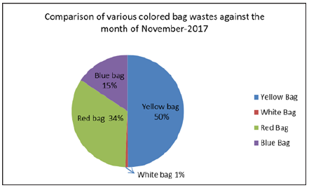

BMW report of November 2017 civil hospital, Barnala

Yellow container - 1042.15kg, Blue container -- 323.57kg

Red container – 696kg, White bag - 13.12kg, Total waste --2066.64kg

Total deliveries in the Gynae Deptt. --495 in number

Human Anatomical waste from Gynae deliveries inkgs - approx 0.5kg per delivery.

Total deliveries=495

So Total Human anatomical waste from gynae depth was 495 x 0.5=247.500kg

Out of 1034.95kg of waste of yellow container, 1042.15-247.500=794.65kg

Means Yellow container contains 247.500kg Human anatomic waste and 794.65kg is other solid waste.

Liquid waste generated after washing instruments and lab containers, test tubes was 300 liters in one-month November 2017.

Discussion of Diagrams

Yellow bag diagram with human anatomical waste with placentas shows

285.25kg and 27% 1st week of November 2017

256.09kg and 25% 2nd week of November 2017

207.66kg and 20% 3rd week of November 2017

293.15kg and 28% 4th week of November 2017

Blue bag diagram with infected plastics, syringes, gloves, plastic tubing’s shows

93.52kg and 29% 1st week of November 2017

51.78kg and 16% 2nd week of November 2017

65.42kg and 20% 3rd week of November 2017

116.09kg and 35% 4th week of November 2017

Red bag diagram with soiled waste, infected dressings, POP casts shows

200.81kg and 29% 1st week of November 2017

199.06kg and 28% 2nd week of November 2017

126.77kg and 18% 3rd week of November 2017

176.99kg and 25% 4th week of November 2017

White bag diagram with sharp needles and cut glasses shows

3.8kg and 23% 1st week of November 2017

3.06kg and 2nd week of November 2017

Nil 3rd week of November 2017

9.86kg and 59% 4th week of November 2017

The idea, to show one-month study and further breaking it into 4 parts (weeks), is to show regularity in the collection of biomedical waste in the hospital.

Treatment and Disposal of BMW

The agency responsible for picking and carrying the biomedical waste in our area is Medicare Environmental Management Pvt. Ltd., Ludhiana. This agency is responsible for treatment and disposal of biomedical waste generated from hospitals. During the study, it has been noted that the hospital is properly following all the protocols in the management of its biomedical waste. The hospital staff segregates the biomedical waste regularly according to the specified categories and color coding. The hospital staff puts sodium hypochlorite 5% into the white container with sharp waste to disinfect the waste. All other color coded Yellow, Red and Blue are handed over to the agency as such. These instructions are being given by the agency to the hospital.

Waste management problem is a “Serious Public Health Threat” , Niti Aayog’s Draft has prepared a 3 year action agenda of April 2017 , plans to set up 100 waste to energy WTE plants to deal with 170000 tons per day of municipal solid waste for some 7935 urban centers. The Action Agenda states that “Incineration or waste to energy is the best option “Even as Annexure A of the Kyoto Protocol marks out waste incineration as a source of greenhouse gases. This protocol remains relevant because the Paris Agreement on climate crises will come into operation only from 2021.

As to implications for public health, a WHO’s publication of 2016 reported an association between birth outcomes (Preterm birth and spontaneous abortion), chronic or acute respiratory effects in children or adults in relation to increased level of exposure to incinerators. It echoes the findings of a judgment of the Delhi High Court which observed, “Residents living within 10 kms of an incinerator, refinery and waste disposal site “Showed” significant increase in laryngeal cancer in men living with closer proximity to the incinerator and other pollution sources.

Results in the Studies

The results obtained showed lack of awareness of bio medical waste management and its handling even in qualified medical staff. The reason being its complex is system and rules that has not become part of daily curriculum till now.

Conclusion

We have studied one-month regular protocol followed for bio medical waste collection by the hospital staff. During the study, we have observed that the hospital is properly managing their biomedical waste. Regularly, the hospital segregates the waste according to the specified categories and color coding. Regarding the capabilities and risks of biomedical waste treatment alternatives, it must be emphasized that the only treatment technologies that are usually used to treat pathological waste are the incineration and mechanical/chemical disinfection systems.

Recommendations

Some suggestions are recommended to the hospital staff, which is well taken and appreciated. The key challenges need to be resolved for biomedical waste management is as under

  • Enhanced awareness about hazardous waste
  • Improving the segregation of medical waste
  • Identifying appropriate treatment alternatives.

One of the most critical issues regarding biomedical waste management is selecting an appropriate treatment. The availability of permitted landfill space and the demographic and geographic factors need to be considered when selecting the most appropriate management strategy. Safety, reliability and cost of alternative treatment methods also affect selection of treatment alternatives.

Regarding the public and environmental issues, a correct Health Care Waste Management (HCWM) will avoid the negative long-term health effects viz., releasing the toxic substances such as dioxin, mercury and others in the environment.

References

  1. Datta P, Mohi GK, Chander J (2018) Biomedical waste management in India: Critical appraisal. J Lab Physicians 10(1): 6-14.
  2. Yadav M (2001) Hospital waste - A major problem. JK Practitioner 8(4): 276-282.
  3. Sharma S (2010) Awareness about bio-medical waste management among health care personnel of some important medical centers in Agra. Int J Environ Sci Dev 1(3): 251-255.
  4. Bhawan P, Nagar A (2016) Revised Guidelines for Common Bio-medical Waste Treatment Facilities i Revised Guidelines for Common Bio-medical Waste Treatment and Disposal Facilities Central Pollution Control Board (Ministry of Environment, Forest and Climate Change), India.
  5. Radha K, Kalaivani K, Lavanya R (2009) A Case Study of biomedical waste management in hospitals. Glob J Health Sci 1(1): 82-88.
  6. Hospitals P (2015) Study of Biomedical Waste Generation and Management by Government and Journal of pharmaceutical and biomedical sciences Study of Biomedical Waste Generation and Management by Government and Private.
  7. Mattoo K, Singh V, Garg R (2014) Are dental training programs heading towards ecological disaster - results from a survey. J Atmos Pollut 2(1): 17-21.

For more articles in Journal of Pharmaceutical Sciences Review and Research
Please click on below link: https://crimsonpublishers.com/cojrr/

Wednesday, March 30, 2022

Photocatalytic Activity of Methylene Blue Using Zinc Nanoparticles Synthesized from Gymnema sylvestre And Antimicrobial Assay_Crimson Publishers

 Photocatalytic Activity of Methylene Blue Using Zinc Nanoparticles Synthesized from Gymnema sylvestre And Antimicrobial Assay by E David in Advancements in Bioequivalence & Bioavailability_Bioequivalence And Bioavailability International Journal


Abstract

Nanoparticles of the Zn were synthesized by Gymnema sylvestre leaf extract characterized by UV (UV-visible spectrum), FT-IR (Fourier transformance infra-red spectroscopy), X-ray diffraction (XRD), DLS (Dynamic light scattering analysis) and scanning electron microscopy (SEM). The XRD patterns of the Zn and nanoparticles could be indexed to hexagonal and Wurtzite phase, respectively. Aggregated nanoparticles of Zn with spherical-like shapes were observed with particle diameter in the range of 200-400μm. These nanoparticles were used for photocatalytic degradation of methylene blue dye under solar light irradiation. The ZnNPs shown very good antimicrobial Assay towards microbes isolated from contaminated dye soil..

Keywords: Gymnema sylvestre; ZnNPs; photocatalytic activity; Dye degradation; Antimicrobial Assay

Abbreviations: UV: UV- Visible Spectrum; FT-IR: Fourier Transformance Infra-Red Spectroscopy; XRD: X-Ray Diffraction; DLS: Dynamic Light Scattering Analysis; SEM: Scanning Electron Microscopy

Introduction

In recent years, the contamination of surface and ground water has increased due to population growth. The main sources of environmental contamination are organic dyes used in the food and textile industries due to their high toxicity and their nonbiodegradability, which have carcinogenic effects on humans. MB dye is used by different industries, for example, as a dye in silk; as a food coloring additive; and as a dye in wool, leather, cotton, jute, and paper [1]. Methylene blue dyes have strong effects on the immune and reproductive systems and exhibit potential carcinogenic and genotoxic effects. Thus, these hazardous dyes must be removed from industrial effluents. Many methods, such as biological treatment, adsorption, and photo catalysis [2], have been used for removal of these dyes from industrial effluents. The use of photo catalysts to degrade organic compounds in contaminated air or water or to convert them into harmless chemicals has been extensively studied to decrease the damage caused by organic dye pollution to the environment and humans [3]. Therefore, heterogeneous photocatalysis is an interesting area of research because the method allows for complete mineralization of these environmentally hazardous dyes Zinc Oxide (Zn) belongs to a category of n-type semiconductors having wide energy band gap of 3.37eV. Recently, Zn has attracted much attention as a promising photocatalytic material for removal of organic pollutants, which present in wastewater, all because of its high catalytic activity, moderate preparation cost and environmentally benign nature [4]. Nanoscale Zn particles possess significant surface area and large number of active sites ensure increased surface catalyzed reaction rates thus promoting photocatalysis [5]. However, the large energy band gap of Zn permits electronic excitations only with photons having energy below 400nm (UV spectrum). High degree of recombination of photogenerated species is another limitation associated with Zn which is responsible for low photocatalytic activity [6,7]. These shortfalls can be remediated by modifying Zn such as to extend its absorption threshold to the visible spectrum and limiting the rate of electron/hole pair recombination. Different attempts were achieved recently to improve the activity of Zn photocatalyst.

Nanotechnology deals with the synthesis of nanoparticles with controlled size, shape, and dispersity of materials at the nanometer scale length and their potential use for human wellbeing. Nanometer sized materials have a high surface area; and a high fraction of surface atoms have been studied because of their exclusive properties such as optic, electronic, and catalytic Among all nanoparticles noble metal nanoparticles have enormous applications in diverse areas such as bioimaging, sensor, diagnosis, and novel therapeutic in biomedical field. Metallic silver and silver nanoparticles were recently applied as antimicrobial agents in various products such as cosmetics, animal feed coating of catheters, wound dressing and water purification with a minimal risk of toxicity in humans. Now a days the biological systems were eagerly used for nanoscale material synthesis and assembly is an alternative method of physical and chemical process. Green approach of nanoparticles synthesis by biological entities has been gaining great advantages which are environmental benign, less toxic, and time consuming and, it is a single step process. Currently, plant and plant derived materials are used for nanoparticles synthesis which is more compatible than the microbe-mediated nanoparticles synthesis process because they eliminate the culture maintenance and are easy to handle. Nanoparticles synthesis by medicinal plants shows more benefit; they may enhance the antibacterial activity of silver nanoparticles, because the medicinally valuable active biomolecule present in the plants may bind on the surface of the nanoparticles and reduce the silver ions to silver nanoparticles [8]. Nanoparticles can be organized into four types namely carbon based, metal based, dendrimers and composites nanoparticles are also referred to as organic nanoparticles. Spherical and ellipsoidal carbon nanoparticles are called as fullerenes, while cylindrical ones are called nanotubes. Quantum dots, nanogold, Nano silver and metal oxides (titanium oxide) are examples of metal-based nanoparticles. Dendrimers are Nano sized polymers which can be synthesized to perform specific chemical functions. Composites are c nanoparticles with other nanoparticles or with larger sized and heavier compounds. Silver nanoparticles are most commonly used in water filters and biosensors [9,10].

Gymnema sylvestre is a perennial woody vine that grows in tropical areas of India, Africa, and Australia and has been used for medicinal purposes in Ayurvedic medicine. Common names include gymnema, Australian cow plant, and Periploca of the woods, and the Hindi term gurmar which means “sugar destroyer”. The leaves and extracts contain Gymnema acids, the major bioactive constituents that interact with taste receptors on the tongue to temporarily suppress the taste of sweetness [11]. Dyes are defined as colored, ionizing and aromatic organic compounds which show affinity towards the substrate to which it is being applied and they are extensively utilized in the textile industry [12]. These nonbiodegradable substances must be removed from the environment and pose to be a dire environmental crisis. Many methods are regularly used for reducing dyes like activated carbon sorption, flocculation, electro coagulation, UV-light degradation and redox treatments [13]. However, due to the ineptitude of these methods in one way or the other, the present situation necessitates bet and improved removal methods. Lately, studies have found that Zn nano particles are good, highly efficient and stable catalysts under ambient temperature with visible light illumination for degrading organic compounds and dyes Hence, the purpose of the present study was to assess the degrading property of the synthesized Zn nanoparticles from Gymnema sylvestre leaves extract towards methylene blue and Antimicrobial Assay.

Materials and Methods

Collection of leaves and preparation of ZnNPs

Leaves of Gymnema sylvestre were collected from the Acharya N G Ranga Agricultural University, Tirupati, Andhra Pradesh, India. 5g wet weight of fresh leaves was cut into fine pieces and washed with distilled water and boiled with 100mL of double distilled water for 15 min at 70 °C. Boiled mixture was filtered through Whatman No. 1 filter paper and collects the supernatant of leaf extract to that 90ml of zinc nitrate solution is added left for overnight when there is a color changes from color less to pale yellow color indicate the formation of ZnNPs and remaining leaf solution stored at 4 °C for further studies.

Collection of dye sample

The Dye sample were collected from Tirupur industry from Tirupur District in Tamilnadu, India. These dye samples were sended to Biotechnology department, Thiruvalluvar University, Vellore and the microbes were collected from dye soil samples through serial dilution and pure microbes were isolated by using streaking method in nutrient agar medium for bacteria and potato dextrose agar for fungi.

Characterization of zinc nanoparticles

The UV-Vis spectrum of this solution was recorded in spectra 50 Analytikjena Spectrophotometer, which was operated in the wavelength range of 400-800 nm. The FT-IR spectrum was taken in the mid IR region of 400-4000cm-1. The spectrum was recorded using attenuated total reflectance technique. The crystalline structure of the nanoparticles was determined using the XRD technique. The XRD pattern was recorded using computer controlled XRD-system, JEOL, and Model: JPX-8030 with Cu Kα radiation (Ni filtered=13,418A°) at the range of 40kV, 20A. The aqueous suspension of the synthesized nanoparticles was filtered through a 0.22lm syringe driven filter unit, and the hydro dynamic diameter (HDD) of the distributed nanoparticles was measured by the principle of dynamic light scattering (DLS) using Nano partica (HORIBA, SZ-100) compact scattering spectrometer, the surface morphological studies of the ZnNPs samples were carried out with scanning electron microscope (SEM) (Hitachi’s SU6600) at magnification ranging from 10 to 600,0000 operated at an accelerating voltage of 30kV.

Photocatalytic degradation of dye

Typically, 10mg of methylene blue dye was added to 1000mL of double distilled water used as stock solution. About 10mg of biosynthesized zinc nanoparticles was added to 100mL of methylene blue dye solution. A control was also maintained without addition of zinc nanoparticles. Before exposing to irradiation, the reaction suspension was well mixed by being magnetically stirred for 40min to clearly make the equilibrium of the working solution. Afterwards, the dispersion was put under the sunlight and monitored from morning to evening sunset. At specific time intervals, aliquots of 5mL suspension were filtered and used to evaluate the photocatalytic degradation of dye. The absorbance spectrum of the supernatant was subsequently measured using UVVis spectrophotometer at the different wavelength. Concentration of dye during degradation was calculated by the absorbance value at 650nm [14].

Antimicrobial activity of plant biosynthesized zinc nanoparticles

The antimicrobial activity of Gymnema sylvestre leaf extract zinc nanoparticles was examined based on colony formation by In-vitro Petri dish assays (disc diffusion). Each fungal and bacterial isolate was cultured on growth media that induced prolific conidia and bacterial production. The fungus isolates were grown on potato dextrose agar medium, and bacterial isolates were grown on nutrient agar medium. Conidia were collected from cultures that were incubated at 37 °C for 10 days (fungi), and bacterial cultures were collected from cultures that were incubated at 37 °C for 2 days for (bacteria) and diluted with sterile, deionized water to a concentration of 106spores ml-1. Aliquots of the conidial suspension and bacterial suspension were mixed with serial concentrations of zinc preparations to a final volume of 1ml and were also mixed with sterile, deionized water as control. A 10μl subsample of the conidia and Gymnema sylvestre zinc mixture stock was taken at 50± 0.8, 100± 1.1 and 170± 1.4 ppm after zinc treatments and diluted 100-foldwith the deionized water. A 10μl aliquot of the diluted spore suspension was spread on PDA (Becton, Dickson and Company, Sparks, MD) medium. Three PDA plates for fungi and three NA plates for bacteria per each combination of exposure zinc concentration were tested. The filter paper disc dipped in different ppm and inserted on mediums (PDA), and then, the plates were incubated at 37 °C for 5days for fungi and 3days for bacteria, respectively. The average number of colonies from zinc-treated spore suspensions (fungi) and (bacteria) was compared with the number on the water control (percent colony formation). The zone size was determined by measuring the diameter of the zone in mm [15].

Statistical analysis

All the data from three independent replicate trials were subjected to analysis using Statistical package for the Social Sciences (SPSS) version 16.0. The data are reported as the mean ± SD.

Results and Discussion

Figure 1:Gymnema sylvestre leaves.


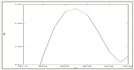
UV-visible spectroscopy characterization method was then used to monitor formation of ZnNPs synthesized from Gymnema sylvestre (Figure 1) and its properties. The absorption of light by the nanoparticles at different wavelengths provides an indication of particle size, while the breadth of the peaks signifies the particle size distribution. The characteristic SPR band of ZnNPs is in the range of 289nm [16] (Figure 2).

Figure 2:UV-Visible spectrum of Gymnema sylvestre leaf extract mediated synthesized zinc nanoparticles.


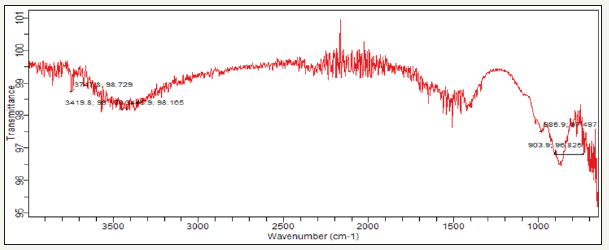
The FT-IR spectra of Gymnema sylvestre leaf extract mediated ZnNPs. From the FT-IR spectra, as seen in (Figure 3), a series of absorption peaks from 500 to 4000cm-1 are found, corresponding to the carboxylate and hydroxyl impurities in materials. A broad band at around 3747cm-1 is assigned to the OH stretching mode of hydroxyl group. The peaks observed at 3419 cm-1 are due to the asymmetrical and symmetrical stretching of the zinc carboxylate, respectively. 985 and 903cm-1 the content of the carboxylate (COO-) and hydroxyl (-OH) groups in the samples decreased. The carboxylate probably comes from reactive carbon containing species during synthesis and the hydroxyl group results from the hygroscopic nature of Zn [17].

XRD of Zn indicates the well-defined hexagonal shape, resulted by calcining the Gymnema sylvestre based Zn. The peaks observed in the diffractogram in (Figure 4) are assigned in terms of the Miller indices (hkl) assuming a hexagonal crystal structure of Zn, Thus, various well-defined dominant diffraction reflections are seen in the observed XRD pattern which are all related and well matched with the diffraction reflections of hexagonal phase Zn. Average crystallite size as measured from XRD using the Debye-Scherrer formula is 80 nm [14]. These D values refer to average values calculated by Δ2θ1/2 in the (100), (002), (101), (102), (110), (112), and (201) etc prominent peaks of the diffractogram.

Figure 3:FT-IR spectrum of Gymnema sylvestre leaf extract mediated synthesized zinc nanoparticles.


Figure 4:XRD spectrum of Gymnema sylvestre leaf extract mediated synthesized zinc nanoparticles.


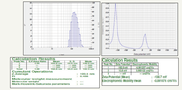
Particle size and zeta potential values were measured using Nano partica SZ-100 (HORIBA). The zeta potential spectra for the Gymnema sylvestre leaf extract nanoparticles were recorded zeta potential verses intensity spectra with zeta potential (mV) on x-axis and intensity (a.u) on y-axis particle size of 100.5 nm and zeta potential of -138.7mV were recorded (Figure 5a,5b), Here the zeta potential value of the zinc nanoparticles indicated good stability with high potential [18].

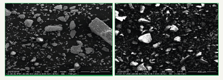
The SEM micrograph of Zn Nano powder prepared by Gymnema sylvestre leaf extract. A very clean surface is seen by the SEM micrograph with the distinct particles [19]. Particles are nearly spherical with hexagonal flakes, spherical shapes and there is some agglomeration of nanoparticles is seen it is due to the presence of proteins present in the Gymnema sylvestre leaf, so particles are very uniformly distributed with an average particle size of 200-400μm (Figure 6).

Photocatalytic degradation of dye

Photocatalytic degradation of methylene blue was carried out by using green synthesized Zn nanoparticles under solar light.

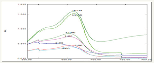
Dye degradation was initially identified by color change. Initially, the color of dye shows deep blue color changed into light blue after the h of incubation with Zn nanoparticles while exposed to solar light (Figure 7). Thereafter light blue was changed into light green. Finally, the degradation process was completed at 7h and was identified by the change of reaction mixture color to colorless. The adsorption of Zn nanoparticles on the methylene blue solution was initially low and further increased with constant increase in time and the percentage of dye degradation. However, at the same time more photo-catalyst would also have induced greater aggregation, which resulted in the decrease in the surface area of the photo-catalyst, making a significant fraction of the catalyst to be inaccessible to adsorbing the dye [20,21]. The experiment was carried out under the following conditions: reaction time of h (10,11,12,1,2,3 and 4) the results show that the photocatalytic activity remained nearly unchanged after five uses, which indicates that the photocatalyst is stable in the photocatalytic oxidation of methylene blue dye solutions. Therefore, this photocatalyst can be separated and recycled while maintaining its stability, making it a promising material for environmental remediation.

Figure 5:DLS spectrum 5a. Particle size 5b. Zeta potential of Gymnema sylvestre leaf extract mediated synthesized zinc nanoparticles.


Figure 6:SEM analysis of Gymnema sylvestre leaf extract mediated synthesized zinc nanoparticles.


Figure 7:Graph showing UV-visible spectrum of different time intervals of Photo-catalytic degradation of methylene blue using Gymnema sylvestre leaf extract mediated synthesized zinc nanoparticles.


Antimicrobial assay

Zn nanoparticles obtained from Gymnema sylvestre leaf extract have shown very strong inhibitory action against fungal Sp, Gram-positive and Gram-negative bacteria (Figures 8 & 9). Three concentrations of ZnNPs (170,100,50ppm) were prepared and were applied against an array of fungal species viz, Aspergillus niger, Rhizopus stolonifer, Aspergillus flavus, Fusarium oxy sporium and bacterial species viz., E. Coli, Bacillus pantothenticus, Pseudomonas fluorescence and Salmonella typhimurium. The higher concentration (170ppm) of leaf mediated synthesized ZnNPs showed significant antimicrobial effect (Tables 1 & 2) compared with other concentrations (100,50ppm) seen in leaf, but when compared to bacterial concentrations fungi shown very good antimicrobial activity towards all concentrations. The inhibitory action of the microbes may be attributed to the loss of replication ability of DNA upon treatment with the Zn ion, besides the fact that expression of ribosomal sub-unit proteins as well as some other cellular proteins and enzymes essential to ATP production becomes inactivated [22]. According to the results, it can be concluded that Zn nanoparticles are effective antibacterial agents both on Gram-positive and Gram-negative bacteria. The same results were confirmed in the study of Supraja et al. [14] in which Gramnegative membrane and Gram-positive membrane disorganization was approved by transmission electron microscopy of bacteria ultrathin sections. To use Zn nanoparticles in in vivo condition, further studies should be performed investigating the toxic effect of Zn nanoparticles on eukaryotic cells.

Table 1:In-vitro antibacterial studies against bacterial pathogens using Gymnema sylvestre leaf extract mediated synthesized zinc nanoparticles.


Table 2:In-vitro antifungal studies against fungal pathogens using Gymnema sylvestre leaf extract mediated synthesized zinc nanoparticles.


Figure 8:Antibacterial activity of different concentrations (50,100,170ppm) of Gymnema sylvestre leaf extract mediated synthesized zinc nanoparticles.


Figure 9:Antifungal activity of different concentrations (50,100,170ppm) of Gymnema sylvestre leaf extract mediated synthesized zinc nanoparticles.


Conclusion

Green synthesis of zinc nanoparticles shows more compatible, ecofriendly, low cost, and less time-consuming process. Herein, the zinc nanoparticles were synthesized by using plant leaf extract of Gymnema sylvestre leaf extract, main absorption peak at 650nm decreased gradually with the extension of the exposure time indicating the photocatalytic degradation of methylene blue dye. The results of photocatalytic studies reveal that the highest photocatalytic activity and stability were obtained for zinc nanoparticle photocatalyst, which can be used to oxidize 94% of methylene blue dye after 7h.

Acknowledgment

Authors are thankful to Acharya N.G. Ranga Agricultural University for providing research facility at institute of Frontier Technology, Regional Agricultural Research Station, Tirupati to carry out this part of the research work.

References

  1. Cheng W, Wang SG, Lu L, Gong WX, Liu XW, et al. (2008) Interaction between congo red and copper in a binary adsorption system: Spectroscopic and kinetic studies. Biochem Eng J 538-546.
  2. Pare B, Sarwan B, Jonnalagadda SB (2011) Photocatalytic mineralization study of malachite green on the surface of Mn-doped BiOCl activated by visible light under ambient condition. Appl Surf Sci 258: 247-253.
  3. Cheng ZJ, Liu TY, Chen XY, Gan HX, Zhang FW, et al. (2012) Study on the electronic structures of the reduced anatase TiO2 by the first-principle calculation. Journal of Physics and Chemistry of Solids 73(2): 302-307.
  4. Yu J, Yu X (2008) Hydrothermal synthesis and photocatalytic activity of zinc oxide hollow spheres. Environ Sci Technol 42(13): 4902-4907.
  5. Sun JH, Dong SY, Wang YK, Sun SP (2009) Preparation and photocatalytic property of a novel dumbbell-shaped ZnO microcrystal photocatalyst. J Hazard Mater 172(2-3): 1520-1526.
  6. Zhou M, Yu J, Cheng B (2008) Preparation, characterization and visible- light-driven photocatalytic activity of Fe-doped Titania nanorods and first-principles study for electronic structures. J Hazard Mater B 137: 1838.
  7. Singhal A, Achary S, Tyagi A, Manna A, Yusuf S (2008) Colloidal Fe-doped ZnO nano crystals: facile low temperature synthesis, characterization and properties. Mater Sci and Eng B 153: 47.
  8. Prasad TNVKV, Elumalai EK (2011) Biofabrication of Ag nanoparticles using Moringa oleifera leaf extract and their antimicrobial activity. Asian Pacific Journal of Tropical Biomedicine 1(6): 439-442.
  9. Patil RS, Kokate MR, Kolekar SS (2012) Bioinspired synthesis of highly stabilized silver nanoparticles using Ocimum tenuiflorum leaf extract and their antibacterial activity. Spectro Chimica Acta A 91: 234-238.
  10. Kumar R, Roopan SM, Prabhakarn A, Khanna VG, Chakroborty S, et al. (2012) Agricultural waste Annona squamosa peel extract: biosynthesis of silver nanoparticles. Spectrochim Acta A Mol Biomol Spectrosc 90: 173-176.
  11. Supraja N, Avinash B, Prasad TNVKV (2017) Green synthesis and characterization of silver nanoparticles from Gymnema sylvestre leaf extract: study of antimicrobial activities. Int J Curr Microbiol App Sci 6(3): 530- 540.
  12. Bonnia NN, Kamaruddin MS, Nawawi MH, Ratim S, Azlina HN, et al. (2016) Green biosynthesis of silver nanoparticles using polygonum hydropiper and study its catalytic degradation of methylene blue. Procedia Chemistry 19: 594-602.
  13. Vanaja M, Paulkumar K, Baburaja M, Rajeshkumar S, Gnanajobitha G, et al. (2014) Degradation of methylene blue using biologically synthesized silver nanoparticles. Bioinorganic Chemistry and Applications 742346: 8.
  14. Supraja N, Prasad TNVKV, Giridhara Krishna T, David E (2016) Synthesis, characterization, and evaluation of the antimicrobial efficacy of Boswellia ovalifoliolata stem bark-extract-mediated zinc oxide nanoparticles. Biochem Biophys Rep 14: 69-77.
  15. Supraja N, Prasad TNVKV, Giridhar Krishna T, David E (2015) Synthesis, characterization and evaluation of the antimicrobial efficacy of boswellia ovalifoliolata stem bark extract mediated zinc oxide nanoparticles. Appl Nanosci 6(4): 581-590.
  16. Karunakaran C, Dhanalakshmi R, Gomathisankar P (2012) Phenol-photodegradation on ZrO2. Enhancement by semiconductors. Spectrochim Acta A Mol Biomol Spectrosc 92: 201-206.
  17. Kansara S, Dhruv D, Joshi Z, Pandya D, Rayaprol S, et al. (2015) Structure and microstructure dependent transport and magnetic properties of sol-gel grown nanostructured La 0.6 Nd 0.1 Sr 0.3 MnO3 manganites: role of oxygen. Appl Surf Sci 356: 1272-1281.
  18. Sri Sindhura K, Prasad TNVKV, Panner Selvam P, Hussain OM (2013) Synthesis and characterization of phytogenic zinc nanoparticles and their antimicrobial activity. Appl Nanosci 4(7): 819-827.
  19. Prabha S, Supraja N, Garud M, Prasad TNVKV (2014) Synthesis, characterization and antimicrobial activity of Alstonia scholars bark-extract- mediated silver nanoparticles. J Nano struct Chem 4(4): 161-170.
  20. Vanaja M, Paulkumar K, Baburaja M, Rajeshkumar S, Gnanajobitha G, et al. (2014) Degradation of methylene blue using biologically synthesized silver nanoparticles Bioinorg Chem Appl 4: 161-170.
  21. Matinise N, Fuku XG, Kaviyarasu K, Mayedwa N, Maaza M, et al. (2017) ZnO nanoparticles via Moringa oleifera green synthesis: physical properties & mechanism of formation. Appl Surf Sci 406: 339-347.
  22. Kouvaris P, Delimitis A, Zaspalis V, Papadopoulos D, Tsipas SA, et al. (2012) Green synthesis and characterization of silver nanoparticles produced using Arbutus Unedo leaf extract. Mater Lett 76: 18-20.

For more articles in Bioequivalence And Bioavailability International Journal
Please click on below link: https://crimsonpublishers.com/abb/

Tuesday, March 29, 2022

Relationship Between Arrhythmias and Level Activity of Athlete’s-Role of HRMs_Crimson Publishers

 Relationship Between Arrhythmias and Level Activity of Athlete’s-Role of HRMs by Robert Gajda in Examines in Physical Medicine and Rehabilitation: Open Access_Rehabilitation medicine journal impact factor

Keywords

Arrhythmias; Exercise; Heart Rate Monitors (HRMs); Endurance sport; Sudden cardiac death

Introduction

Among all papers devoted to the relationship between arrhythmias and endurance training, the study Andersen et al. is particular value [1]. The study includes all Swedish citizens completing the famous Swedish skiing event “Vasaloppet,” enduring 90 kilometers of strenuous cross-country skiing. The participants are ranging from elite to recreational athletes and their training status (measured as maximal oxygen consumption) is closely related to their finishing time. The authors completed data about more than 4,400 participants of this strenuous ski marathon during the period 1989-1998.

Accounting for age, socioeconomic status and education, they observed a higher incidence of arrhythmias in cross-country skiers with a long history of endurance training [1]. Compared to those who had completed only one single race, those who had completed 7 or more races had a 29% higher risk of a subsequent arrhythmia. Further, elite athletes finishing at 100- 160% of the winning time had a 37% higher risk of arrhythmias than recreational athletes finishing at more than 241% of the winning time. The associations were mainly driven by the most common types of arrhythmia: atrial fibrillation and brady arrhythmias. The authors did not find any significantly increased incidence of potential lethal ventricular arrhythmias with any of the exposures.

In another paper published in 2015 in BMJ, Andersen and co-workers presented a very unique analysis about the associations of exercise capacity and muscle strength in late adolescence with the risk of vascular disease and arrhythmia based on long-term observation of 1.1 million young Swedish men who participated in mandatory military conscription between 1972 and 1995. During a median follow-up of 26 years, more than 26,000 vascular disease events and more than 17,300 arrhythmia events were recorded. The study revealed that exercise capacity and muscle strength in late adolescents are independently associated with a lower risk of vascular disease and arrhythmia. Exercise capacity had a U-shaped association with risk of arrhythmia driven association with risk of atrial fibrillation and a U-shaped with bradyarrhythmia. Thus, the benefit of a lower risk of vascular events with higher exercise capacity (hazard ratio for vascular events of 0.67) was not outweighed by a higher risk of arrhythmia [2, 3].

Other risk factors of arrhythmias in athletes comprise age, exercise intensity and above all-exercise duration. A threshold for the increased risk of atrial fibrillation is 1500 hours of exercise per life. Atrial volume is a strong predictor of AF (athletes with AF have a larger leftatrial volume than those without) in older athletes (39±9 years old) and in veterans [4-7].The etiology and pathophysiology of AF in athletes are not clear. Increased activity of the parasympathetic vagal system is the most important modulator and trigger of AF in athletes, whereas a substrate has still been a subject of debate. Structural remodeling of atrial myocardium in response to permanent pressure and volume overloading (“overtraining syndrome”), inflammation and fibrosis are the most probable causative factors [8].

Ventricular tachyarrhythmias, identified in elite athletes without cardiovascular abnormalities, are frequent expressions of the “Athlete’s Heart Syndrome” (up to 70%), though they are not related to the presence or magnitude of training-induced LV hypertrophy [9]. Brief deconditioning (5-6 months) usually results in the resolution of arrhythmias in athletes without organic heart disease [9]. Many authors of older and recently published original papers and reviews agreed that Supra Ventricular Tachyarrhythmias (SVT) in sportsmen are rare and usually benign. This kind of benign arrhythmia may appear with palpitations, weakness, lightheadedness, and even syncope. Frequent SVT may impair athletic performance. Sinus node reentry tachycardia is an uncommon finding in athletes. The average heart rate is between 130 and 140bpm. Atrial tachycardia is extremely rare in athletes. The atrial rate is generally between 150 and 200bpm [10-12].

The widespread use of sport Heart Rate Monitors (HRMs) contributes to the “catching” of an increasing number of tachyarrhythmias among both symptomatic competitive athletes and amateurs [13]. Especially in the case of “professionals” it can be a life-saving factor [14]. Although heart rate monitors do not recognize the type of arrhythmia, their worrying indications in conjunction with clinical symptoms allow for a fairly preliminary diagnosis [15]. In the future, the technological development of sport heart rate monitors will definitely provide further solutions for athletes allowing to determine the type of arrhythmias if they appear in sports training [16].

References

  1. Andersen K, Farahmand B, Ahlbom A, Held C, Ljunghall S, et al. (2013) Risk of arrhythmias in 52 755 long-distance cross-country skiers: A cohort study. Eur Heart J 34(47): 3624-3631.
  2. Andersen K, Rasmussen F, Held C, Martin N, Per T, et al. (2015) Exercise capacity and muscle strength and risk of vascular disease and arrhythmia in 1.1 million young Swedish men: Cohort study. BMJ 351: h4543.
  3. Hoogsteen J, Bennekers JH, Wall EE, Hemel NM, Wilde AA, et al. (2004) Recommendations and cardiological evaluation of athletes with arrhythmias: Part 1. Neth Heart J 12(4): 157-164.
  4. Nielsen JR, Wachtell K, Abdulla J (2013) The relationship between physical activity and risk of atrial fibrillation-A systematic review and meta-analysis. J Atr Fibrillation 5(5): 789.
  5. Everett BM, Conen D, Buring JE, Moorthy MV, Lee IM, et al. (2011) Physical activity and the risk of incident atrial fibrillation in women. Circ Cardiovasc Qual Outcomes 4(3): 321-327.
  6. Eijsvogels TM, Fernandez AB, Thompson PD (2016) Are there deleterious cardiac effects of acute and chronic endurance exercise?. Physiol Rev 96(1): 99-125.
  7. Hong KL, Glover BM (2018) The impact of lifestyle intervention on atrial fibrillation. Curr Opin Cardiol 33(1): 14-19.
  8. Olshansky B, Sullivan R (2014) Increased prevalence of atrial fibrillation in the endurance athlete: Potential mechanisms and sport specificity. Phys Sportsmed 42(1): 45-51.
  9. Biffi A, Maron BJ, Giacinto B, Porcacchia P, Verdile L, et al. (2008) Relation between training-induced left ventricular hypertrophy and risk for ventricular tachyarrhythmias in elite athletes. Am J Cardiol 101(12): 1792-1795.
  10. Biffi A, Maron BJ, Verdile L, Fernando F, Spataro A, et al. (2004) Impact of physical deconditioning on ventricular tachyarrhythmias in trained athletes. J Am Coll Cardiol 44(5): 1053-1058.
  11. Viitasalo MT, Kala R, Eisalo A (1984) Ambulatory electrocardiographic findings in young athletes between 14 and 16 years of age. Eur Heart J 5(1): 2-6.
  12. Verdile L, Maron BJ, Pelliccia A, Spataro A, Santini M, et al. (2015) Clinical significance of exercise-induced ventricular tachyarrhythmias in trained athletes without cardiovascular abnormalities. Heart Rhythm 12(1): 78-85.
  13. Gajda R (2019) Extreme bradycardia and brady arrhythmias at athletes. What will technology development bring as a help to diagnosis them? Res Inves Sports Med 5(4).
  14. Gajda R, Biernacka EK, Drygas W (2018a) Are heart rate monitors valuable tools for diagnosing arrhythmias in endurance athletes?. Scand J Med Sci Sports 28(2): 496-516.
  15. Gajda R, Biernacka EK, Drygas W ( 2019) Atrial Fibrillation in athletes-easier to recognize today? Res Inves Sports Med 5(4).
  16. Gajda R, Biernacka EK, Drygas W (2018b) “The problem of arrhythmias in endurance athletes: are heart rate monitors valuable tools for diagnosing arrhythmias?”, Horizons in World Cardiovascular Research. Nova Science Publishers, New York, USA 15: 1-64.

For more articles in Rehabilitation medicine journal impact factor
Please click on below link: https://crimsonpublishers.com/epmr/


Friday, March 25, 2022

Effect of HER2 Receptor Expression on Cardiac Ejection Fraction in Cancer Patients Treated with Trastuzumab_Crimson Publishers

 Effect of HER2 Receptor Expression on Cardiac Ejection Fraction in Cancer Patients Treated with Trastuzumab by Mariah Obino in Open Journal of Cardiology & Heart Diseases_American Journal of Cardiology


Abstract

Background: Human epidermal growth factor receptor 2 (HER2) over expression has been identified as an important drug target for the treatment of many cancers. Trastuzumab (Herceptin, Genentech, CA, USA) is an anti-HER2 monoclonal antibody effective at inducing cell death in patients who overexpress this gene. The degree of HER2 expression is a determining factor in the decision to treat patients with trastuzumab. At Froedtert & the Medical College of Wisconsin, a dual probe fluorescent in situ hybridization is completed and the decision to treat is then made based on the results which are reported as a ratio of HER2 gene signals relative to those of chromosome 17 (CEP17). A patient may qualify for treatment by meeting the minimum threshold requirement of HER2 expression (HER2:CEP17 ratio ≥2 or if HER2:CEP17 ratio <2 and ≥6 HER2 signals/cell), while other patients may far surpass the minimum level. Trastuzumab contains a warning for possible cardiomyopathy associated with symptomatic and asymptomatic reductions in left ventricular ejection fraction (LVEF). There has been no investigation as to whether the degree of HER2 expression is related to the patient’s risk for developing trastuzumab-induced cardiotoxicity.

Methods: We performed a retrospective cohort review of patients having received trastuzumab within Froedtert & the Medical College of Wisconsin Health System in an effort to identify and describe the relationship between degree of HER2 expression and incidence of cardiotoxicity in patients receiving trastuzumab therapy. The primary objective of this study was to identify the correlation between HER2 receptor expression and cardiotoxicity in patients treated with trastuzumab. The secondary objectives were to identify the correlation between HER2 expression and absolute change in LVEF and time to cardiotoxicity in patients treated with trastuzumab. For the purposes of this study, cardiotoxicity was defined as a decrease in LVEF ≥ 16% from baseline or a LVEF <50% and ≥10% decrease from baseline. Change in global peak systolic longitudinal strain of ≥15%, grade ≥3 left ventricular systolic dysfunction as defined by the National Cancer Institute Common Terminology Criteria for Adverse Events v 4.03 (NCI CTCAE), or treatment discontinuation due to trastuzumab cardiotoxicity as determined by physician was also used to meet criteria for primary end point of cardiotoxicity. Froedtert & the Medical College of Wisconsin IRB approved completion of this study.

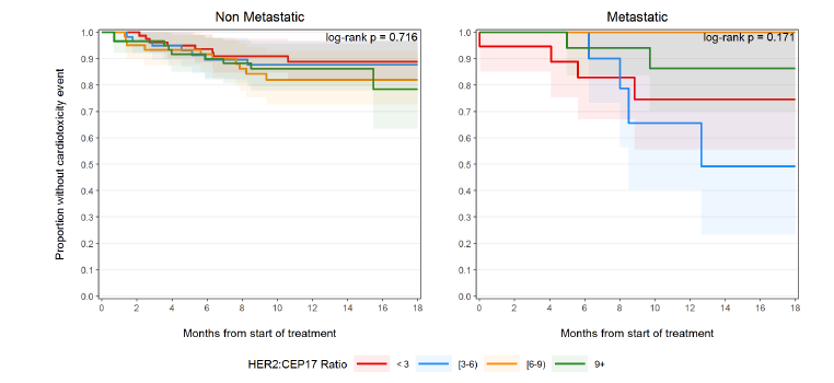
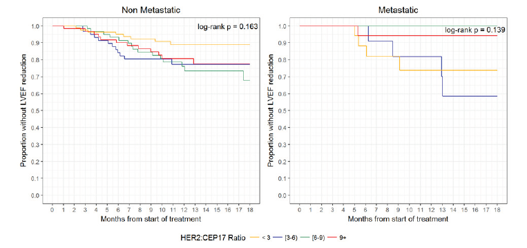
Results: There were 44 (34 non-metastatic and 10 metastatic) patients with cardiotoxicity event(s) and 11 deaths without cardiotoxicity (all metastatic patients) within 18 months of follow-up. Overall one-year incidence of cardiotoxicity was 14.2% [10.7%, 18.9%], with incidences of 13.6% [9.9%, 18.8%] in non-metastatic patients and 16.7% [9.1%, 30.5%] in metastatic patients. The median survival time (time at which half of all patients are expected to experience cardiotoxicity) was not reached within 18 months. There was no significant difference in survival from cardiotoxicity between HER2:CEP17 groups (<3, 3-6, 6-9, 9+) among non-metastatic patients (log-rank test p=0.72) or metastatic patients (log-rank test p=0.17). The HER2:CEP17 ratio (continuous) was not significantly associated with cardiotoxicity in non-metastatic patients (aHR 1.00 [0.94, 1.07], p=0.95) or metastatic patients (aHR 0.88 [0.72, 1.07], p=0.19) after adjusting for baseline age, comorbidities, radiation exposure, and anthracycline exposure. In the multiple regression analysis of non-metastatic patients, anthracycline exposure (aHR 2.73 [1.28, 5.81], p=0.009), baseline comorbidities (aHR 2.41 [1.14, 5.09], p=0.021), and younger age at first dose (aHR 0.95 [0.92, 0.98], p=0.002) were significantly associated with increased hazard of cardiotoxicity. There were 56 (47 non-metastatic and 9 metastatic) patients with LVEF reduction. Overall one-year incidence of LVEF reduction was 17.4% [13.5%, 22.3%], with incidences of 18.6% [14.2%, 24.4%] in non-metastatic patients and 12.3% [6.2%, 24.8%] in metastatic patients. The median time to LVEF reduction was not reached within 18 months. The HER2:CEP17 ratio (continuous) was not significantly associated with LVEF reduction in non-metastatic patients (aHR 1.02 [0.97, 1.07], p = 0.53) or metastatic patients (aHR 0.81 [0.62, 1.05], p = 0.12) after adjusting for age, baseline comorbid conditions, radiation exposure, and anthracycline exposure (Tables 1-3). Only anthracycline exposure was significantly associated with increased hazard of LVEF reduction (aHR 2.37 [1.27, 4.43], p = 0.007) in non-metastatic patients.

Conclusion: Tumor cell HER2:CEP17 ratio does not predict incidence or time to onset of cardiotoxicity associated with trastuzumab.

Keywords: Trastuzumab; Cardiotoxicity; HER2

Abbreviations: HER2: Human Epidermal Growth Factor Receptor 2; LVEF: Left Ventricular Ejection Fraction; IHC: Immunohistochemistry; CEP17: Chromosome 17; FISH: Fluorescent Insitu Hybridization; MUGA: Multigated Acquisition Scan; NCI CTCAE: National Cancer Institute Common Terminology Criteria for Adverse Events

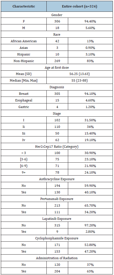
Table 1:Baseline demographics.


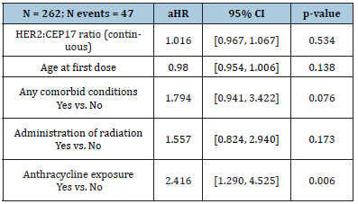
Table 2:Association with LVEF reduction in Non-Metastatic Patient Population


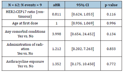
Table 3:Association with LVEF reduction in Metastatic Patient Population


Introduction

Human epidermal growth factor receptor 2 (HER2) overexpression has been identified as an important drug target for the treatment of certain malignancies. Once activated, HER2 is responsible for a signaling cascade associated with proliferation and cell survival. In 1998, trastuzumab (Herceptin®, Genentech, CA, USA) an anti-HER2 monoclonal antibody, was developed and found to be effective at inducing cell death in patient’s tumor cells which overexpress this gene [1-3]. Trastuzumab is FDA approved for use in HER2 positive breast cancers as well as in HER2 positive metastatic gastric cancer. Trastuzumab contains a black boxed warning for possible cardiomyopathy associated with reductions in left ventricular ejection fraction (LVEF). Research has shown HER2 receptor mediated signaling pathways control critical cellular functions related to cardiac myocyte survival and hypertrophy. In animal models, HER2 knockout mice developed cardiomyopathy within 12 weeks of life and adapted poorly to cardiac stressors [4]. The disruption of this pathway is believed to be the primary means of trastuzumab mediated cardiotoxicity however the entire mechanism has yet to be fully elucidated [5].

It is unknown if there is a correlation between degree of HER2 expression and degree of cardiotoxicity resulting from HER2 inhibition. Trastuzumab-associated cardiotoxicity was most commonly seen in patients receiving concurrent anthracyclinetrastuzumab based regimens. A phase III trial reported 27% of patients developed some grade of left ventricular dysfunction [3]. Due to the concern for development of congestive heart failure, cardiac monitoring guidelines were developed and subsequently adopted as standard of care for patients receiving this therapy. Additional risk factors have been identified for the development of cardiotoxicity including age, female sex, high dose radiation therapy, smoking, hypertension, diabetes, and dyslipidemia. The current trastuzumab monitoring recommendations indicate that LVEF monitoring should occur at baseline and at 3-month intervals during treatment [6]. Inconsistencies related to adherence to cardiac monitoring guidelines often occur in clinical practice due to an absence of data on if this rigorous monitoring is justified for all patients or if only certain populations necessitate increased monitoring [1,3,7].

There is no universal definition of cardiotoxicity. However, most clinical trials define cardiotoxicity by a serial decline in LVEF. The American Society of Echocardiography consensus document defines cardiotoxicity as LVEF drop ≥10% to a value of <53%. In the phase 3 Herceptin Adjuvant (HERA) trial, the cardiac review and evaluation committee defined cardiotoxicity as an asymptomatic reduction in LVEF ≥10% from baseline to below 50% or if patients experienced severe (New York Heart Association functional class III or IV) or symptomatic heart failure [8,9]. Additional trials and the trastuzumab package insert, have defined cardiotoxicity as a decrease in LVEF of ≥16% from baseline or LVEF below normal limits and ≥10% decrease from baseline. More recently myocardial strain has been proposed as a potential means of identifying subclinical myocardial injury and may help predict impending changes in LVEF [1,4,10-13]. A relative reduction of global peak systolic longitudinal strain of 10-11% at 3 or 6 months during trastuzumab treatment predicts future cardiotoxicity in women receiving trastuzumab for breast cancer. Additionally, the American Society of Echocardiography has included a decline in global peak systolic longitudinal strain of >15% during cancer treatment in their definition of cardiotoxicity. With no overtly clear definition of cardiotoxicity, clinicians may need to utilize a combination of expert guidance recommendations [14,15].

While it is known that trasuzumab can induce cardiotoxicity, the degree of HER2 expression and its correlation to the development of cardiotoxicity remains unknown. The degree of HER2 expression is an important factor in the decision to treat patients with trastuzumab. The American Society of Clinical Oncology/College of American Pathologists has released guideline recommendations for HER2 testing to improve its accuracy and reliability. These guidelines recommend at least one tumor sample be tested for either HER2 protein expression via immunohistochemistry (IHC) or HER2 gene expression via fluorescent in situ hybridization (FISH) reported as a ratio of HER2 gene signals relative to those of chromosome 17 (CEP17). A patient may qualify for treatment by meeting the minimum threshold requirement of HER2 gene expression (HER2:CEP17 ratio ≥2 or if HER2:CEP17 ratio <2 and ≥6 HER2 signals/cell), while other patients may far surpass the minimum expression required for treatment [15]. The purpose of our study was to identify and describe the relationship between degree of HER2 expression and incidence of cardiotoxicity in patients receiving trastuzumab therapy.

Methods

Patients receiving trastuzumab therapy at Froedtert & the Medical College of Wisconsin from May 1st, 2004 to July 1st, 2017 were evaluated after receiving IRB approval via retrospective chart review. Eligible patients carried a diagnosis of metastatic or nonmetastatic breast, metastatic gastric, or metastatic esophageal cancer. Patients included in the study had received at least one dose of trastuzumab, had a baseline LVEF measured prior to receiving trastuzumab therapy, at least one LVEF measurement for comparison while receiving trastuzumab, and a reported FISH HER2:CEP17 ratio. Measurement of LVEF could occur via multigated acquisition scan (MUGA) or echocardiogram (ECHO). Patients who were less than 18 years of age or had insufficient information in their medical record were excluded from the study. Multiple data points were collected for the analysis including patient demographic information, cardiac comorbidities, cancer diagnosis and stage, trastuzumab dose, dosing interval, dates of administration, number of doses received, FISH HER2:CEP17 ratio, and IHC result if available. Information regarding the method of LVEF measurement, LVEF value at baseline and while receiving trastuzumab therapy, global peak systolic longitudinal strain measurements, and number of trastuzumab doses prior to experiencing cardiac toxicity were also collected. Cardiac comorbidities as well as exposure to cardiotoxic therapies, such as previous exposure to radiation, anthracyclines, and other anti-HER2 therapies were also collected to adjust for confounding variables.

The primary objective of this study was to describe the relationship between HER2 receptor expression and cardiotoxicity in patients treated with trastuzumab. The secondary objectives were to identify the association between HER2 expression and absolute change in LVEF and time to cardiotoxicity in patients treated with trastuzumab. For the purposes of this study, cardiotoxicity was defined as a decrease in LVEF ≥ 16% from baseline or a LVEF <50% and ≥10% decrease from baseline. Change in global peak systolic longitudinal strain of ≥15%, grade ≥3 left ventricular systolic dysfunction as defined by the National Cancer Institute Common Terminology Criteria for Adverse Events v 4.03 (NCI CTCAE), or treatment discontinuation due to trastuzumab cardiotoxicity as determined by physician was also used to meet criteria for primary end point of cardiotoxicity. Froedtert & the Medical College of Wisconsin IRB approved completion of this study.

Statistics

Study variables were summarized using the mean, median, standard deviation, and range for continuous variables and frequency and percentage for categorical variables. There were no missing values among analytic variables. The primary predictor, HER2 receptor expression, was evaluated as both a continuous variable and as categories grouped by the median observed value (< 6 and 6+) and approximate quantiles (< 3, [3-6), [6-9), and 9+). Patient demographics and baseline clinical characteristics were compared between HER2 groups (< 6 and 6+) using Wilcoxon rank-sum tests for continuous variables and Fisher’s exact tests for categorical variables. All analyses were stratified by cancer stage (metastatic vs. non-metastatic) due to inherent differences in and length of trastuzumab treatment and dosing.

The primary outcome, cardiotoxicity, was analyzed using survival analysis methods. The survival time was taken as the time from start of therapy to the first observed cardiotoxicity event. Patients were censored at the last known follow-up or death up to 18 months. Kaplan-Meier methods were used to plot the (unadjusted) survival distributions of each HER2 group (median and quantile groupings) and the log-rank test was used to compare them. Multivariable Cox proportional hazards regression were used to compare time to cardiotoxicity by HER2 expression adjusting for age at first dose, presence of any comorbid conditions, concurrent radiation, and anthracycline exposure; two models were produced to assess HER2 as a linear predictor and alternatively using the median categories. The secondary outcome, reduction in LVEF, was analyzed similarly using survival methods. The event was defined as the first reduction from baseline LVEF of at least 16% or at least 10% with resulting LVEF below 50%. All statistical analyses were performed using R version 3.3.4 (R Foundation for Statistical Computing, http://www.R-project.org). All p-values were 2-sided and p < 0.05 was considered statistically significant.

Results

There were 44 (34 non-metastatic and 10 metastatic) patients with cardiotoxicity event(s) and 11 deaths without cardiotoxicity (all metastatic patients) within 18 months of follow-up; the median follow-up among patients without cardiotoxicity or death was 11.7months (range, 9.0-16.2 months). Overall one-year incidence of cardiotoxicity was 14.2% [10.7%, 18.9%], with incidences of 13.6% [9.9%, 18.8%] in non-metastatic patients and 16.7% [9.1%, 30.5%] in metastatic patients. The median survival time (time at which half of all patients are expected to experience cardiotoxicity) was not reached within 18 months. There was no significant difference in survival from cardiotoxicity between HER2:CEP17 groups (<3, 3-6, 6-9, 9+) among non-metastatic patients (log-rank test p = 0.72) or metastatic patients (log-rank test p = 0.17) (Figure 1). The HER2:CEP17 ratio (continuous) was not significantly associated cardiotoxicity in non-metastatic patients (aHR 1.00 [0.94, 1.07], p = 0.95) or metastatic patients (aHR 0.88 [0.72, 1.07], p = 0.19) after adjusting for baseline age, comorbidities, radiation exposure, and anthracycline exposure. In the multiple regression analysis of non-metastatic patients, anthracycline exposure (aHR 2.73 [1.28, 5.81], p = 0.009), baseline comorbidities (aHR 2.41 [1.14, 5.09], p = 0.021), and younger age at first dose (aHR 0.95 [0.92, 0.98], p = 0.002) were significantly associated with increased hazard of cardiotoxicity.

Figure 1:Survival from cardiotoxicity


There were 56 (47 non-metastatic and 9 metastatic) patients with LVEF reduction and 12 deaths without cardiotoxicity (all metastatic patients) within 18 months of follow-up; the median follow-up among patients without LVEF reduction or death was 12.4 months (range, 9.6-16.8 months). Overall one-year incidence of LVEF reduction was 17.4% [13.5%, 22.3%], with incidences of 18.6% [14.2%, 24.4%] in non-metastatic patients and 12.3% [6.2%, 24.8%] in metastatic patients. The median time to LVEF reduction was not reached within 18 months. There was no significant difference in survival from LVEF reduction between HER2:CEP17 groups (<3, 3-6, 6-9, 9+) among non-metastatic patients (log-rank test p = 0.16) or metastatic patients (log-rank test p = 0.14) (Figure 2). The HER2:CEP17 ratio (continuous) was not significantly associated with LVEF reduction in non-metastatic patients (aHR 1.02 [0.97, 1.07], p = 0.53) or metastatic patients (aHR 0.81 [0.62, 1.05], p = 0.12) after adjusting for age, baseline comorbid conditions, radiation exposure, and anthracycline exposure (Tables 2 & 3). Only anthracycline exposure was significantly associated with increased hazard of LVEF reduction (aHR 2.37 [1.27, 4.43], p = 0.007) in non-metastatic patients.

Figure 2:Survival from LVEF reduction


Discussion

Our evaluation of this retrospective cohort of 324 patients with HER2 positive breast and gastric cancer, followed up to 13 years demonstrated no association between the HER2:CEP17 ratios and the incidence of cardiotoxicity. Furthermore, our study revealed that >90% of cardiotoxicity events occurred during the first year in both the non-metastatic and metastatic patient population. We therefore limited statistical analysis to 18 months. The rarity of events beyond 1 year of therapy in the non-metastatic setting supports a lack of long-term toxicity once therapy is complete. Within the metastatic setting, trastuzumab therapy was tolerated remarkably well and supports a lack of cumulative dose related toxicity which may allow for less frequent cardiac monitoring in metastatic patient receiving greater than one year of therapy. These data represent a real-world cohort of patients with pre-exiting cardiac comorbidities which adds to the external validity of our findings.

To our knowledge, this is the first investigation as to whether the degree of HER2 gene expression is related to patients’ risk for developing trastuzumab-induced cardiotoxicity. Clinical prediction tools have been published which attempt to identify patients at highest risk for cardiotoxicity in the setting of HER2 positive breast cancer treated with trastuzumab. Romond et al. [1] developed a prediction tool, which revealed age and baseline LVEF played the biggest role in cardiotoxicity development. A second prediction tool was developed in 2014 by Ezaz et al. [17] which revealed age, adjuvant chemotherapy, history of cardiac disease, and cardiac risk factors to be predictive of cardiotoxicity [16]. Currently, no prediction tool has incorporated biomarkers or diagnostic tests and imaging into their model [17]. It is important to note that our study utilizes HER2:CEP17 ratios from a patient’s tumor cells. Without matched cardiac tissue testing for HER2 expression it is not possible to ascertain whether there is a correlation between tumor cell expression of HER2 and cardiac myocyte expression. Therefore, it is possible HER2:CEP17 ratios directly from cardiac tissue could be predictive of trastuzumab related cardiotoxicity, however obtaining these samples without another compelling indication is largely impractical.

Cardiotoxicity is an important potential cause of treatment delay and early discontinuation of treatment which ultimately results in sub-optimal treatment of the patient’s malignancy. Despite longterm follow-up of patients treated with trastuzumab and years of research on the prediction of cardiotoxicity, there is currently no standardized model available to guide risk stratification for trastuzumab related cardiotoxicity in today’s clinical practice. With the continued use of anti-HER2 targeted therapies, as well as other anti-cancer agents with cardiotoxicity potential, further research is needed to identify patients who are at highest risk for therapy related toxicity. Our study captured a heterogeneous population of patient receiving trastuzumab, both those that are healthy and those that have comorbidities. Although it was retrospective single center study, we were able to follow our patients for up to thirteen years after receiving therapy which allowed optimal time to capture cardiotoxicity events.

Conclusion

In this retrospective study, tumor cell HER2:CEP17 ratio did not predict incidence or time to onset of cardiotoxicity associated with trastuzumab. With the growing number of patients living longer with cancer, it is important to assess a patient’s risk for cardiac dysfunction as a result of chemotherapy treatment. Further research is needed to find novel patient characteristics to create standardized risk stratification tools and monitoring guidelines for patients receiving trastuzumab in an attempt to achieve a high level of cardiac safety without sacrificing outcomes or incurring unnecessary medical expense and patient inconvenience.

Declarations

Ethics approval and consent to participate. This study was approved by the Froedtert & the Medical College of Wisconsin IRB.

Availability of Data and Materials

The datasets during and/or analyzed during the current study available from the corresponding author on reasonable request.

Author Contributions

CL was principal investigator of the study and lead author of the manuscript. CO served in an advisory role in study design and implementation with expertise in trastuzumab use in GI malignancy. She provided guidance in data analyzation as well as key edits and discussion points to the manuscript.

EW served in an advisory role in study design and implementation with expertise in trastuzumab use in breast cancer. She provided guidance in data analyzation as well as key edits and discussion points to the manuscript. AU served in an advisory role in study design and implementation with expertise in clinical trials. BA significantly contributed to data collection as well as authored the methods portion of the manuscript. LR provided statistical analysis and guidance in study design as well as authored the statistics and results section of the manuscript. SK served in an advisory role with expertise in the treatment of breast cancer. As our physician champion she provided key guidance in study design, manuscript edits and discussion contributions.

References

  1. Romond EH, Perez EA, Bryant J, Suman VJ, Geyer CE, et al. (2005) Trastuzumab plus adjuvant chemotherapy for operable HER2-positive breast cancer. N Engl J Med 353(16): 1673-1684.
  2. Goldhirsch A, Gelber RD, Piccart-Gebhart MJ, Procter M, Suter TM, et al. (2013) Herceptin adjuvant (HERA) trial study team: 2 years versus 1 year of adjuvant trastuzumab for HER2-poositive breast cancer (HERA): An open-label, randomized controlled trial. Lancet 382(9897): 1021-1028.
  3. Slamon DJ, Leyland-Jones B, Shak S, Fuchs H, Paton V, et al. (2001) Use of chemotherapy plus a monoclonal antibody against HER2 for metastatic breast cancer that overexpresses HER2. N Engl J Med 344: 783-792.
  4. Seidman A, Hudis C, Pierri MK, Shak S, Paton V, et al. (2002) Cardiac dysfunction in the trastuzumab clinical trials experience. J Clin Oncol 20(5): 1215-1221.
  5. Zhao YY, Sawyer DR, Baliga RR, Opel DJ, Han X, et al. (1998) Neuregulin’s promote survival and growth of cardiac myocytes: Persistence of ERbB2 and ErbB4 expression in neonatal and adult ventricular myocytes. J Biol Chem 273: 10261-10269.
  6. G Curigliano, D Cardinale, T Suter, G Plataniotis, E de Azambuja, et al. (2012) Cardiovascular toxicity induced by chemotherapy, targeted agents and radiotherapy: ESMO clinical practice guidelines. Annals of Oncology 23(7): vii155-vii166.
  7. Tan-Chiu E, Yothers G, Romond E, Geyer CE, Ewer M, et al. (2005) Assessment of cardiac dysfunction in a randomized trial comparing doxorubicin and cyclophosphamide followed by paclitaxel, with or without trastuzumab as adjuvant therapy in node-positive, human epidermal growth factor receptor 2-overexpressing breast cancer. J Clin Oncol 23(31): 7811-7819.
  8. Dang CT, Yu AF, Jones LW, Liu J, Steingart RM, et al. (2016) Cardiac surveillance guidelines for trastuzumab-containing therapy in early-stage breast cancer: Getting to the heart of the matter. J Clin Oncol 34(10): 1030-1033.
  9. Plana JC, Galderisi M, Barac A, Ewer MS, Ky B, et al. (2014) Expert consensus for multimodality imaging evaluation of adult patients during and after cancer therapy: A report from the American society of echocardiography and the European association of cardiovascular imaging. J Am Soc Echocardiogr 27(9): 911-939.
  10. Ozcelik C, Erdmann B, Pilz B, Wettschureck N, Britsch S, et al. (2002) Conditional mutation of the ErbB2 (HER2) receptor in cardiomyocytes leads to dilated cardiomyopathy. Proc Natl Acad Sci U S A 99(13): 8880-8885.
  11. Joensuu H, Kellokumpu-Lehtinen PL, Bono P, Alanko T, Kataja V, et al. (2006) Adjuvant docetaxel or vinorelbine with or without trastuzumab for breast cancer. N Engl J Med 354: 809-820.
  12. Perez EA, Suman VJ, Davidson NE, Gralow JR, Kaufman PA, et al. (2011) Sequential versus concurrent trastuzumab in adjuvant chemotherapy for breast cancer. J Clin Oncol 29(34): 4491-4497.
  13. Herceptin®(trastuzumab) (2017) Genentech Inc South, San Francisco, USA.
  14. Negishi K, Negishi T, Hare JL, Haluska BA, Plana JC, et al. (2013) Independent and incremental value of deformation indices for prediction of trastuzumab-induced cardiotoxicity. J Am Soc Echocardiogr 26(5): 493-498.
  15. Wolff AC, Hammond MEH, Hicks DG, Dowsett M, McShane LM, et al. (2013) Recommendations for human epidermal growth factor receptor 2 testing in breast cancer: American society of clinical oncology/college of american pathologists clinical practice guideline update. J Clin Oncol 31(31): 3997-4013.
  16. Romond EH, Jeong JH, Rastogi P, Swain SM, Geyer CE, et al. (2012) Seven-year follow-up assessment of cardiac function in NSABP B-31, a randomized control trial comparing doxorubicin and cyclophosphamide followed by paclitaxel (ACP) with ACP plus trastuzumab as adjuvant therapy for patients with node-positive, human epidermal growth factor receptor 2–positive breast cancer. J Clin Oncol 30(31): 3792-3799.
  17. Ezaz G, Long JB, Gross CP, Chen J (2014) Risk prediction model for heart failure and cardiomyopathy after adjuvant trastuzumab for breast cancer. J Am Heart Assoc 3(1): e000472.

For more articles in American Journal of Cardiology
Please click on below link: https://crimsonpublishers.com/ojchd/

A Close Look at the Application of the Yin-Yang- Based Acupoint Pairs_Crimson Publishers

A Close Look at the Application of the Yin-Yang- Based Acupoint Pairs by Tong Zheng Hong in Advancements in Bioequivalence & Bioavailabi...