Crimson Publishers High Impact Journals

Showing posts with label Journal of Arthritis Bone Disease. Show all posts

Tuesday, November 9, 2021

Rheumatoid Arthritis in the Cervical Spine_Crimson Publishers

 Rheumatoid Arthritis in the Cervical Spine by Tanaka M in Researches in Arthritis & Bone Study_Journal of Arthritis Bone Disease

Abstract

1. Abstract

Rheumatoid Arthritis (RA) is a chronic, systematic, and progressive autoimmune disorder. More than two-thirds of all RA patients show symptomatic cervical spine lesions. RA in the cervical spine is categorized into three types: Atlantoaxial Subluxation (AAS), Vertical Subluxation (VS), and Sub axial Subluxation (SS). For the diagnosis of RA, plain radiographs are the basis of radiographic evaluation. Computed Tomography (CT) and Magnetic Resonance Imaging (MRI) are important for the further examination. The significantly better results can be achieved with surgical treatment than with conservative treatment. However, the Indications for transoral decompression are limited. For most of the condition, the posterior atlantoaxial fixation can achieve a good result.

2. Keywords: Rheumatoid Arthritis; cervical spine; radiographs; surgical treatment

Introduction

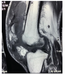
Rheumatoid Arthritis (RA) is a chronic, systematic, and progressive autoimmune disorder, with an incidence of 1-3% [1]. This condition can occur at any age, especially during the fourth or fifth decade of life. The most common sites are the metacarpophalangeal and metatarsophalangeal joints (Figure 1). More than two-thirds of all RA patients show symptomatic cervical spine lesions [2,3], but the lumbar spine is rarely involved. The inflammation in RA causes synovitis, then loosening of the ligaments and capsules, and finally bony erosion, vertebral collapse, and subluxation.

Figure 1:


Clinical presentation

RA in the cervical spine is categorized into three types: atlantoaxial subluxation (AAS), Vertical Subluxation (VS), and Sub axial Subluxation (SS).

Atlantoaxial subluxation (AAS)

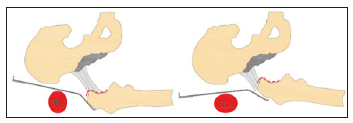
Rheumatoid synovitis at the atlantoaxial joints and around the stabilizing transverse, alar and apical ligaments leads to atlantoaxial instability. Disintegration of the transverse ligament causes anterior AAS. The reported prevalence of anterior AAS was 10-55% [4,5]. Compression of the cervical cord is usually from posteriorly, leading to demyelination of the posterior fascicle. Posterior AAS is less frequent (less than 1%) due to odontoid destruction or fracture [6].

Vertical subluxation

Destruction of the lateral atlantoaxial joint leads to joint depression and upward movement of the axis. Pathologically, this destruction spreads to the occipitoaxial joint [7].

Sub axial subluxation

Destruction of the sub axial joints and dysfunction of the posterior ligaments leads to SS. Common sites are C4/5 and C5/6. The prevalence of SAS is 13% among all RA patients [8]. This condition is associated with AAS and onset is usually late.

Plain radiography

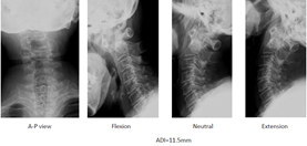
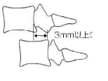
Plain radiographs are the basis of radiographic evaluation for RA in routine screening of the wider population. Anterior AAS may be assessed on a lateral cervical spine radiograph by measuring the Anterior Atlantoaxial Interval (AADI) from the middorsal margin of the anterior ring of C1 to the ventral surface of the odontoid (Figure 2). AADI more than 3mm in an adult or 4mm in a child is considered abnormal [9] (Figure 3). The interval is accentuated on a flexion view if the AAS is present. Posterior AAS may be assessed by measuring the Posterior Atlantoaxial Interval (PADI) from the dorsal aspect of the odontoid to the ventral margin of the C1 lamina.

Figure 2:Atlantoaxial subluxation (AAS).


Figure 3:Radiologic evaluation (Anterior subluxation).


VS can be measured by McRae’s line, Chamberlin’s line or McGregor’s line (Figure 4). McRae’s line connects the front and back of the foramen magnum. If the odontoid process exceeds this line, this condition is regarded as VS. Chamberlin’s line connects the dorsal margin of the hard palate to the dorsal margin of the foramen magnum. If the tip of the odontoid projects more than 6mm, this condition is also regarded as VS. On the other hand, McGregor’s line is easier to use, as it is drawn from the posterior margin of the hard palate to the most caudal point of the occiput. The tip of the odontoid should not project more than 4.5mm.

Figure 4:76 Women, RA, Anterior atlantoaxial subluxation.

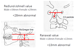
The tip of the odontoid is sometimes difficult to identify because of erosion. In such situations, the Redlund-Johnnell [10] method and Ranawat [11] method are very useful to diagnose VS. The Redlund-Johnnell [10] value is measured as the distance between McGregor’s line and the vertebral body of the axis (Figure 4). A value less than 34mm in men and less than 29mm in women is defined as VS. Ranawat value is measured as the distance between McGregor’s line and the center of the C2 pedicle. A value less than 15mm in men and less than 13mm in women is defined as VS (Figure 5 & 6).

Figure 5:Redlund Johnell method.
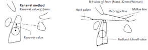

Figure 6:Radiologic evaluation (Vertical subluxation).

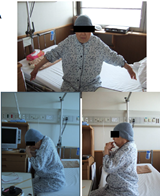
SAS is defined as a slip of more than 3mm in the sub axial vertebra (Figure 7). For RA patients, the lack of osteophyte formation represents a unique feature compared with OA patients (Figure 8). The Space Available for the Cord (SAC) is related to the onset of myelopathy and surgical results [12]. RA patients need to hyper-flex the neck during meals because of ROM (Range of Motion) disturbance of the upper extremities (Figure 9).

Figure 7:79 Women, RA, Vertical subluxation.
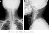

Figure 8:79 Women, RA, Vertical subluxation, CT.
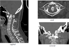

Figure 9:Sub axial Subluxation (SAS).

Computed tomography (CT)

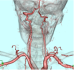
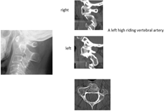
CT is preferred for evaluating bony destruction and ruling out anatomical anomalies. Multiplanar Reconstruction (MPR) provides a three-dimensional evaluation of the cervical spine. Sagittal reconstruction images allow clear visualization of AADI, odontoid erosion, and high-riding vertebral artery (Figures 10-13).

Figure 10:71 Women, RA, Sub axial subluxation.
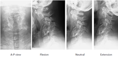

Figure 11:67 Women, RA, Subaxial subluxation.


Figure 12:76 Women, RA.
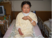

Figure 13:74 Women, RA.

Magnetic resonance imaging (MRI)

As plain radiograms cannot evaluate soft tissues, MRI is very important to assess pannus, cord compression, myelomalacia, and soft tissue (Figures 14-17). MRI in flexion is used to check compression of the spinal cord in AAAS patients [13]. MRI can be used to evaluate the corticomedullary angle. An angle less than 135° is correlated with neurological compression of the medulla [14]. Magnetic resonance angiography reveals a vertebral artery abnormality in 34% of RA patients [15].

Figure 14:Acetabular
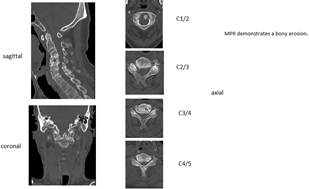

Figure 15:75 Woman, RA.
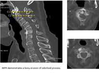

Figure 16:75 Woman, RA.


Figure 17:75 Woman, RA.

Natural history

The prevalence of cervical subluxation in RA patients is 43-86% [16]. The most common form of subluxation is AAAS. Fujiwara et al. identified AAAS in 31% of RA patients, and reducible AAAS progresses to irreducible AAAS, then to VS (Figure 18) [14]. Approximately 20% of subluxations in RA patients involve VS. Neurological deficits are reported in only 7-58% of RA patients [17,18]. Once paralysis develops, the natural history seems to be one of progressive neurological deterioration. Sudden death occurs in as many as 10% of RA patients due to medullary compression [16].

Figure 18:79 Women, RA.

Symptoms

Local symptoms: Neck pain from RA is very common and is caused by inflammation present in the joint when the disease is active. Pain at rest indicates inflammation, but pain during motion indicates instability of the joint.

3.9.2. Neurological deficit: Neurological examinations may be difficult to interpret in RA patients, as subtle neurological changes may be masked. Manual muscle testing is difficult to perform in patients with multiple joint destruction. Peripheral nerve entrapment syndrome modifies the neurological status. Deep tendon reflex is relatively useful to evaluate myelopathy, even in patients with multiple joint destruction. VS patients may show symptoms of medullary compression or lower cranial nerve symptoms.

Vertebral Artery (VA) insufficiency syndrome: VA insufficiency syndrome may occur in patients with AAAS, especially rotatory subluxation. These patients complain of vertigo, tinnitus, nausea, vomiting, and faintness during neck motion. Approximately 20% of RA patients experience occlusion of a VA [19].

3.9.4. General symptoms: RA patients typically complain of fatigue before other symptoms become obvious. Morning stiffness is often an early sign of RA, followed by joint pain, joint swelling, and loss of joint range of motion. When accompanied by other symptoms, low-grade fever can also be a symptom of RA.

Non-surgical treatment: Marks reported significantly better results can be achieved with surgical treatment than with conservative treatment [20]. Among RA patients with myelopathy who refuse surgical treatment, 76% become bedridden within 3 years, and mean survival is 2.3 years [21]. Mainstay nonsurgical treatments for PA patients are physical therapy, patient education, and medication. Non-steroidal anti-inflammatory drugs, steroids, and new immune-modulatory biologic medications are the cornerstones of pharmacotherapy for RA.

Initial pharmacological therapy typically includes methotrexate at 7.5-15mg/week. Corticosteroids are powerful drugs useful for achieving rapid relief from pain. A soft or hard collar can be used to limit anterior subluxation and improve discomfort.

Surgical treatment: Surgery is indicated for RA patients with intractable pain or progressive neurological deficits. The goals of surgery include pain relief, neurological decompression, and prevention of further neurological deterioration. Some patients require awake intubation because of severe cervical cord compression. Preoperative skeletal traction with a halo ring or halo jacket should be considered for patients with severe myelopathy (Figure 19). Somatosensory and motor-evoked potentials are important for monitoring spinal cord function during positioning and surgery.

Figure 19:67 Woman, RA.
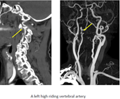

Transoral odontoidectomy: Indications for this technique are limited, because RA patients can experience difficulty opening the mouth completely due to temporomandibular joint disturbances. A distance of at least 25mm between the front teeth is needed for this procedure. As RA patients already have some degree of immunodeficiency, surgical-site infection may be another problem associated with this procedure [22] (Figure 20).

Figure 20:79 Women, RA.
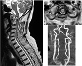

Posterior atlantoaxial fixation: In AAS patients, an AADI of 9mm or more, a SAC of less than 14 mm, or vertebral artery insufficiency are candidates for atlantoaxial arthrodesis surgery [23]. In the historical background, this technique was described Gallie [24] and Brooks & Jenkins [25] (Figures 21 & 22). C2 laminar screw and C1 lateral mass screw fixation, regarded as the Wright technique, is also a safer than Magerl [26] method (Figures 23 & 24). Three famous methods are available to put screws into the C1 lateral mass; Goel, Tan, and Notch methods (Figures 25 & 26). A disadvantage of these techniques is a high rate of nonunion due to reduced stability [27]. Magerl [28] reported trans-articular screw fixation, as the strongest anchor for the upper cervical spine (Figures 27 & 28). However, this procedure has a potential for VA injury in the presence of a high-riding VA [29,30] (Figure 29). Goel reported safer, more rigid fixation using a C1 lateral mass screw and C2 pedicle screw [31] (Figures 30-32). The most important advantage of this technique is that after placement of four screws, AAS can be reduced using screws and rods.

Figure 21:60 Men, RA.
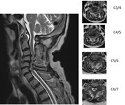

Figure 22:76 Woman, RA.
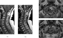

Figure 23:67 Woman, RA.
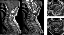

Figure 24:
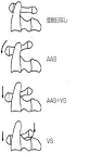

Figure 25:
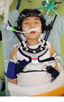

Figure 26:Transoral adenoidectomy.
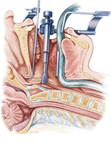

Figure 27:Gallie fusion.
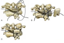

Figure 28:Brooks-Jenkins Fusion.
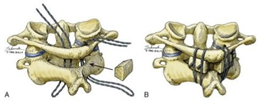

Figure 29: C2 laminar screw (Wright 2004).

Posterior occiput cervical fusion: This technique is mainly indicated for patients with irreducible AAAS and VS. For VS patients, odontoid migration cranial to McGregor’s line of at least 5mm or an abnormal Redlund-Johnnell [10] or Ranawat [11] value are indications for occipitoaxial (OC2) fusion surgery [32]. A major complication for this procedure is dysphagia (Figures 33-36), so OC2 angle should be considered before surgery. An angle of approximately 20° is ideal for OC2 fusion [33]. The navigation is very useful for identifying the thickest part of the occiput and screwing pedicle screws into the cervical spine [34] (Figures 37-44).

Figure 30:52 Men, RA, AAAS, Wright method.
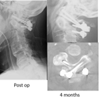

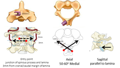
Figure 31:Three methods of C1 LMS placement.
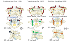

Figure 32:C1 LMS technique.
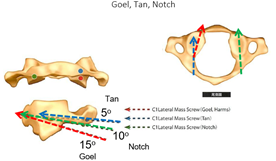

Figure 33:C1/2 transarticular screw (Magerl 1987).
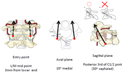

Figure 34:62 Woman, RA, AAAS, Magerl fixation.
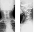

Figure 35:Iatrogenic VA Injury with Magerl Fixation.
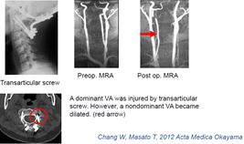

Figure 36:Goel method (C1 LMS & C2 Pedicle screw).
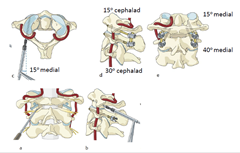

Figure 37:73 Woman, RA, AAAS, Goel fixation.
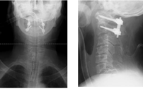

Figure 38:C2 pedicle screw
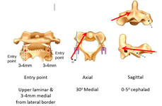

Figure 39:C2 pars screw (Judet 1962).


Figure 40:Dysphagia after OC2 Fusion.
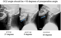

Figure 41:75 Woman, RA, AAAS.
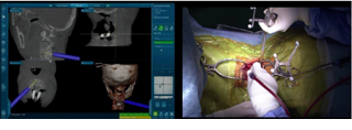

Figure 42:75 Woman, RA, AAAS.
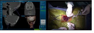

Figure 43:75 Woman, RA, AAAS.
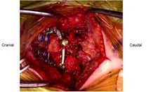

Figure 44:75 Woman, RA, AAAS.


Conclusion

The goal in management of RA cervical spine lesion is to prevent severe neurological deficit. The early diagnosis is very important because myelopathy is masked with peripheral joint problems. Among several imaging, the lateral flexion radiogram and MRI are very useful to diagnose cervical subluxation. For the surgery, the posterior fixation with instrumentation is the standard treatment. However, the surgeon should be reminded several severe complications. When the solid bony fusion is obtained, the pain relief and neurological recovery of the RA patient are expected.

References

    1. Helmick CG, Felson DT, Lawrence RC, Gabriel S, Hirsch R et al. (2008) National arthritis data workgroup, Estimates of the prevalence of arthritis and other rheumatic conditions in the United States. Part I Arthritis Rheum 58(1): 15-25.

    2. Papadopoulos SM, Dickman CA, Sonntag VK (1991) Atlantoaxial stabilization in rheumatoid arthritis. J Neurosurg 74(1): 1-7.

    3. Delamarter RB, Bohlman HH (1994) Postmortem osseous and neuropathologic analysis of the rheumatoid cervical spine. Spine (Phila Pa 1976) 19(20): 2267-2274.

    4. Conlon PW, Isdale IC, Rose BS (1966) Rheumatoid arthritis of the cervical spine: an analysis of 333 cases. Ann Rheum Dis 25(2): 120-126.

    5. Park WM, O Brien W (1969) Computer-assisted analysis of radiographic neck lesions in chronic rheumatoid arthritis. Acta Radiologicaa Diagnosis 8(6): 529-34.

    6. Joaquim AF, Appenzeller S (2014) Cervical spine involvement in rheumatoid arthritis-A systematic review. Autoimmun Rev 13(12): 1195-202.

    7. Eulderink F, Meijers KA (1976) Pathology of the cervical spine in rheumatoid arthritis: a controlled study of 44 spines. J Pathol 120(2): 91-108.

    8. Zhang T, Pope J (2015) Cervical spine involvement in rheumatoid arthritis over time: results from a meta-analysis. Arthritis Res Ther 17(1): 148.

    9. Rojas, CA, Bertozzi JC, Martinez CR, Whitlow J (2007) Reassessment of the craniocervical junction: Normal values on CT. American Journal of Neuroradiology. 28(9): 1819-1823.

    10. Redlund-Johnell I, Pettersson H (1984) Vertical dislocation of the C1 and C2 vertebrae in rheumatoid arthritis. Acta Radiol Diagn (Stockh) 25(2): 133-141.

    11. Ranawat CS, O Leary P, Pellicci P, Tsairis P, Marchisello P et al. (1979) Cervical spine fusion in rheumatoid arthritis. J Bone Joint Surg Am 61(7): 1003-1010.

    12. Boden SD, Dodge LD, Bohlman HH, Rechtine GR (1993) Rheumatoid arthritis of the cervical spine. A long-term analysis with predictors of paralysis and recovery. J Bone Joint Surg Am 75(9): 1282-1297.

    13. Reynolds H, Carter SW, Murtagh FR, Silbiger M, Rechtine GR (1987) Cervical rheumatoid arthritis: value of flexion and extension views in imaging. Radiology 164(1): 215-218.

    14. Narváez JA, Narváez J, Serrallonga M, De Lama E, de Albert M, et al. (2008) Cervical spine involvement in rheumatoid arthritis: correlation between neurological manifestations and magnetic resonance imaging findings. Rheumatology (Oxford) 47(12): 1814-1819.

    15. Zenmyo M, Ijiri K, Sasaki H, Sakakima H, Taketomi E, et al. (2010) Magnetic resonance angiography for vertebral artery evaluation in rheumatoid arthritis patients. Neurosurgery 66(6): 1174-1180.

    16. Rasker JJ, Cosh JA (1978) Radiological study of cervical spine and hand in patients with rheumatoid arthritis of 15 years' duration: an assessment of the effects of corticosteroid treatment. Ann Rheum Dis 37(6): 529-535.

    17. Fujiwara K, Owaki H, Fujimoto M, Yonenobu K, Ochi T (2000) A long-term follow-up study of cervical lesions in rheumatoid arthritis. J Spinal Disord 13(6): 519-526.

    18. Conaty JP, Mongan ES (1981) Cervical fusion in rheumatoid arthritis. J Bone Joint Surg Am 63(8): 1218-1227.

    19. Sherk HH (1978) Atlantoaxial instability and acquired basilar invagination in rheumatoid arthritis. Orthop Clin North Am 9(4): 1053-1063.

    20. Marks JS, Sharp J (1981) Rheumatoid cervical myelopathy. Q J Med 50(199): 307-319.

    21. Sunahara N, Matsunaga S, Mori T, Ijiri K, Sakou T (1997) Clinical course of conservatively managed rheumatoid arthritis patients with myelopathy. c Spine (Phila Pa 1976) 22(22): 2603-2607.

    22. Crockard HA, Calder I, Ransford AO (1990) One-stage transoral decompression and posterior fixation in rheumatoid atlanto-axial subluxation. J Bone Joint Surg Br 72(4): 682-685.

    23. Oda T, Yonenobu K, Fujimura Y, Ishii Y, Nakahara S et al. (2009) Diagnostic validity of space available for the spinal cord at C1 level for cervical myelopathy in patients with rheumatoid arthritis. Spine (Phila Pa 1976) 34(13): 1395-1398.

    24. Gallie WE (1939) Fractures and dislocations of cervical spine. Amj Surg 46(3): 495-499.

    25. Brooks AL, Jenkins EB (1978) Atlantoaxial arthrodesis by the wedge compression method. J Bone Joint Surg Am 60(3): 279-284.

    26. Wright NM, Lauryssen C (1998) Techniques of posterior C1-C2 stabilization. Tech in Neurosurg 4: 286-297.

    27. Grob D, Crisco JJ, Panjabi MM, Wang P, Dvorak J (1992) Biomechanical evaluation of four different posterior atlantoaxial fixation techniques Spine 17(5): 480-490.

    28. Grob D, Magerl F (1987) Surgical stabilization of C1 and C2 fractures. Orthopade 16(1): 46-54.

    29. Neo M, Matsushita M, Yasuda T, Sakamoto T, Nakamura T (2002) Use of an aiming device in posterior atlantoaxial transarticular screw fixation. Technical note. J Neurosurg. 97(1 Suppl): 123-127.

    30. Zhang W, Tanaka M, Sugimoto Y, Ikuma H, Nakanishi K et al. (2012) Dominant vertebral artery injury during posterior atlantoaxial transarticular screw fixation in a juvenile rheumatoid arthritis patient with atlantoaxial subluxation. Acta Med Okayama 66(1): 77-81.

    31. Goel A, Laheri V (1994) Plate and screw fixation for atlanto-axial subluxation. Acta Neurochir (Wien) 129(1-2): 47-53.

    32. Riew KD, Hilibrand AS, Palumbo MA, Sethi N, Bohlman HH (2001) Diagnosing basilar invagination in the rheumatoid patient. The reliability of radiographic criteria. J Bone Joint Surg Am 83(2): 194-200.

    33. Izeki M, Neo M, Takemoto M, Fujibayashi S, Ito H et al. (2014) The O-C2 angle established at occipito-cervical fusion dictates the patient's destiny in terms of postoperative dyspnea and/or dysphagia. Eur Spine J 23(2): 328-336.

    34. Krauss WE, Bledsoe JM, Clarke MJ, Nottmeier EW, Pichelmann MA (2010) Rheumatoid arthritis of the craniovertebral junction. Neurosurgery 66(3 Suppl): 83-95.

    https://crimsonpublishers.com/rabs/fulltext/RABS.000522.php

    Crimson Publishers: https://crimsonpublishers.com/

    For more articles in Journal of Arthritis Bone Disease
    Please click on below link: https://crimsonpublishers.com/rabs/    

Thursday, August 12, 2021

Different Approach to Arthritis_ Crimson Publishers

 Different Approach to Arthritis by Maria Kuman* in Researches in Arthritis & Bone Study_ Journal of Arthritis Bone Disease


Abstract

At the age of 60, I started experiencing arthritis-kind of pain - very step was painful and I couldn’t open a jar. The lawnmower was so heavy that I barely walk after it. I would do barely ½ of the yard and leave the rest for another day. I decided I needed to do something about it. I read about a Chinese man, who lived to be 256 years old. He received from the Chinese government awards for long living three times: at ages 150, 200, and 250. When they asked him, what made him live so long, his answer was simple: inner peace and periodic cleansing.

So, I decided to do cleansing-to flush my body clean. It made sense to me. We regularly change the air and gas filters of our cars knowing that if we don’t do it, the car efficiency will decrease and finally the car will stop working. We change or flush regularly the air filters of our air-conditioning system in the house knowing that if we don’t do it, the efficiency of our air conditioning will decrease and with time it would stop working. The same should be true for our body, but somehow, we expect it to keep working with the same efficiency and never to fail working without any cleansing.

https://crimsonpublishers.com/rabs/fulltext/RABS.000521.php

Crimson Publishers: https://crimsonpublishers.com/

For more articles in Journal of Arthritis Bone Disease,
Please click on below link: https://crimsonpublishers.com/rabs/

Tuesday, May 18, 2021

The Direct Anterior Approach for Total Hip Arthroplasty Without Specific Table: Surgical Approach and Our Seven Years of Experience_ Crimson Publishers

 The Direct Anterior Approach for Total Hip Arthroplasty Without Specific Table: Surgical Approach and Our Seven Years of Experience by Klemen Bedencic* in Researches in Arthritis & Bone Study_ Journal of arthritis bone disease


Abstract
Background: The Direct Anterior Approach (DAA) for Total Hip Arthroplasty (THA) has been gaining popularity in recent years because it is the only approach performed in both the intermuscular and the interneural planes. The number of academic articles about DAA is increasing, but still a number of authors continue to question the safety of this approach and its utilization on a day-to-day basis as a standard approach, especially because of the associated steep learning curve. In this article we describe our surgical technique and present lessons learnt from seven years of uniformly excellent experience with DAA.

Materials and Methods: The key technical steps of our surgical technique are described below: namely, surgical exposure, acetabular preparation, femoral preparation, trial reduction and definite prosthesis insertion, wound closure, and rehabilitation. We present DAA with the patient in the supine position on a regular orthopaedic table, both legs are draped free. For femoral exposure, the leg operated upon is placed in the “Figure 1” position.

Result: Between November 2010 and June 2017, we have performed 1528 THA using DAA. In the last year 400 cases, there were 379 primary THA and 21 revisions. Patients are mobilized the same day and the average length of hospitalization is three days. We observe on average ten calcar fractures per year, which can either be merely kept under observation or fixed with a single cortical screw. The rate of prosthesis dislocation is significantly below 0.5% and the rate of deep infection remained as low as with the previous approaches used.

Conclusion: In our opinion, DAA is the best approach for THA because it is the only truly minimally invasive one. It causes fewer dislocations, minimizes postoperative pain and enables faster rehabilitation without the stricter limitations when compared to other approaches

https://crimsonpublishers.com/rabs/fulltext/RABS.000520.php

Crimson Publishers: https://crimsonpublishers.com/

For more articles in Journal of arthritis bone disease,

Please click on below link: https://crimsonpublishers.com/rabs/

Thursday, March 4, 2021

Knee Dislocation and Patellar Tendon Disruption: A Case Report_ Crimson Publishers

 Knee Dislocation and Patellar Tendon Disruption: A Case Report by Rodrigo KP* in Researches in Arthritis & Bone Study_ Journal of Arthritis Bone Disease


Abstract

Dislocation of the knee is a serious event, often caused by a violent trauma. Patellar tendon rupture cases are described in patients with trauma and even with no trauma at all in some chronic systemic diseases. This is a case report of these two events together in the same patient. A 56 years-old man with rheumatoid arthritis had knee dislocation and patellar tendon disruption after a small trauma. Due to local cellulitis, the surgery was postponed. We decided to perform reconstruction of the patellar tendon only because of the patient’s comorbidity and because the knee was stable. The patient resumed his job after six months of physical therapy. In this report, we describe a very uncommon case of knee dislocation and patellar tendon disruption together. Although the surgery is currently the choice to treat knee dislocation, this patient had only the patellar tendon reconstruction due the rheumatoid arthritis. This very uncommon case, with a surgery treatment, reminds us to be always careful when examining a patient with knee dislocation. We should not forget to examine the patellar height.

https://crimsonpublishers.com/rabs/fulltext/RABS.000519.php

Crimson Publishers: https://crimsonpublishers.com/

For more articles in Journal of Arthritis Bone Disease,
Please click on below link: https://crimsonpublishers.com/rabs/

Tuesday, February 23, 2021

Aquatic Exercise for Patients with Osteoarthritis_ Crimson Publishers

Aquatic Exercise for Patients with Osteoarthritis by Michaels NN* in Researches in Arthritis & Bone Study_ Journal of Arthritis Bone Disease


Abstract
Osteoarthritis (OA), also known as degenerative joint disease, is the most common type of arthritis, affecting over 30 million people in the United States alone. The joint pain experienced by patients with OA often results in decreased mobility and function which contributes to the weight gain, joint swelling, muscle weakness, and depression often observed in this population. Exercise in the water offers a low impact option for exercise, with the soothing effects of buoyancy. This paper discusses the use of aquatic exercise as a non-pharmacological option to improve mobility and reduce pain for people with OA.

Introduction
Osteoarthritis (OA), also known as degenerative joint disease, is the most common type of arthritis, affecting over 30 million people in the United states alone [1]. OA affects both men and women is listed as one of the most frequent causes of physical disability in the older adult population [2]. The joint pain experienced by patients with OA often results in decreased mobility and function which contributes to the weight gain, joint swelling, muscle weakness, and depression often observed in this population [3]. As a physical therapist (PT) and an occupational therapist (OT), we have both been exposed to many interventions supported in the literature to reduce pain and improve mobility in individuals with OA, including pharmacological, non-pharmacological, and surgical interventions [4].

This paper discusses the use of aquatic exercise as an option to improve mobility and reduce pain for people with OA. OT and PT both fall within the non-pharmacological intervention category. According to Woods et al., the only non-pharmacological interventions offered by the European League Against Rheumatism (EULAR) for knee OA are the use of appliances like braces and insoles, and appropriate footwear, although these researchers found transcutaneous electrical nerve stimulation (TNS) to be an effective option. Aquatic exercise was not mentioned, and yet, this is a good alternative to pharmacological measures in the reduction of pain from OA

https://crimsonpublishers.com/rabs/fulltext/RABS.000518.php

Crimson Publishers: https://crimsonpublishers.com/

For more articles in Journal of Arthritis Bone Disease,

Please click on below link: https://crimsonpublishers.com/rabs/

Monday, September 14, 2020

Simultaneous Oblique Lumbar Interbody Fusion (OLIF) and Lateral Percutaneous Pedicle Screw Fixation (Lateral PPS): A Technical Note_ Crimson Publishers

 Simultaneous Oblique Lumbar Interbody Fusion (OLIF) and Lateral Percutaneous Pedicle Screw Fixation (Lateral PPS): A Technical Note by Tanaka M* in Researches in Arthritis & Bone Study_ Journal of Arthritis Bone Disease


Abstract

Purpose: Due to its distinct advantages, lateral approach to the lumbar spine with insertion of percutaneous pedicle screws is increasingly used to treat lumbar degenerative pathologies. We describe a novel technique of simultaneous Oblique Lumbar Interbody Fusion [OLIF] and Percutaneous Pedicle Screw [PPS] insertion in single lateral position with O-arm based navigation system. To the best of our knowledge, this technique has not yet been described in the literature.

Methods: The authors report clinical and radiological results of a 73-year-old lady treated for degenerative lumbar pathology. The authors performed C-arm free OLIF and PPS in single lateral decubitus position simultaneously and describe the technique in detail.

Result: The patient was successfully relieved of her pre-operative symptoms with no neurological sequelae. At 18 months follow up, she maintained good sagittal alignment and balance.

Conclusion: Simultaneous insertion of OLIF cage and PPS in lateral position is possible with O-arm based navigation technique. This significantly reduces operating time and radiation exposure without compromising the results.

https://crimsonpublishers.com/rabs/fulltext/RABS.000517.php

For more Open access journals in Crimson Publishers,
Please click on the link: https://crimsonpublishers.com/

For more articles in Journal ofArthritis Bone Disease,
Please click on below link: https://crimsonpublishers.com/rabs/

Follow On Publons: https://publons.com/publisher/6342/crimson-publishers/
Follow On Linkedin: https://www.linkedin.com/company/crimsonpublishers/

High impact journals in Crimson Publishers?
https://www.quora.com/What-are-the-high-impact-journals-in-Crimson-Publishers

Crimson Publishers Journals
https://crimsonpublishers.com/

Visual-cognitive Skills and Physical Qualities in Elite Soccer: Practical Considerations for Training and Return-to-Play Protocols: Crimson Publishers

Visual-cognitive Skills and Physical Qualities in Elite Soccer: Practical Considerations for Training and Return-to-Play Protocols by Lukasz...