Crimson Publishers High Impact Journals

Thursday, June 30, 2022

Sexual Abuse Identified after Suicide: Case Report_Crimson Publishers

 Sexual Abuse Identified after Suicide: Case Report by Kenan Kaya in COJ Nursing & Healthcare_research in nursing and health


Abstract

Child abuse is a maltreatment of a child under the age of 18 by his or her parent, carer, someone living in their home, or someone working with children. Abuse of a child is anything that causes injury or puts the child in danger of physical injury. Child abuse can be physical, sexual or emotional. Sexual abuse has been reported to be the largest component of suicide attempts in childhood. Between 1950 and 1993, children under the age of 15 had a 4-fold increase in suicide-related deaths among causes of death. In 1994, 6 percent of deaths between 5 and 14 years of age were found to be suicidal. Case; on a 14-year-old girl’s body’s external examination, where dead stiffness continued and purple-colored dead stains began to appear in unprinted areas. It was recorded that the telem was viewed as a parchmented area starting from the middle line, 10cm long and 1.5cm wide in the thickest area. On the outer side of the left knee there was a 3x2cm scar tissue. It was observed that the hymen was 2cm wide and had a notched structure and a partial tear at the level of 7 o’clock. There was no ecchymosis and hemorrhage around the anus and hymen. On the internal examination, a 2.5 centimeter fetus was detected in the uterus between 6-8 weeks of gestation. Only male DNA profiles were detected from the pregnancy material. Toxicological analyzes result in no detectable substance. It was determined that death was caused by mechanical asphyxia, the result of hanging. A 14-year-old girl who has been diagnosed with suicide mortality and pregnancy after autopsy will be examined in the light of literature in terms of child abuse and pregnancy.

Keyword: Death; Suicide; Sexual abuse; Child; Pregnancy

Introduction

Child abuse is the maltreatment of a child under the age of 18 by his or her parent, carer, someone living in their home, or someone working with children. Abuse of a child is anything that causes injury or puts the child in danger of physical injury. Child abuse can be physical, sexual or emotional. The World Health Organization refers to child abuse as “all of the behaviors of an adult, society or state, which harm the general values of the child’s life, health, development, trust, responsibility and skills” [1].

It is stated that child abuse is an important part of sexual abuse among the causes [2]. Between 1950 and 1993, children under the age of 15 had a 4-fold increase in suicide-related deaths among causes of death, and in 1994, 6% of deaths between 5 and 14 years of age were suicide-related [3]. De Wilde and colleagues reported that people who had sexually abused adolescents constituted a high risk group for suicide [4]. According to a study conducted in the United States on the Youth Health Behavior, the proportion of high school students who have experienced sexual harassment has reached 20.9% in Oregon and high-risk behaviors and suicide attempts rates have been found to increase in the group of sexual exploitation, especially during adolescence [5]. In the case report, we aimed to analyze the cause of pregnancy and sexual abuse detected after the autopsy of a 14-year-old girl.

Case Report

On a 14-year-old girl’s body’s external examination, where dead stiffness continued and purple-colored dead stains began to appear in unprinted areas. It was recorded that the telem was viewed as a parchmented area starting from the middle line, 10cm long and 1.5cm wide in the thickest area. On the outer side of the left knee there was a 3x2cm scar tissue. It was observed that the hymen was 2cm wide and had a notched structure and a partial tear at the level of 7 o’clock. Postmortem dilatation was detected in the anal examination. Ecchymosis and hemorrhage were not observed around the anus and hymen. On the internal examination, the stomach was seen as empty, and a 2.5centimeter fetus was detected in the uterus. In the toxicological analysis of the blood taken during the autopsy, there were no foreign substances in the blood, It was determined that the fetus was between 6-8 weeks of gestation, that the pregnancy material was compatible with the male DNA profile, and that the death was caused by mechanical asphyxia the result of hanging. According to the statements taken in the forensic investigation; the girl was hanging in her uncle’s house, the girl was already dead when the rope was cut off, the girl did not finish her primary school, the girl went to work in the cotton field 2 months ago and the girl tried to commit suicide by taking medication a month ago.

Discussion

Childhood suicides have increased in recent years. Sexual abuse of the child has an important place among causes of childhood suicide attempts. In a study conducted in 1993, 177 adolescent cases were examined and a significant relationship was found between physical or sexual abuse and suicide attempt. These cases have been reported to have recurrent suicide attempts [2]. In our case, the child had a history of suicide attempts a month ago, and the child has not been sufficiently observed, and has not been investigated for pregnancy. This shows that child abuse does not take place in the differential diagnosis. Suicide attempts affect family and society negatively; but it should not be forgotten that suicide is a call for help.

In a survey conducted by the National Society for the Prevention of Cruelty to Children (NSPCC), it was determined that the average age of sexually abused children was 10.2 and that 80 percent of these children were girls [6]. The concept of death under the age of ten is sophisticated and undefined, so there is less suicide attempt. It is believed that the developmental process of the concept of death was created at the age of 7-8 [7].

Many studies of sexual trauma show that the rate of exposure of girls to sexual assault is very common. A prevalence study has shown that 12 percent of adolescents are exposed to lifetime sexual abuse and / or assault [8]. In another literature, 76 percent of suicide attempts have been reported to occur in the home environment [9].

A child who has been sexually abused may face different psychosexual problems that affect him/her throughout his/her life. It has been reported that psychological damage caused by abuse causes the self-respect of the child to diminish in some way, and that social relations deteriorate following self-harm [10]. Kilpatrick and colleagues note that the anxieties and fears of sexual abuse (AIDS, sexually transmitted diseases, pregnancy, witnessing in court) are experienced by all victims [11]. The unacceptable nature and sudden onset of your activity lead to excessive fear, lack of control, and increased trauma response [12]. It should be kept in mind that the 14-year-old girl’s pregnancy is interpreted as child abuse, and the reasons for not telling the pregnancy to the family are embarrassment, social and traditional pressure.

In such deaths, hair and similar evidence on the body and clothing in the acute process is important for the identification of the attacker. It is also suggested that (especially if genital, perianal, and anal regions have traumatic symptoms) investigate traumatic symptoms; oral, rectal, and vaginal swabs taken for DNA analysis and antigenic seminal fluid speciation, searching for vaginal and rectal impairments etc. [13,14]. However, in our case about the presence of a 6-8 week fetus, DNA is the most important evidence for finding the identity of the attacker.

Conclusion

According to the law, a girl aged 14 years is guilty of sexual intercourse with her own discretion. Providing adequate support for the child’s protection, protecting the child from the family or community (ethical) pressures associated with possible pregnancies, and building confidence in this issue should be one of the government’s most important tasks.

References

  1. WHO (1999) Guidelines for medico-legal care for victims of sexual violence. Geneva, Switzerland.
  2. Shaunescy K, Cohen JL, Plummer B, Bcrmaii A (1993) Suicidally in hospitalized adolescental relationship tp prior abuse. Am J Orthopsychiatry 63(1): 113-119.
  3. Anonymous (1997) Rates of homicide, suicide and fire-arm related death among children 26 industrialized countries. MMWR Morb Mortal Wkly Rep 46(5): 101-105.
  4. De Wilde, Kienhorst EJ, Diekstra CW, Wolters RF (1994) Social support, life events and behavioral characteristics of psychologically distressed adolescents at high risk for attempted suicide. Adolescence 29(113): 49- 60.
  5. Nelson DE, Higginson GK, Grant Wiley WA (1994) Using the youth risk behavior survey to estimate prevalance of sexual abuse among oregon high school students. J School Health 64(10): 413-416.
  6. Surrey J, Sweet C, Michaels A, Levin S (1990) Reported history of physical and sexual abuse and severity of symptomatology in women psychiatric outpatients. Am J Orthopsychiatry 60(3): 412-417.
  7. Ozdogan B (1988) Child and Play Therapy. Ankara, Turkey.
  8. Council on Scientific Affairs, American Medical Association (1992) Violence against women: relevance for medical practitioners. JAMA 267(23): 3184-3189.
  9. Cirit H, Eğilmez A, Kültür S (1990) Investigation of psychiatric symptomatology and family characteristics in adults whoare attempting suicide. XXVI Scientific Studies of National Congress of Psychiatry and Neurological Sciences. İzmir, Turkey, pp. 547-553.
  10. Zciller B (1982) Physical and psychological abused and delinquent adolescents. Child Abuse Neglet 6(2): 207.
  11. Moscarello R (1990) Psycological management of victims of sexual assault. Canadian Journal of Psychiatry 35(1): 25-30.
  12. Davidson JRT, Smith RD (1990) Traumatic experience in psychiatric outpatients. Journal of Traumatic Stress 3(3): 459-476.
  13. Yorulmaz C, Bütün C, Dokgöz H, Citici I, Altun G (2003) Death associated with sexual abuse of children. Forensic Sci İnt 136(Suppl 1): 397-398.
  14. Polat O, Yaycı N (2007) Approach to cases of death due to abuse. Child Abuse by All Dimensions. Prevention and Rehabilitation 2: 221-256.

For more articles in research in nursing and health
Please click on below link: https://crimsonpublishers.com/cojnh/

Wednesday, June 29, 2022

Evaluation of Leg Length Discrepancy (LLD) after Unilateral Total Knee Arthroplasty (TKA) Done for Varus Knee Deformity_Crimson Publishers

Evaluation of Leg Length Discrepancy (LLD) after Unilateral Total Knee Arthroplasty (TKA) Done for Varus Knee Deformity by Ahmed Khalifa A in Researches in Arthritis & Bone Study_Arthritis Open Access Journals


Abstract

Leg Length Discrepancy (LLD) is a major concern in patients undergoing lower limb total joint arthroplasty, it is commonly studied in Total Hip Arthroplasty (THA) literature, however, few studies evaluated this issue in Total Knee Arthroplasty (TKA) patients. In this mini-review, we will mention in brief how to evaluate the LLD associated with LLD as well as reviewing what had been reported about its incidence and the possible effect on clinical outcomes.

Introduction

Varus deformity associated with knee osteoarthritis considered to be the most commonly encountered deformity in patients undergoing Total Knee Arthroplasty (TKA), which is usually associated with varying degrees of medial soft tissue structures contractures, laxity of the lateral soft tissue structures, flexion deformity and medial knee compartment bone erosions [1]. The challenges in performing TKA in a varus arthritic knee include the restoration of mechanical limb alignment after balancing the medial and lateral soft tissue tension by equalizing the flexion and extension gaps [2]. Limb Length Discrepancy (LLD) less than 2cm is usually not noticeable and does not require treatment, however, if the LLD is over 2cm, it is usually noticed by the patients with a tendency to perform a self-compensation mechanism such as walking on the ball of the foot (toe down) or by tilting the pelvis and curving the spine, which eventually may lead to lower back pain, gait abnormalities and even can lead to hip arthritis [3-5]. LLD and its effects on patient function have been discussed in depth in the Total Hip Arthroplasty (THA) literature, although it had been reported with TKA, however, few studies handled this issue with its effects on the clinical outcomes [6,7].

How to assess LLD associated with TKA

Clinically: Apparent or functional leg length (which can be affected due to other causes rather than the TKA such as pelvis or spine problems) can be measured using measuring tape from a fixed point (usually the umbilicus) to a point represented bilaterally in both limb (usually the medial malleolus) [5,8,9], while the true or the anatomical leg length (which represents the actual bony length of the lower limb) can be measured from a separate points on each limb independently (which usually the anterior superior iliac spine “ASIS” proximally and the medial malleolus distally)

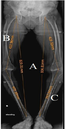
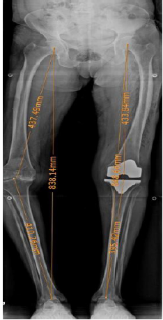
Radiologically: Using a full-length (hip to ankle) standing anteroposterior (AP) radiographs of the bilateral lower extremities as a routine during pre- and postoperative evaluation of TKA patients [10-12]. A marker is used to digitally scale the radiograph and the functional length is performed by measuring the distance from the center of the head to a point located at the center of the tibial plafond (Figure 1A) for both sides, while to determine the anatomical length of the limb, the femur and the tibia are measured separately. Anatomical length of the femur is determined by the length of a line connecting the center of the femoral head to the center of the roof of the intercondylar notch (Figure 1B), while the anatomical length of the tibia is determined by the length of a line connecting the center of the tibial plateau to the base of the tibial plafond (Figure 1C). Obtaining these measurements can guide the surgeon to detect if there is a pre- or post TKA LLD, and if present, it will determine its source (Figure 2).

Figure 1:Preoperative radiological measurement of leg length of both lower limbs on long film AP radiograph.
A. Functional length of both limbs.
B. Anatomical length of the femur.
C. Anatomical length of the tibia.


Figure 2:Postoperative long film AP radiograph showing the LLD between the operated and nonoperated limb after TKA.


Will the LLD after TKA affect the clinical outcomes

As we mentioned earlier that the data regarding the effect of LLD on patients having THA is huge but deficient regarding its incidence and secondary effect in the case of patients having TKA, however, few studies discussed this issue. Lang et al. [3] in their study in 2010 on 102 knees performed over a period of seven months, they found that 85 (83.3%) knees showed increase in the limb length after TKA and the average lengthening was 6.3mm (SD: 6.85mm; range, -11.0 to 24.0mm), however, they didn’t correlate this LLD with patients clinical outcomes. A study by Vaidya et al. [13] who reported that LLD after unilateral, not bilateral TKA done for varus knees with osteoarthritis significantly affected the functional outcomes. However, they also noted that 83.3% of the patients in the unilateral group and 46.6% in the bilateral group had LLD. Another study by Tipton et al. [14] in 2015 including 203 patients underwent TKA, 59.1% of the patients included in this study experienced an increase in limb length with an average increase of 0.438cm.
The authors didn’t report on the clinical outcomes as well as the previous study. A recent study by Kim et al. [15] including 148 patients reviewed retrospectively which was divided into two groups with one group including Eighty-one knees having a <15mm LLD, and the other group including 67 knees found to have more than a 15mm LLD, they evaluated the radiographic outcomes, clinical outcomes, patients satisfaction, and perception of LLD were also evaluated. They found a significant difference in the Knee Society function score and the score for the difficulty with ascending the stairs in the Western Ontario and McMaster Universities score between both groups, however, there was no difference in the results of their satisfaction questionnaires. They concluded that the functional outcomes of more than 15mm post-operative LLD after TKA were lower than those of the <15mm LLD. Thus, the reduced post-operative LLD should be considered to improve the functional outcomes of primary TKA.

Conclusion

Although not much studied, LLD related to TKA can occur and surgeons should keep it into account while performing TKA. More studies should be performed to assess the incidence of LLD following TKA as well as its effect on clinical as well as radiological outcomes and if it has any impact on implant survival and rate of revision.

References

  1. Mihalko WM, Saleh KJ, Krackow KA, Whiteside LA (2009) Soft tissue balancing during total knee arthroplasty in the varus knee. J Am Acad Orthop Surg 17(12): 766-774.
  2. Mullaji, Arun B, Shetty, Gautam (2014) Deformity correction in total knee arthroplasty. Asian Heart Institute and Research Center, India.
  3. Lang JE, Scott RD, Lonner JH, Bono JV, Hunter DJ, et al. (2012) Magnitude of limb lengthening after primary total knee arthroplasty. J Arthroplasty 27(3): 341-346.
  4. Khalifa AA (2017) Leg length discrepancy: Assessment and secondary effects. Ortho & Rheum Open Access 6(1): 001-005.
  5. Clark CR, Huddleston HD, Schoch EP, Thomas BJ (2006) Leg-length discrepancy after total hip arthroplasty. J Am Acad Orthop Surg 14(1): 38-45.
  6. Konyves A, Bannister G (2005) The importance of leg length discrepancy after total hip arthroplasty. J Bone Joint Surg Br 87(2): 155-157.
  7. Edeen J, Sharkey P, Alexander AH (1995) Clinical significance of leg-length inequality after total hip arthroplasty. Am J Orthop (Belle Mead NJ) 24(4): 347-351.
  8. McCaw S, Bates B (1991) Biomechanical implications of mild leg length inequality. Br J Sports Med 25(1): 10-13.
  9. Beard D, Andrew JG, Jeya P, John FN (2008) Incidence and effect of leg length discrepancy following total hip arthroplasty. Physiotherapy 94(2): 91-96.
  10. Murray KJ, Azari MF (2015) Leg length discrepancy and osteoarthritis in the knee, hip and lumbar spine. J Can Chiropr Assoc 59(3): 226-237.
  11. Sabharwal S, Kumar A (2008) Methods for assessing leg length discrepancy. Clin Orthop Relat Res 466(12): 2910-2922.
  12. Harvey WF, Yang M, Cooke TD, Segal NA, Lane N, et al. (2010) Association of leg-length inequality with knee osteoarthritis: A cohort study. Ann Intern Med 152(5): 287-295.
  13. Vaidya SV, Patel MR, Panghate AN, Rathod PA (2010) Total knee arthroplasty: Limb length discrepancy and functional outcome. Indian J Orthop 44(3): 300-307.
  14. Tipton S, Sutherland, Schwarzkopf R (2015) Change in limb length after total knee arthroplasty. Geriatr Orthop Surg Rehabil 6(3): 197-201.
  15. Kim SH, Rhee SM, Lim JW, Lee HJ (2016) The effect of leg length discrepancy on clinical outcome after TKA and identification of possible risk factors. Knee Surg Sports Traumatol Arthrosc 24(8): 2678-2685.

For more articles in Arthritis Open Access Journals
Please click on below link: https://crimsonpublishers.com/rabs/

Tuesday, June 28, 2022

Endovascular Treatment of Acute Pulmonary Artery Trunk Embolism Following Thrombosis of Double Inferior Vena Cava_Crimson Publishers

Endovascular Treatment of Acute Pulmonary Artery Trunk Embolism Following Thrombosis of Double Inferior Vena Cava by Tao Zhang Z in Developments in Anaesthetics & Pain Management_Journal of Research and Review


Abstract

A double Inferior Vena Cava (IVC) is a rare disease but can cause venous thrombosis and pulmonary embolism. We herein describe a patient who was admitted for evaluation of syncope. A severe pulmonary trunk embolism was found by computed tomography pulmonary angiography, but ultrasound showed no deep vein thrombosis in the lower extremity. A double IVC accompanied by floating thrombi was found during IVC angiography, and the double IVC converged above the renal veins. A temporary filter was implanted through the right internal jugular vein, and catheter-directed thrombolysis in both the pulmonary artery and IVC was performed. The patient recovered smoothly.

Conclusion: For patients with pulmonary embolism and no thrombosis in the deep veins of the lower extremities, the source of the thrombi should be clarified and the possibility of IVC malformation should be considered.

Keywords: Double inferior vena cava; Vessel anomalies; Pulmonary embolism; Catheter-directedthrombolysis

Introduction

Inferior Vena Cava (IVC) malformations are not common, but they should be carefully identified prior to IVC surgery and retroperitoneal surgery to avoid serious complications. A double IVC is a common type of IVC anomaly. A double IVC can cause anatomical and blood flow abnormalities that lead to thrombosis and secondary Pulmonary Embolism (PE). We herein report our experience with the diagnosis and treatment of a patient with acute pulmonary artery trunk embolism following thrombosis of a double IVC.

Case Report

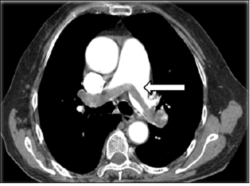
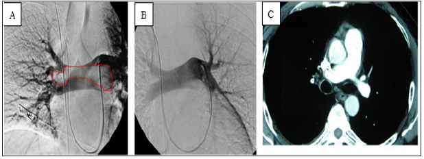
A previously healthy 46-year-old man was admitted because of sudden-onset syncope 8 hours previously. The patient had suddenly fainted 8 hours prior to presentation with no known cause and lost consciousness; however, he had no convulsions of any limbs and no fecal or urinary incontinence. The syncope lasted for about 3 minutes, and the patient gradually regained his consciousness accompanied by shortness of breath, chest distress, coughing, and expectoration. Examination upon admission revealed type I respiratory failure, and Computed Tomography (CT) pulmonary angiography showed an embolism in the main pulmonary artery; however, ultrasound examination showed no thrombus in the lower extremities (Figure 1). On physical examination, the patient’s blood pressure was 98/45mmHg, his lips were cyanosed, his heart rate was 115beats/min with a regular rhythm, and his respiratory rate was rapid (about 30breaths/min) with coarse respiratory sounds in the bilateral lungs. No swelling was seen in the lower limbs. After admission, blood gas analysis (mask-delivered oxygen at 5L/min) showed the following: pH, 7.30; PO2, 45.40mmHg; PCO2, 36.50mmHg; and SO2, 85.8%. His D-dimer concentration was 3.75mg/L. After treatment with oxygen inhalation, the patient was able to assume a supine position. His PE severity index was 126 points (grade V is defined as >125 points, which indicates very high 30-day mortality risk of (10.0-24.5) %.

Figure 1:Preoperative computed tomography showed thromboembolism in the bilateral main pulmonary arteries (arrow).


Preoperative Analysis

Peripheral thrombolysis or Catheter-Directed Thrombolysis (CDT) after IVC filter placement

The patient had acute pulmonary arterial embolism, and the thrombi were relatively large. They were considered to have been caused by the detachment of large thrombi, but no thrombus was found on an ultrasound examination of the bilateral lower extremities. Because the source of the thrombi was not clear, we were concerned that peripheral thrombolysis may result in a new thrombus that could readily detach and aggravate the PE. Therefore, the patient was scheduled to undergo IVC angiography followed by pulmonary angiography and pulmonary artery CDT.

Source of the thrombi

CT pulmonary angiography showed PE, but no swelling in the lower limbs. The ultrasound examination showed no thrombosis in the deep veins of the lower limbs. The source of the thrombi could not be identified. IVC angiography was therefore performed to determine the source of the thrombi. Because thrombi are usually seen in the IVC system, IVC angiography was the first-choice procedure. If thrombi were present, a filter could be implanted through the superior vena cava.

Operative procedure

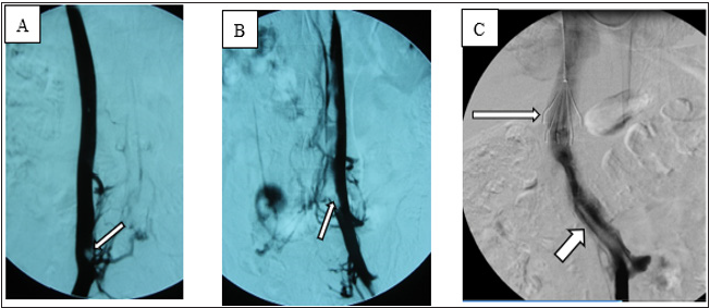
Local anesthesia was administered, and the angiography procedure was performed via puncture of the right femoral vein. Angiography showed that the IVC was relatively small in diameter and contained a filling defect. After puncture via the left femoral vein, angiography showed a double IVC (Figure 2A) and thrombosis (floating thrombi) within the IVC. The left and right IVCs merged into one branch above the renal vein. Puncture was performed via the right jugular vein, and a temporary vena cava filter was implanted (Tempofilter II; B. Braun, Melsungen, Germany) (Figure 2B & 2C). The filter was located above the junction of the two IVCs. Pulmonary artery angiography (Figure 3A) and pulmonary artery CDT were performed through the right subclavian vein. IVC CDT was performed through the left femoral vein. The thrombolysis catheter used in this patient was a Uni Fuse (AngioDynamics, Latham, NY, USA).

Figure 2: Angiography showed a double IVC with thrombosis (arrows) in both IVCs.
(A) Angiography via right femoral vein.
(B) Angiography via left femoral vein.
(C) Junction of left and right IVCs, IVC filter (long arrow) and floating thrombi in IVC (short arrow).


Postoperative treatment

After pulmonary arterial CDT and IVC filter placement, the patient was continuously infused with 500,000 IU of urokinase for 10 hours and administered low-molecular-weight dextran daily. Additionally, low-molecular-weight heparin (nadroparin at 100IU/ kg) was injected subcutaneously for anticoagulation treatment. The patient’s respiratory condition gradually improved, and his shortness of breath and chest distress disappeared. After 3 days, his blood pressure was 120/75mmHg, heart rate was 90beats/ min, and blood oxygen saturation returned to 95% to 100%. His oxygen partial pressure was normal on repeated blood gas analysis. Additionally, repeat angiography showed that the pulmonary artery thrombi had disappeared, and his blood flow was smooth (Figure 3B). After 1 month, the IVC filter was removed.

Figure 3: (A) Pulmonary angiography showed a filling defect of the main pulmonary artery (circle).
(B)After thrombolysis, the thrombi in the pulmonary arteries disappeared.
(C)Reexamination by computed tomography pulmonary angiography of the pulmonary arteries at 3 months postoperatively showed no abnormalities.


Follow-up visits

After discharge, the patient received oral warfarin as long-term anticoagulant therapy. CT pulmonary angiography was performed again 3 months after the operation (Figure 3C). The patient was followed up for 2 years, during which time he developed no symptoms such as chest distress or shortness of breath and exhibited no lower limb venous insufficiency.

Discussion

Choice of treatment method

Generally, PE with syncope as the main manifestation is relatively severe. This patient had a massive thromboembolism in the main pulmonary arteries of the bilateral lungs, his medical condition was critical, and the source of the thrombi was unclear. Peripheral thrombolysis may result in a new thrombus in such cases, and peripheral thrombolysis therapy alone may not completely dissolve the thrombus. In our patient, angiography of the IVC was performed first, and a double IVC containing floating thrombi was found. Implantation of an IVC filter was required before thrombolysis therapy to avoid recurrence of the PE. The patient had a double IVC characterized by the joining of two IVCs between the hepatic veins and the renal veins, and thrombi had formed in the bilateral IVCs. The filter was implanted into the trunk of the IVC, and this needed to be performed via the internal jugular vein. Because the patient was young, a temporary filter was required. The Tempofilter II met these requirements.

Pulmonary arterial CDT

The treatment of acute PE includes anticoagulant therapy, thrombolytic therapy, catheter interventional surgery, surgical thrombectomy, implantation of IVC filters, and possibly other measures. The main purpose of percutaneous catheter interventional therapy is to quickly reduce the pulmonary artery pressure, restore right ventricular function, and increase systemic perfusion. In 2014, the European Society of Cardiology (ESC) guideline recommended that CDT [1] may be the first-choice treatment for patients without absolute contraindications for thrombolysis [1]. In 2019, the ESC guideline stated that surgical thrombectomy or CDT could be considered as an alternative to thrombolysis rescue, and the recommendation level was increased from IIb to IIa [2]. In the present case, the patient underwent pulmonary artery angiography via the right subclavian vein and received thrombolysis therapy by means of thrombolysis catheter implantation. After the treatment, his respiration was significantly improved, and his oxygen partial pressure returned to normal as shown by blood gas examination. After 3 days, repeat angiography showed that the pulmonary artery thrombi had disappeared and that the blood flow in the pulmonary arteries was unobstructed. The effect of thrombolysis was satisfactory in this case for two reasons: first, the CDT allowed the drugs to act quickly and intensively, and second, the CDT was conducted within 24 hours of symptom onset.

IVC malformations

The IVC can be divided into four segments: the hepatic segment, suprarenal segment, renal segment, and infrarenal segment. IVC malformations can be divided into three types according to the lesion characteristics:
1. Infrarenal: left-sided IVC, duplication, pre-aortic IVC, and absence of the infrarenal IVC
2. Renal: left retroaortic renal vein and left circumaortic renal collar, accessory left renal vein
3. Suprarenal: absent hepatic IVC with azygos continuation, congenital caval membranes, congenital IVC stenosis or atresia [3,4].

IVC malformations are relatively rare, with an incidence of about 0.3% to 0.6% in the general population [5,6]. Although a large collateral circulation is present, IVC abnormalities can cause obstructions of venous reflux, leading to venous hypertension and blood flow stagnation, eventually resulting in venous thrombosis [3,7]. Some patients visit the hospital because of chronic venous insufficiency and are further diagnosed with an IVC malformation [8,9]. It is generally accepted that congenital IVC malformations should be considered in young patients with spontaneous bilateral deep venous thrombosis, and most such patients have subclinical symptoms [3,6,10]. Contrast-enhanced CT is very effective in the examination of IVC abnormalities. It can reveal the venous extension and the presence of thrombi [6,11]. Duplication is a common IVC anomaly. Notably, embryonic development of the IVC is complicated. There are three pairs of embryonic veins (postcardinal, subcardinal, and supracardinal) that form a complex anastomosis among one another, gradually regressing thereafter to form the final IVC. Failure of regression of both supracardinal veins and failure of formation of adequate connections between the primitive veins are considered reasonable explanations of double IVC formation [6,12,13]. In most cases, the abnormal left IVC is located at the level of the left renal vein, spans the aorta, and connects to the right IVC [14], and it sometimes directly connects to the left renal vein [15]. Double IVCs can be divided into asymmetric and symmetric duplication. In most cases, the right IVC is still the dominant reflux channel [6]. The clinical manifestations of a double IVC include lumbago, thrombosis, and other conditions; some patients are asymptomatic [16]. In patients with a double IVC, clinicians must be vigilant before retroperitoneal surgery; avoid massive hemorrhage, thrombosis, and other complications; determine whether a double IVC is present before IVC filter implantation; and avoid filter-related complications [6,13,17]. In the present case, two IVCs had joined above the renal vein with floating thrombi, and severe PE was present; however, the deep veins of the lower limbs were free of thrombi, and the patient had no symptoms of venous insufficiency, considering that the thrombosis was related to the venous malformation.

Conclusion

Although a double IVC with floating thrombi is rare, clinicians must still be vigilant of the possibility, and it is helpful to formulate a treatment plan. For patients with PE and no swelling in the lower limbs or thrombosis in the deep veins of the lower limbs as shown by ultrasound examination, the source of the thrombi should be clarified. Both the IVC and iliac vein should be considered as potential sources.

References

  1. Konstantinides SV (2014) ESC Guidelines on the diagnosis and management of acute pulmonary embolism. Eur Heart J 35: 3145-3146.
  2. Konstantinides SV, Meyer G, Becattini C (2019) ESC Guidelines for the diagnosis and management of acute pulmonary embolism developed in collaboration with the European Respiratory Society (ERS). Eur Heart J 41: 543-603.
  3. McAree BJ, O Donnell ME, Fitzmaurice GJ (2013) Inferior vena cava thrombosis: A review of current practice. Vasc Med 18(1): 32-43. 
  4. Alkhouli M, Morad M, Narins CR, Raza F, Bashir R (2016) Inferior vena cava thrombosis. JACC Cardiovasc Interv 9(7): 629-643.
  5. Ordóñez SF, Carrasco GJC, Recio BFJ, Aguilar RC, López TF, et al. (1998) Absence of the inferior vena cava causing repeated deep venous thrombosis in an adult-A case report. Angiology 49(11): 951-956.
  6. Spentzouris G, Zandian A, Cesmebasi A, Kinsella CR, Muhleman M, et al. (2014) The clinical anatomy of the inferior vena cava: A review of common congenital anomalies and considerations for clinicians. Clin Anat 27(8): 1234-1243.
  7. Fuster GMJ, Forner MJ, Lorente B, Soler J, Campos S (2006) Inferior vena cava malformations and deep venous thrombosis. Rev Esp Cardiol 59(2): 171-175.
  8. Sitwala PS, Ladia VM, Brahmbhatt PB, Jain V, Bajaj K (2014) Inferior vena cava anomaly: A risk for deep vein thrombosis. N Am J Med Sci 6(11): 601-603.
  9. Baeshko AA, Zhuk GV, Orlovskii Iu N, Ulezko EA, Savitskaia TV, et al. (2007) Congenital anomalies of the inferior vena cava: Diagnosis and medical treatment. Angiol Sosud Khir 13(1): 91-95.
  10. Chee YL, Culligan DJ, Watson HG (2001) Inferior vena cava malformation as a risk factor for deep venous thrombosis in the young. Br J Haematol 114(4): 878-880.
  11. Gayer G, Luboshitz J, Hertz M, Zissin R, Thaleret M, et al. (2003) Congenital anomalies of the inferior vena cava revealed on CT in patients with deep vein thrombosis. AJR Am J Roentgenol 180(3): 729-732.
  12. Shaw MB, Cutress M, Papavassiliou V, White S, Thompson M, et al. (2003) Duplicated inferior vena cava and crossed renal ectopia with abdominal aortic aneurysm: Preoperative anatomic studies facilitate surgery. Clin Anat 16(4): 355-357.
  13. Natsis K, Apostolidis S, Noussios G, Papathanasiou E, Kyriazidou A, et al. (2010) Duplication of the inferior vena cava: Anatomy, embryology and classification proposal. Anat Sci Int 85(1): 56-60.
  14. Sartori MT, Zampieri P, Andres AL, Prandoni P, Motta R, et al. (2006) Double vena cava filter insertion in congenital duplicated inferior vena cava: A case report and literature review. Haematologica 91(Suppl): ECR30.
  15. Kapetanakis S, Papadopoulos C, Galani P, Dimitrakopoulou G, Fiska A (2010) Anomalies of the inferior vena cava: A report of two cases and a short review of the literature. Folia Morphol (Warsz) 69(3): 123-127.
  16. Di Nicolò P, Zanoli L, Figuera M, Granata A (2016) An unusual cause of lumbar pain after physical exercise: Caval vein duplicity and its detection by ultrasound. J Ultrasound 19(4): 289-293.
  17. Katyal A, Javed MA (2016) Duplicate inferior vena cava filters: More is not always better. Am J Emerg Med 34(1): 115.e1-4.

For more articles in Journal of Research and Review
Please click on below link: https://crimsonpublishers.com/dapm/

Monday, June 27, 2022

Mass Disabilities as a Result Pellet Guns in (IAK) Indian Administered Kashmir-An Opinion Paper_Crimson Publishers

Mass Disabilities as a Result Pellet Guns in (IAK) Indian Administered Kashmir-An Opinion Paper by Navshad Ahmad Wani in Degenerative Intellectual & Developmental Disabilities_Journal of Intellectual Disabilities   

Opinion

The horror of pellet guns in the Indian Administered Kashmir from 2016 in mass people uprising has rendered a huge population blinded both partially and completely. The independent sources put forth a figure of 800 to 1200 completely blinded and around 10000 people partially blinded in such mass usage of pellet guns in the widespread unrest of Kashmir valley. The use of deadly pellet guns in addition to tear smoke shells and other riot control measures by Indian forces have proven very fatal in the sociopolitical context of Indian Administered Kashmir because of its large scale psychological significance in the context of disability studies. The affected people are mostly young boys and girls who are participants of street protests. The protests are being witnessed in IAK-Indian Administered Kashmir since the mass armed uprising started in 1989 with the outbreak of violent militancy yet the use of pellets is a new phenomenon.

Context

The state of Jammu and Kashmir has been the source of age old animosity between India and Pakistan since 1947. The disputed territory of Kashmir which got bifurcated in 1947 following the partition of Indian Sub-continent and a major part of Kashmir went under illegal control of India. The people of IAK have been demanding ‘Right to self determination’ from Indian control. It is also imperative to mention that half of the Kashmir has been under the illegal occupation of Pakistan also who happen to be the party to the conflict as there are numerous United Nations Organizations’resolutions on Jammu and Kashmir. With the violent outbreak of militancy a large section of Hindu minority known as Kashmiri Pundits have also left the valley for security to the other division, a Hindu dominated Jammu division. The Kashmiri population has been the victim of unending political hostility between India and Pakistan. Thousands of people have died, jailed and even rendered disable over the thirty years of death and destruction. The resistance from the Kashmiri people has changed many shapes since 1989; the recent one has been the street protests with occasional stone pelting on armed forces, which usually have protective equipment in comparison to the protestors. The protestors become easily the victims of the measures taken by the government forces. It is also pertinent to mention that in IAKIndian administered the use of tear gas deadly pellet guns and even live ammunition has been used to control the unarmed protestors. There has been no use of water cannons, rubber bullets or other less deadly arms.

The use of pellet guns has serious psychological consequences. The serious concerns have also been raised by Human Right groups both locally and internationally yet there have been no effect on the ground as the young people are being continuously rendered blind due to the use of pellet guns. The Wikipedia makes a reference to the pellet guns, “a pellet gun is a non-spherical projectile designed to be shot from an air gun”. Indian popular newspaper, ‘The Hindu’ in its news report by Vijaiyta Singh of 21 July, 2018 has explained that the pellet guns are a form of non lethal crowd control methods used by police in addition or in special casesof riot control through water cannon, pepper spray, taser guns etc. are also popular in hunting”.She also reports that these guns are intended to injure individuals and cause pain and can be effective over short ranges up to 500 yards but when fired from close quarters can be highly lethal”. The pellets penetrate soft tissues when hit and if hit at a very close range above the waist height can prove fatal by penetrating deep into the vital organs of the body. This has been the case with most of the pellet injuries in Kashmir which are mostly above waist height and that become the reasons for such widespread blindness of youth due to pellets.

Psychological Consequences

The rampant use of pellet guns may have a prime concern to induce a fear among the masses but its continuous use has different consequences too. The pellet guns have rendered a large section of population disable physically but the psychological scars are far more dreadful and annoying. The suffrage of Post Traumatic Stress Disorder due to the exposure to the pellets and such direct confrontations seem to be cause of many mental disorders usually caused by the exposure to violent conflicts. The trauma exposure, depression, anxiety, chronic anhedonia and most importantly body dysmorphic disorders among affected youthresulting out of the loss of eyes and other vital parts of body due to the pellet guns among young Kashmiri people is a matter of serious concern. A lot of research is required to know as to how pellet guns have rendered many people disable.

Scope for Rehabilitation and Psychological Intervention

A famine, a deadly disease or health vulnerability may be the focus of much international attention but the silent suffering of the people of Kashmir in the form of mass disabilities as a result of use of pellet guns remains unnoticed. Due to such havoc provoking circumstances in Kashmir there is a lot of scope for research on such new and challenging psychological areas of interest in the context of disability studies for researchers who are interested in Rehabilitation Psychology or the Psychology of Conflict. There is scope for a separate naming of such a disability which results as an induced disability due to pellet guns. The rehabilitation psychology has a lot of scope in this context and more specifically in this part of globe. I am hopeful of a serious interest from the researchers to address and focus on such issues. Further a lot of insights are required into the specialized intervention strategies for the professionals in the field of counseling and psychotherapy.

https://crimsonpublishers.com/didd/fulltext/DIDD.000521.php


Please click on below link: https://crimsonpublishers.com/didd/

Friday, June 24, 2022

Nanodrugs to End Viral Infection in Pancreatic β-Cell and Pyknosis_Crimson Publishers

Nanodrugs to End Viral Infection in Pancreatic β-Cell and Pyknosis by Rajiv Kumar in Determinations in Nanomedicine & Nanotechnology_Nanotechnology open access journals


Keywords

Nanoplatforms; Viral infection; Pancreatic β cell; Pyknosis

Introduction

Diabetes mellitus, a metabolic disorder wherein the natural ability of the insulin to hold surfaces well tumbledown and because of it, the surface receptors turn erroneous in the creation of insulin. Thus, the pancreatic β-cell initiates mass reduction lead to types 1 and 2 diabetes [1,2]. This is a nonstop unanimity of lowering the secretion of insulin from top to bottom trigger numerous negative inferences, reparations, and dysfunction of organs (eyes, kidneys, blood vessels, nerves, and heart) [3]. In diabetes, the etiology of β-cell death and ongoing multifaceted mechanisms of mitochondria uphold precarious implications. Wantonness in the routine of the progressive β-cells, hyperglycemia and chronic inflammatory itineraries lead more imperfections in the secretion of insulin caused unpredictable dysfunction (improper functioning of granulocytes, monocyte/macrophages, natural killer cells, dendritic cells, T-cells, B-cells, and destroy cytokine signaling by producing of IL1β mediator) in nearby cells and organelles too. Later on, it initiates inflammation and amplified oxidative stress.

Such annoying happenings assassinate glutathione efficiency which safeguards DNA, proteins, and lipid membranes on or after radical outbreaks. Such transgressions doings augment the level of 3-nitrotyrosine, a key factor to initiate diabetes and pyknosis. Enteroviruses, (Cytomegalovirus, Epstein-Barr virus, Mumps virus, Rotavirus, Parvovirus B19, and Human Endogenous Retroviruses) infections highlighted as the foremost cause for fiascoes in the routine functioning of pancreatic β-cells. The pathways of progressive autoimmune-mediated catalysis answerable for amplified biological routes (pathology of pancreatic β-cell mass lose, apoptosis, immunological implications, and enteroviral tropism). These viruses infect and alter the islets of nondiabetic donors, as well as gene expression profiles of the virus strain. Such transformations instigate upregulation of pro-inflammatory cytokines interleukin 1α (IL-1α), IL-1β, viral inclusions, and coeval, enrich apoptosis, and proliferate pyknosis [4,5].

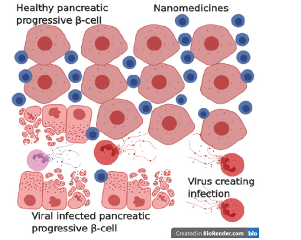
Cellular permissiveness to viral infection is modulated by kick off innate antiviral ripostes from the poles apart cells. Usually, pancreatic islet α cells trigger antiviral response proficiently to do battle with these diabetogenic viruses to a certain degree do β-cells to get rid of viral infections intuitively without undergoing apoptosis. Nanoparticles of albumin (non-toxic, non-immunogenic, biocompatible, and biodegradable) emerged as a versatile source to incorporate with macromolecular protein as therapeutic and theranostic resources to cure unhealthy and infected pancreatic β-cell and to stop pyknosis. These nanoparticles are impasse with bioactive molecules (chemicals, proteins/peptides, and oligonucleotides) and macromolecules having better-quality of pharmacokinetics to enhance longer circulation time, disease-specific buildup and easy to use a carrier for antiviral drugs across molecular barriers of cells deftly (Figure 1).

Figure 1: Schematic representation of the mechanism of nanomedicine’s impact on various stages of pancreatic β-cell, prevention routes, therapeutics, riposte for destroying viruses, and other virus creating the infection.


Conclusion

These innovations (glucose nanosensors, carbon nanotubes, quantum dots, nanopumps, oral insulins, microspheres, and artificial pancreas) in nanotechnology renewing cure preferences for the bloodbath of infected pancreatic β-cell. Further discoveries on the routes and types of machinery of vital diabetogenic viruses responsible for insulitis may be helpful in the innovation of novel nanovaccines to end metabolic virus’s blameworthiness liable for crafting infection in pancreatic β-cell and catalyzing pyknosis.

References

  1. Kahn SE (2003) The relative contribution of insulin resistance and beta‐cell dysfunction to the pathophysiology of type 2 diabetes. Diabetologia 46: 3-19.
  2. American Diabetes Association (2015) 2. Classification and diagnosis of diabetes. Diabetes Care 38: S8-S16.
  3. Alicic, RZ, Tuttle, KR (2014) Novel therapies for diabetic kidney disease. Adv Chronic Kidney Dis 21(2): 121-133.
  4. Antje P, Michele S, Klaus-Peter K (2015) Mechanisms of beta cell dysfunction associated with viral infection. Current Diab Rep 15(10): 73.
  5. Lopes M, Shrestha N, Correia A, Shahbazi MA, Sarmento B, et al. (2016) Dual chitosan/albumin-coated alginate/dextran sulfate nanoparticles for enhanced oral delivery of insulin. J Control Release 232: 29-41.

For more articles in Nanotechnology open access journals
Please click on below link: https://crimsonpublishers.com/dnn/

Thursday, June 16, 2022

Evidences of Large-Scale Shearing from South-Eastern Extension of The Mahakoshal Belt, Covering Parts of Sidhi & Singrauli Districts of Madhya Pradesh and Sonbhadra District, Uttar Pradesh, India_Crimson Publishers

Evidences of Large-Scale Shearing from South-Eastern Extension of The Mahakoshal Belt, Covering Parts of Sidhi & Singrauli Districts of Madhya Pradesh and Sonbhadra District, Uttar Pradesh, India by Banerji DC in Integrative Journal of Conference Proceedings_integrative journal of conference proceedings impact factor


Abstract

The WNW-ESE trending arm of the Mahakoshal belt, extending towards south-east in the state of Jharkhand, through U.P, exposes variety of structural features those are uncommon in main Mahakoshals having ENE-WSW trend. The turbidites, said to be characteristic of the Parsoi Formation of the WNW trending arm, appear to be shear generated structures, having imprints across the length and width of the arm. Evidences of bulk flow along the pervasive foliation make it a mylonitic foliation, restricting possibility of survival of primary structures.

Overwhelming presence of anticlockwise rotated fabric elements, producing S-C structure in phyllite/ phyllonite and in meta-greywacke, along with frequent presence of ‘alpha’ and ‘delta’ type structures, strongly asymmetric ‘S’ and at places ‘Z’ shaped folds and the sheath folds suggest that the ductile shearing with a sinistral shear sense is spread over the entire WNW trending sinuous arm of Mahakoshals. This belt has a strong manifestation on the imagery, maintaining a cross cutting relationship with the ENEWSW trending fabric of the main Mahakoshals and appears to be superposed over it. Evidences suggest it to be a wide and extensive shear zone.

Keywords: Shears; Progressive shearing; Sinistral shear sense; Rotation; Mylonitic Foliation; Turbidites

Introduction

The Mahakoshals have a WNW-ESE trending arm in the south-eastern corner, especially south of Chitrangi. East of Chitrangi, the main Mahakoshals, continuing with an ENE-WSW trend from Narsighpur district of Madhya Pradesh, continues within the state of Uttar Pradesh and abuts against the Son River (Figure 1). The WNW-ESE trending arm, however, continues much to ESE and traceable even within the state of Jharkhand after crossing over the state of Uttar Pradesh.

Figure 1:Geological sketch map of parts of Sidhi and Singrauli districts of M.P. and Sonbhadra district of U.P.


Explanation to this change of trend from ENE to WNW, and the nature of contact between the two arms of Mahakoshals, is barely available in the existing literature. Banerji [1,2] reported large scale non-coaxial shearing of Mahakoshal rocks, resulting into a WNWESE trending sinuous belt, having imprints of superimposition over the ENE trending main Mahakoshals. The superimposition of the WNW fabric is well supported by the satellite image of the area. Sharma [7], however, has reported the presence of turbidites restricted within the WNW trending arm of the Mahakoshals.

Present work further analyses the ground evidences to ascertain if the differently oriented arm of Mahakoshals is a result of ductile flow, producing a WNW trending mylonitic foliaton.

Geologic setting

The ENE-WSW trending narrow belt of Mahakoshal rocks extends along the Narmada and Son rivers between Barmanghat (Narsinghpur, Madhya Pradesh) in the west to Palamau (Jharkhand) in the east. This belt is represented by rocks like quartzite, phyllite, dolomite, meta basics and banded ferruginous chert. The lower division of the Mahakoshals, occupying the western and central part of the belt is known as Agori Formation and is represented by a sequence of basal quartzite interbedded with meta basics and overlained by dolomites with chert inter bands. The eastern part of the belt is dominated by meta basics, banded ferruginous chert, phyllite and meta-greywacke with near absence of carbonates. Nair et al. [3] have considered the meta basics / ultramafic flows occurring near Chitrangi to be the lowermost part of Mahakoshals and named them ‘Chitrangi Formation’. Sharma et al. [8] however, have considered these meta basics / ultramafic flows as Post Jungel igeous suit. The argillite-meta greywacke association occupying central part of the WNW trending arm of the Mahakoshals (Figure 1) is flanked on either side by rocks of Agori Formation and has been considered {Nair et al. [3]} as uppermost ‘Parsoi Formation’. The Parsoi rocks also include turbidites, restricted only to this part of the belt [7]. The southern margin of the eastern Mahakoshal belt is affected by intrusions of granitic to granodioritic and syenitic plutons [9]. These igneous rocks are generalized as Dudhi granitoid in the existing geological maps.

Banerji [2] reported the presence of a major shear zone in the south-eastern part of Mahakoshal belt and considered the WNWESE trending tract of Agori and Parsoi Formation of the south-east as mylonites, representing his ‘Dudhmania Shear Zone’. Ductile shearing along the southern boundary of the WNW trending Mahakoshal belt of south-east, is also discussed by Roy & Devrajan [6].

Structural features

In the WNW trending arm of the Mahakoshals, the trend of the foliation is N80° W-S80° E in the western end, swerving to N60° W-S60° E in the central part and again swinging to N80° W-S80° E in the eastern end of the belt. This change in the trend of foliation imparts a sinuous appearance to the belt. The dip of the foliation is sub-vertical.

Northern margin of Dudhi Granitoid, located south of the Songarh-Kasar-Dudhi (SKD) fault bordering WNW trending belt of Mahakoshals, record ample evidences of ductile shearing. The rock locally acquires gneissosity because of alignment of stretched feldspars alternating with layer lattice minerals. The granitoid shows imprints of two obliquely running planes, having an angle of about 100 to 120 between them (Figure 2). While, the feldspar phenocrysts are extremely stretched along pervasive ‘C’ planes representing mylonitic foliation, the obliquely oriented grains along ‘S’ planes show regular offsest by the mylonitic foliation, imparting an ophthalmic shape to the phenocrysts. Crude rotation of the ‘S’ planes gives impression of a strain sensitive fabric.

Figure 2:Sheared granitoid (pink gneiss) from Kanhana, north of Singrauli.
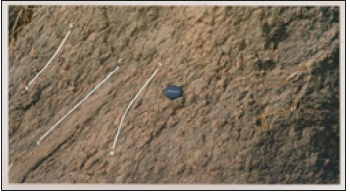

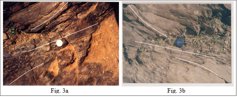
North of the SKD fault, exposures of greywacke/phyllite/ phyllonite are common, representing the WNW trending Mahakoshal belt. Development of a strain sensitive fabric with ‘S’ planes rotated both clockwise and anticlockwise (Figure 3a & 3b) directions, in mesoscopic scale, is common all along the rocks of bordering zone. The ‘C’ planes define mylonitic foliation, running parallel to the boundary of the WNW trending Mahakoshal belt. The angle between these two planes, near the flank, is about 15°. This type of rotational feature persists across the length and width of the WNW trending Mahakoshal belt. The angle between the two planes reduces to less than 10° in the central part of the belt.

Figure 3:Strain sensitive fabric developed in phyllite/phyllonite. The coin is placed on ‘C’ plane dominated area, while above and below the coin ‘S’ planes show rotation.

Also, the ubiquitously present Quartz veins and the chert bands of the peripheral area exhibit variety of shortening and extensional features. The bands which are oriented parallel to the pervasive foliation (‘C’ planes) show stretching and boudinaging. Boudins, thus created within the stretched part, lying parallel to the flow planes, are further rotated during the progressive movement and play the role of excellent shear sense indicators (Figure 4a & 4b). On the contrary, veins/bands having oblique orientation to the flow plane display shortening by compression, producing short hinges. With the progressive movement, the limbs of the short hinges acquire parallelism with the flow direction and get considerably stretched. This phenomenon is quite corroborative with the instantaneous extensional and shortening regimes, conforming flow along mylonitic foliation. The extent of stretching of the limbs along the ‘C’ planes, during the progressive deformation, also indicates the amount of flow involved.

Figure 4: Shortened and boudinaged (extended) quartz veins oriented oblique and parallel to mylonitic foliation respectively. Note the anticlockwise rotation of the boudins, and extended limbs and short hinges in obliquely oriented quartz veins. The extent of stretching of the limbs in Figure b is also noteworthy. Sense of shear is sinistral


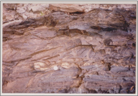
Formation of ‘alpha’ and ‘delta’ structures (Figure 5a & 5b), within the stretched bands are again a common phenomenon throughout the belt. The boudins formed during stretching are rotated either clockwise or anticlockwise, depending upon the prevailing dextral or sinistral movement. In most of the cases, however, the shear sense deduced from such rotated boudins indicate a sinistral sense of movement. One of the chert bands from east of Dudhmania exhibit a complex structure, which appears to have resulted from change in direction of non-coaxial movement, from dextral to sinistral, and consequent reverse rotation of the boudins (Figure 6).

Figure 5:(a) Quartz vein showing ‘alpha’ structure. (b) Quartz vein showing ‘delta’ structure. Sense of shear is sinistral.
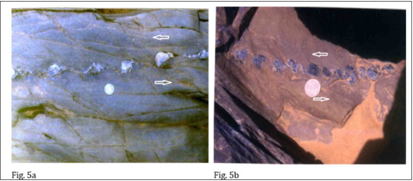

Figure 6:Complex structure showings reverse rotation in pre-existing rotated boudins. The over tightening is caused probably by reverse rotation of the earlier rotated boudins in chert band.

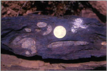
The over tightening of ‘Z’ fold, represented by a chert band observed near Dudhmania and its reverse rotation (Figure 7) also suggest that initial sense of asymmetry of folds (i.e. dextral as indicated by ‘Z’ folds) was subjected to reverse shearing (sinistral) during the progressive non-coaxial deformation [5].

Figure 7:Compex structure produced by reverse rotated ‘Z’ fold in the chert band. Because of reverse rotation, the lower hinge of ‘Z’ fold is rotated upward, and the upper hinge has come closer to the lower hinge. The extended / boudinaged fragments of the upper and lower limbs have moved inward (towards center).

Chert bands of this area also preserve a variety of folds, tight to isoclinal and asymmetric in nature. Both ‘S’ and ‘Z’ shaped folds are represented; however, ‘S’ shaped folds predominate over the other type. These mesoscopic folds have low (<30°) easterly plunges. However, steep (50°-70°) easterly plunges are also observed in the obliquely trending (NNE-SSW to NNW-SSE) shortened chert bands. Anticlockwise rotation of the axial planes of near isoclinal folds and their amplification (Figure 8) is also a regular phenomenon in this part of the belt. Sheath folds with an eyed outcrop pattern and the textbook type plane non-cylindrical sheaths (Figure 9) are also represented in the area having proximity to the central belt of Parsoi Formation of Nair et al. [3].

Figure 8:Anticlockwise rotated axial planes of near isoclinal ‘S’ folds in chert band. Shear sense is sinistral.
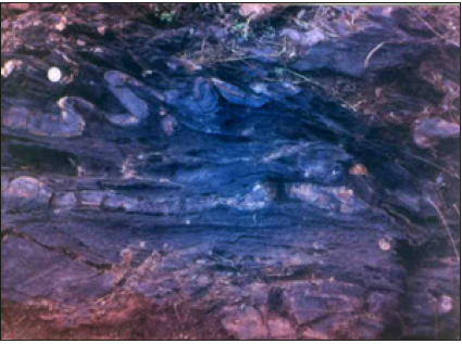

Figure 9:Sheath fold. Of the two white colored chert bands, the one present to the right of the coin has produced plane non cylindrical sheath fold.
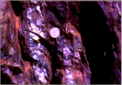

Further towards the central part of the belt, i.e. near the belt of Parsoi rocks of Nair et al. [3], the greywacke exhibit presence of numerous, thin, siliceous lamellae, concentrated as discontinuous bands. Extent of such bands is restricted to a few tens of meters, tapering at either ends before disappearance. These siliceous lamellae, in fact, get dissipated within the greywacke, leaving behind still traceable faint markings. Within the bands, the lamellae are stretched, rotated and shortened and displaced along the long axis, depending upon the orientation of the lamellae with respect to the direction of flow. The rotational component of flow is exhibited by rotation of the siliceous lamellae having obliquity to the flow plane (Figure 10). Rotation of these quartzose lamellae very often produce a pseudo cross bedding. Nevertheless, these pseudo cross bedded structures occur in close association with tight rootless miniature folds (Figure 11), indicating that such structures are the products of intense deformation [4]. These structures are likely to be mistaken for turbidites. However, as discussed earlier, evidences of bulk flow along the pervasive foliation, ubiquitously present in the belt, reduces the possibility of existence of any primary structure. Such structures, therefore, represent secondary structures, produced by progressive shearing of the rocks.

Figure 10: Anticlockwise rotation of siliceous lamellae having obliquity to the flow plane (bottom right) in metagreywacke. Note the systematic partitioning of the rotational component by discrete shear planes and presence of rootless folds (below coin). Shear sense is sinistral.
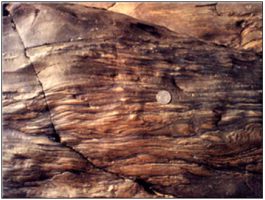

Figure 11: Pseudo cross bedding associated with rootless folds (above and below the coin). Shear sense is sinistral.
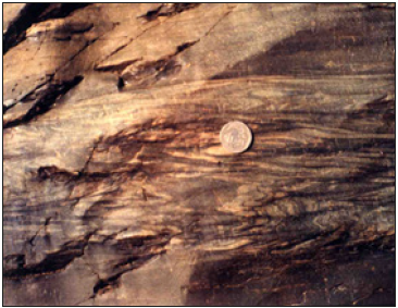

Close to the central belt of Parsoi Formation, these siliceous lamellae are present only as small discontinuous patches within the meta-greywacke, mostly as hinges of small folds with markedly oblique relationship with the flow plane. Continuity of these obliquely oriented and highly folded lamellae are conspicuously broken along the discrete planes representing foliation. The rock itself acquires a crudely banded nature with alternate light and darker bands (Figure 12).

Figure 12:Crudely banded greywacke with traces of obliquely running siliceous lamellae.
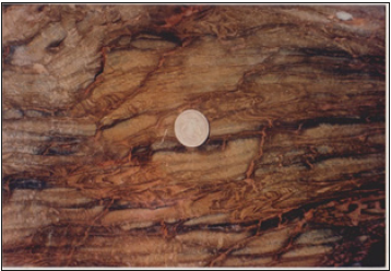

Remarkably, adjacent to Parsoi belt, the chert bands, both ferruginous and nonferrous types, drastically reduce in number. They, however, continue displaying rotation of the axial planes by a dominant sinistral flow (Figure 13). Greywacke of this zone attains a well-defined banded nature, very likely to be confused with turbidites. The alternate light and dark bands are arranged parallel to the foliation and show shortening features, wherever there is an obliquity to the flow plane, differentiating them from the turbidites, the rock becomes a classic banded mylonite (Figure 14), with a marked sinistral shear sense.

Figure 13: Banded ferruginous chert showing anticlockwise rotation.
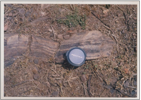

Figure 14:Banded mylonite. Alternate light and dark bands are arranged parallel to the foliation and show shortening/rotational features, wherever there is an obliquity.
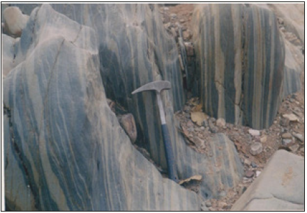

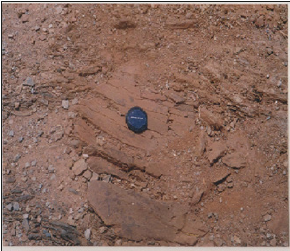
In the central belt of Parsoi Formation, the banded mylonite passes into ultra mylonite with still retaining the banded nature. Rocks are friable and show faster denudation. The alternate dark and light bands, however, become thinner with minimum obliquity between them. Rotational features continue as pressure shadows (Figure 15), very helpful in differentiating them from a turbidite. A very strong and close spaced flow plane representing mylonitic foliation is preserved all through the Parsoi belt. Near-complete transposition and rotation of oblique planes produce localized pseudo cross bedding (Figure 16). However, without a holistic approach, differentiating them from a normal turbidite is difficult. Rarely, small, rounded, relatively stiff mass of rock (isotropic) appear as rotated segments wrapped within the intensely foliated mass. These rounded segments show a crude sigmoidal trail of the internal foliation, oblique to the external foliation i.e. the foliation present in the wrapping mass (Figure 17). This represents the presence of a component of simple shear flow along the foliation present in the wrapping mass, causing rotation to the relatively stiff and isotropic material.

Figure 15:Layered ultramylonite with rotated pressure shadow (near left margin).
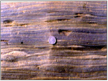

Figure 16:Near complete transposition and rotation of planes / siliceous lamellae in phyllite/ phyllonite. Note the presence of pseudo cross bedding (below lens cap).


Figure 17: Small, rounded, relatively stiff mass, present as rotated segment, wrapped within the intensely foliated ultramylonite. The rounded segment shows a crude sigmoidal trail of the internal foliation, oblique to the external foliation. Shear sense is sinistral.

The Parsoi belt, having a width of about 2km, is remarkably devoid of chert bands. Innumerable thin quartz veins, sub-parallel to the mylonitic foliation, however, traverse this belt. These quartz veins are also affected by shearing and rendered as quartz mylonites.

The commonest micro shape fabric in meta-graywacke, near the margin of the WNW trending arm of Mahakoshals, is a porphyroclastic texture where the ‘porphyroclasts’ are wrapped around by quartz ribbons and muscovite flakes (Figure 18). Quartz veins present in the entire belt are rendered quartz mylonite showing a micro fabric predominantly of S-C type (Figure 19). Presence of synthetic fractures/ slips in chloritoid porphyroclast of phyllonite/ultramylonite (Figure 20) also represents a component of simple shear (sinistral) affecting the rocks.

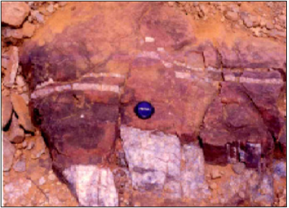
Minor faults with sinistral shift of the trail of vein quartz boudins/blocks (Figure 21), present near the southern margin, further confirm continuity of shear deformation till much late, even after hardening of the deforming mylonite.

Figure 18: Quartz porphyroclast wrapped by quartz ribbon and muscovite flakes. Crossed nicols, 32.
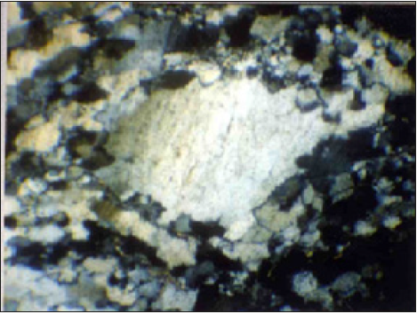

Figure 19:S-C fabric shown by quartz mylonite. Crossed nicols, 3X. Shear sense is sinistral.
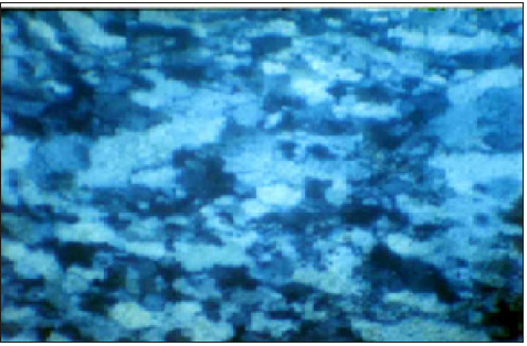

Figure 20:Synthetic fracture/slip in chloritoid porphyroclast within phyllite. Crossed nicols, 3X. Shear sense is sinistral.
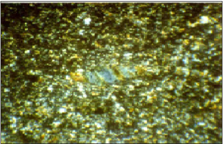

Figure 21: Fault affecting the quartz vein resulting in trail of quartz boudins/blocks. A sinistral shift.

Supportive evidences from remote sensing

In satellite imagery, the WNW trending arm of the Mahakoshal is represented as a sinuous belt hitting against the ENE-WSW trending arm of the main Mahakoshals. The belt has a characteristic broom stick fabric. This uncommon, very close spaced, fabric is different from rest of the Mahakoshals. The close spaced fabric has a cross cutting relationship with the ENE-WSW trending fabric of the main Mahakoshals, as is seen west of Obra (Figure 22). In the strike continuity, south of Chitrangi, the WNW-ESE fabric of the sinuous belt abuts / merges into the main Mahakoshal belt. This fabric appears to be superposed over the fabric present in the northern main Mahakoshals.

Figure 22: Satellite image showing the sinuous belt of the Sidhi and Singrauli districts of M.P. and Sonbhadra district of U.P. The northern Obra-Amsi-Jiawan (OAJ) fault (F) and the southern Songarh-kasar-Dudhi (SKD) fault is marked with broken white lines.
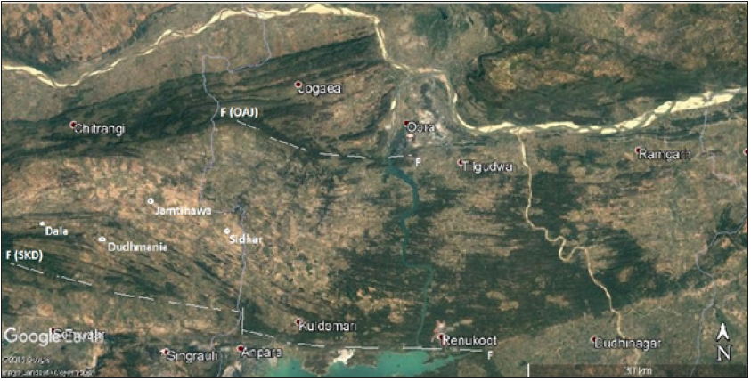

Discussion and Conclusion

The main belt of Mahakoshal, extending between Narsinghpur in the west to Sidhi in east, has a dominant ENE-WSW trend, representing the axial traces of D1 and D2 structures. However, the southeastern extension of the belt, lying between Obra-Amsi- Jiawan fault (OAJ) and the Songarh-Kasar-Dudhi (SKD) fault, shows a WNW-ESE trend. This trend clearly is superimposed on the main Mahakoshal trend, well depicted in imagery, and does not appear to have resulted by folding.

The overwhelming presence of rotation of fabric elements all along the length and width of the WNW trending arm, frequent presence of ‘alpha’ and ‘delta’ type structures, strongly asymmetric ‘S’ and at places ‘Z’ shaped folds and the sheath folds suggest that the belt has undergone strong ductile deformation. Transposition of fabric along the WNW-ESE trending foliation is also amply clear all through the belt. This foliation, therefore, represents a ‘mylonitic foliation’. The stretching and boudinaging of chert bands and quartz veins along this foliation and shortening (tight folding) of the obliquely oriented features, is suggestive of bulk flow along the foliation. Rotation of the stretched boudins and of the axial planes of near isoclinal folds, however, indicates that the deformation was non-coaxial and progressive in nature. The dominance of anticlockwise rotated fabric and shape symmetry indicate existence of a sinistral sense of movement, though, at places, clockwise rotated fabric along with ‘Z’ folds are also observed.

Finally, it is obvious that the shear generated structures present in the sinuous belt were mistaken for turbidites in earlier literature. Close association of these structures with shear sense indicators further helps in differentiating them from normal syn-sedimentary structures (turbidites) formed in tectonically active basins. Overwhelming presence of rotational features in chert bands and in quartz veins, further ascertains that the terrain is representative of a flow regime. Splendid representation of the belt in satellite imagery, clearly showing a superposed trend, leaves no ambiguity in mind in differentiating the sinuous belt from the ENE-WSW trending main Mahakoshals. The name DUDHMANIA SHEAR ZONE {Banerji [1]} was proposed for this remarkable belt.

References

  1. Banerji DC, Prasad A (1997) A report on specialized thematic mapping of the mahakoshal group of rocks around Chakaria Kalan. Unpub Prog Rep Geol Surv Ind.
  2. Banerji DC (2011) A discussion on structural aspects of gold bearing belt of Singrauli and Sidhi districts of Madhya Pradesh and Sonbhadra district, Uttar Pradesh. Journal of Economic Geology and Geo resource Management 8(1-2): 85-96.
  3. Nair KK, Jain SC, Yedekar DB (1995) Stratigraphy, structure and geochemistry of the mahakoshal greenstone belt. GSI Mem 31: 403-432.
  4. Passchier CW, Myors JS, Kroner A (1991) Field geology of high-grade gneiss terrains. Narora publishing House, New York, USA.
  5. Ramsay JG, Casey M, Kligfield R (1983) Role of shear in development of the halvetic fold-thrust belt of Switzerland. Geology 11(8): 439-442.
  6. Roy A, Devrajan MK (2000) A reappraisal of the stratigraphy and tectonics of the palaeo-proterozoic mahakoshal supracrustal belt, Central India. Geol Surv Ind 57: 79-97.
  7. Sharma RS (2009) Cratons and Fold Belts of India, lecture notes in earth sciences. Berlin Heidelberg, Germany.
  8. Sharma DP, Sinha VP, kannadasan T, Khan MA, Mehrotra RD, et al. (2000) Gold mineralization in eastern part of son valley greenstone belt, Sidhi and Sonbhadra districts. Geol Surv Ind 57: 271-278.
  9. Soni MK, Jha DK (2001) Mahakoshal greenstone belt and associated gold mineralization. Journal of Geological Society India 64: 317-326.

For more articles in integrative journal of conference proceedings impact factor
Please click on below link: https://crimsonpublishers.com/icp/


A Close Look at the Application of the Yin-Yang- Based Acupoint Pairs_Crimson Publishers

A Close Look at the Application of the Yin-Yang- Based Acupoint Pairs by Tong Zheng Hong in Advancements in Bioequivalence & Bioavailabi...