Crimson Publishers High Impact Journals

Tuesday, December 21, 2021

New Discoveries in Medicine and Physiology Originated in Urology_Crimson Publishers

New Discoveries in Medicine and Physiology Originated in Urology by Ahmed N Ghanem in Surgical Medicine Open Access Journal_Surgery Journal Impact Factor


Abstract

Objective: To report new discoveries made by a single investigator working solo.

Methods: The discoveries were reported. All the investigations were self-financed done over a career life. The discoveries originated in Urology but cover specialties that include Physics, Physiology, Medicine, Nephrology, Cardiovascular, respiratory medicine and Surgery.

Result: The discoveries include 2 new vascular shocks, namely Volumetric Overload Shocks (VOS) that complicate fluid therapy in hospitals. Proving a physiological law of Starling wrong and providing the correct replacement which is the hydrodynamic of the porous orifice (G) tube. Revealing many errors and misconceptions on fluid therapy and reporting its corrections. It all started with an investigation aimed at understanding a rare obscure syndrome that kills patients in Urology; the Transurethral Resection of The Prostate (TURP) syndrome. The patho-etiology of the Loin Pain Haematuria Syndrome (LPHS) was discovered revealing its link with Symptomatic Nephroptosis (SN) and reporting a new curative surgery for its treatment- the Renal Sympathetic Denervation and Nephropexy Surgery (RSD&N). A new point of technique on the surgery for cancer bladder, namely capsule sparing cystoprostadenectomy that make the surgery easier and shorter while avoiding incontinence, impotence and urethral anastomosis was reported.

Conclusion: The discoveries started in urology but extended to Physics, Physiology, Medicine, Nephrology, Cardiovascular, respiratory medicine and Surgery. It includes 2 new shocks of VOS, proving a physiological law of Starling wrong and providing the correct replacement: the hydrodynamic of the G tube. It resolves the puzzles of TURP syndrome, hyponatraemia, the acute respiratory distress syndrome and LPHS.

Keywords: Shock; Hyponatraemia; The TURP syndrome; Bladder cancer surgery

Abbreviations: HN: Hyponatraemia; VOS: Volumetric Overload Shock; VOS1: Volumetric Overload Shock Type 1; VOS2: Volumetric Overload Shock Type 2; TURP: The Transurethral Resection of the Prostate; ARDS: Acute Respiratory Distress Syndrome; MVOD/F: Multiple Vital Organ Dysfunction/Failure; HST: Hypertonic Sodium Therapy; ARF: Acute Renal Failure; NaCl: Sodium chloride; NaCo3: Sodium Bicarbonate; G tube: Porous Orifice Tube; LPHS: Loin Pain Haematuria Syndrome; SN: Symptomatic Nephroptosis; RSD&N: Renal Sympathetic Denervation and Nephropexy; IVU: Intravenous Urography; IVU-E: Intravenous Urography Erect Film; RGP: Retrograde Pyelography

Introduction

Reported here is a summary of the new discoveries in medicine and physiology originated in urology made by a single investigator working solo. All the research was self-financed without any external funds done over a career life of 32 years. The discoveries expanded to cover many specialties that include Physics, Physiology, Medicine, Nephrology, Cardiovascular, respiratory medicine and Surgery.

Method

The new discoveries were reported and are based on the following investigations: For the discovery of VOS and relation to the path-etiology of the TURP syndrome, dilution Hyponatraemia (HN) and the Acute Respiratory Distress Syndrome (ARDS) as well as phenomenon of G tube as replacement for the wrong Starling’s law the following studies were done.

A. Physics study on the hydrodynamic of the porous orifice tube comparing it to Poiseuille’s tube- based on which Starling’s law attributes capillary filtration to hydrostatic pressure.

B. Physiological study on the hind limb of sheep to determine if the capillary works as G tube not Poiseuille’s tube.

C. Prospective clinical study on 100 consecutive TURP patients among whom 10 developed the TURP syndromes randomized into treatments comparing hypertonic sodium therapy (HST) with the conservative treatment of using volume expansion for treating the new shock of VOS.

D. Case series of 23 patients who suffered the TURP syndrome.

E. Critical comprehensive literature review of the literature on the capillary physiology, the TURP syndrome, ARDS and fluid therapy in hospital.

For the discovery of LPHS patho-etiology and its successful therapy the following studies were done:

A. Prospective study on 190 patients presenting with Symptomatic Nephroptosis (SN) among whom 36 (18.9%) developed the Loin Pain Haematuria Syndrome (LPHS).

B. Critical comprehensive literature review of the literature on SN and LPHS.

Result

The physics study demonstrated that the hydrodynamic of the porous orifice (G) tube is totally different from Poiseuille’s tube. The side pressure in Poiseuille’s tube is positive all along the entire length of the tube causing “filtration”. Thus, Starling proposed the oncotic pressure of plasma proteins as opposing force to induce “absorption”. The side pressure of the G tube induces pressure gradient which is negative near the inlet and turns positive near the exit. This produces a dynamic magnetic field-like fluid circulation between the G tube lumen and a surrounding fluid compartment which is autonomous in inducing both “absorption and filtration”.
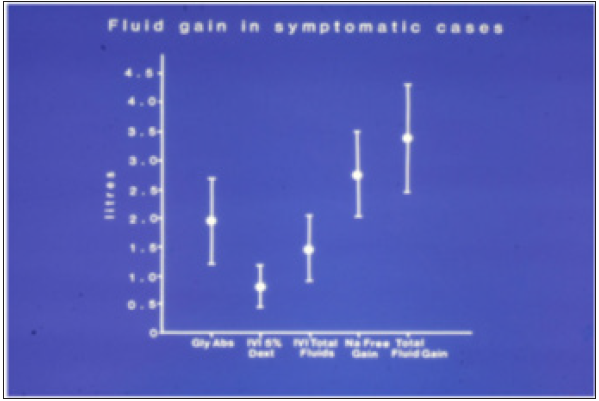
Literature review demonstrated that the oncotic pressure of plasma proteins does not work. The physiological evidence proves that the capillary works as G tube not Poiseuille’s tube. Thus Starling’ law is proved wrong on both forces. The correct replacement is the hydrodynamic of G tube (Figure 1). The prospective study demonstrated that Volumetric Overload (VO) is the most significant factor in inducing the vascular shock of TURP syndrome (Figure 2; Table 1). This shock is recognized as VOS. The correct lifesaving treatment for VOS of the TURP syndrome is Hypertonic Sodium Therapy (HST) of 5%NaCl or 8.4%NaCo3.

Figure 1: Shows Diagram of the porous orifice (G) tube enclosed in chamber (C) based on several photographs demonstrating the magnetic field-like G-C circulation phenomenon. The proximal inflow (arterial) pressure:
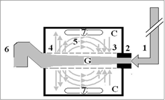
1. Pushes fluid through the orifice.
2. Creating fluid jet in the lumen of the G tube. The fluid jet creates negative side pressure gradient causing suction maximal over the proximal half of the G tube near the inlet.
3. That sucks fluid into lumen. The side pressure gradient turns positive pushing fluid out of lumen over the distal half maximally near the outlet.
4. Thus the fluid around G tube inside C moves in magnetic field-like fluid circulation.
5. Taking an opposite direction to lumen flow of G. tube. The inflow (arterial) pressure (1) and orifice (2) induce the negative side pressure energy creating the dynamic G-C circulation phenomenon that is rapid, autonomous and efficient in moving fluid out from the G tube lumen at (4), irrigating C at (5), then sucking it back again at (3), maintaining net negative energy pressure (7) inside C. The distal outflow (venous) pressure.
6. Enhances outflow at (4) and its elevation may turn the negative energy pressure.
7. Inside C into positive, increasing volume and pressure inside C chamber.


Figure 2:shows the means and standard deviations of volumetric overload in 10 symptomatic patients presenting with shock and hyponatremia among 100 consecutive patients during a prospective study on transurethral resection of the prostate. The fluids were of Glycine absorbed (Gly abs), intravenously infused 5% Dextrose (IVI Dext) Total IVI fluids, Total Sodium-free fluid gained (Na Free Gain) and total fluid gain in liters.

The case series of 23 patients demonstrated that mistaking VOS for one of the recognized shocks such as septic or hemorrhagic shock and treating it with further volumetric expansion caused death of the first 3 patients. Diagnosing VOS correctly and treating it with prompt HST saved the lives of 20 patients. VOS is of two types depending on the type of fluid inducing it. Sodium-free fluid such as 1.5% Glycine and 5% Glucose induce VOS1. The TURP syndrome and dilution Hyponatraemia (HN) represent VOS1. Sodium-based fluid such as Normal Saline, Hartmann’ solution, plasma, plasma substitute and/or blood induce VOS2. Both VOS are always mistaken for one of the recognized shocks and wrongly treated with volume expansion using isotonic sodium-based fluids causing ARDS and death. Discovery of VOS has resolved the puzzles of TURP syndrome, HN and ARDS discovering the exact patho-etiology and successful lifesaving therapy.

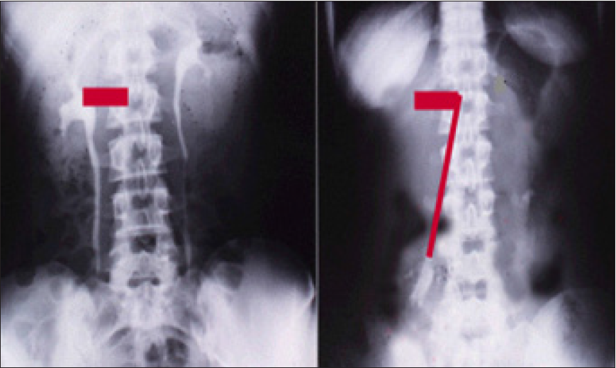
Discovering the link of LPHS with SN has revealed the exact patho-etology and allowed a successful curable surgery of the renal sympathetic denervation and nephropexy. The new IVU 7 sign demonstrated stretch of renal pedicle caused vessels stenosis inducing renal ischemia with medullary renal damage causing LPHS. Sympathetic neuropathy also occurs. The surgery was successfully curable in 18 patients as it addressed both issues of the patho-etiology.

Table 1:Shows the multiple regression analysis of total per-operative fluid gain, drop in measured serum osmolality (OsmM), sodium, albumin, Hb and increase in serum glycine occurring immediately post-operatively in relation to signs of the TURP syndrome. Volumetric gain and hypo-osmolality are the only significant factors. The significance of volumetric overload is remarkable.


Discussion

Volumetric Overload Shocks (VOS) are iatrogenic complications of fluid therapy in hospitals [1-3]. It affects urological, surgical and obstetric patients of men, women and children undergoing surgery. It is new discoveries in medicine and physiology [4] that originated in urology. The objective of this article is to bring these new discoveries into the attention of readers, particularly surgeons, anesthetists and urologists as these conditions concern them most. The scientific discoveries include 2 VOS1-3, proving the physiological law of Starling for the capillary-interstitial fluid transfer wrong and finding a new correct replacement which is the hydrodynamic of the porous orifice (G) tube (Figure 1); [5- 7]. Starling’s law being wrong has resulted in many errors and misconceptions on fluid therapy [8], during prolonged surgery and the resuscitation of shock and the acutely ill patients. This misleads physicians into giving too much fluid which induces VOS, causing cardiac or respiratory arrest or both “cardiopulmonary arrest” immediately in theatre [9] or the acute respiratory distress syndrome (ARDS) later [10].
VOS are two types depending on the type of fluid: VOS1 is induced by sodium-free fluid such as 5% Glucose and/or 1.5% Glycine used as irrigating fluid during the Transurethral Resection of the Prostate (TURP) surgery. It is known in urology as the TURP syndrome [11] or hyponatraemic shock [12]. This VOS1 is induced by 1.5% Glycine absorption and 5% glucose infusion of about 3.5- 5 liters or >5% of body weight and is characterized with dilution Hyponatraemia (HN) [13,14]. It has 2 nadirs and 2 paradoxes15 making it dynamic and illusive16. The 2 nadirs are: The immediate drop of serum sodium level as result of dilution of the extra-cellular fluid that occurs during or immediately after surgery. The second nadir is that occurring later within 24 hours after water shift into the intracellular compartment causing spontaneous elevation of serum sodium level towards normal, yet the clinical picture gets worse due to generalized cellular edema. This cellular edema manifests as the multiple vital organ dysfunction/ failure (MVOD/F) syndrome. The 2 paradoxes are: A pathological volumetric overload induces hypotensive shock of VOS and Acute Renal Failure (ARF) which is paradoxical to the response of physiological volume replacement that treats hypotensive shock and induces diuresis [14-16].
VOS1 currently has a lifesaving therapy of Hypertonic Sodium Therapy (HST) of 5% NaCl or 8.4% Co3 [17]. It may present with cardiopulmonary arrest [9], or one or more of the other manifestations of MVOD/F syndrome- being the new name for ARDS10. The clinical manifestations include in addition to cardiorespiratory features: coma, ARF and hepatic dysfunction. It also causes coagulopathies and excessive bleeding at the surgical site. VOS1 affects women too during the trans-cervical resection of endometrium due to Glycine absorption, and during Cesarean section due to excessive 5% Glucose infusion [14]. VOS1 initially presents with cardiovascular hypotensive shock to anesthetists and surgeons in the operating theatre characterizes not only with HN but also with bradycardia. Other dysrhythmia may occur up to cardiac arrest. Respiratory arrest or both “cardiopulmonary arrest” may occur. Survived patients of HN present to physicians by the next morning after surgery with encephalopathy coma, paralysis and convulsions.
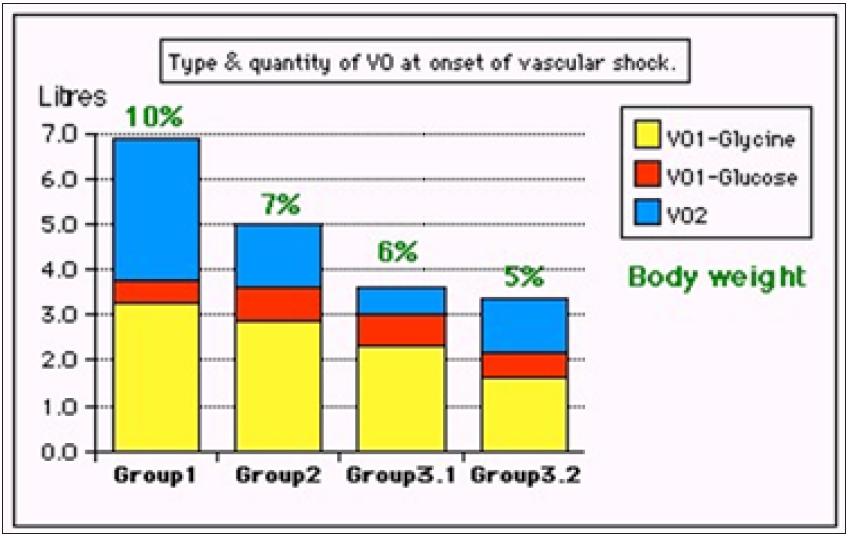
In theatre VOS is always mistaken for one of the recognized shocks such as hemorrhagic and septic shocks then wrongly treated with further volume expansion using sodium-based isotonic fluids. This induces VOS2 and cardiopulmonary arrest that has no serum markers of HN2 and causes ARDS in patients who survive a little longer [8,9]. Multiple regression analysis has proved that volumetric overload is the most significant factor in causing the clinical picture of VOS of the TURP syndrome (Figure 2 & 3; Table 1) [3,11]. Volumetric Overload Shock Type 2 (VOS2)1- 3,10 is induced by massive infusion of sodium-based fluids such as normal saline, Hartmann, plasma, plasma substitutes and blood or a combination of it. VOS2 may complicate VOS1 or is induced by sodium-based fluid during fluid therapy for resuscitation of shock and the critically ill and prolonged surgery and presents with ARDS later. Volumetric gain of 12-14 liters of sodium-based fluids reported in the first article on ARDS [18], which is the only article in the whole literature, other than the articles of mine some of which are referenced here, that documents the volume of retained fluid in ARDS.

Figure 3: shows volumetric overload (VO) quantity (in litres and as percent of body weight) and types of fluids. Group 1 was the 3 patients who died in the case series as they were misdiagnosed as one of the previously known shocks and treated with further volume expansion. Group 2 were 10 patients from the series who were correctly diagnosed as volumetric overload shock and treated with Hypertonic Sodium Therapy (HST). Group 3 were 10 patients who were seen in the prospective study and subdivided into 2 groups; Group 3.1 of 5 patients treated with HST and Group 3.2 of 5 patients who were treated with guarded volume expansion using isotonic saline

On another subject, this article [19], reports the overlooked link of Loin Pain Hematuria Syndrome (LPHS) with Symptomatic Nephroptosis (SN) and the Results of a new curative surgery; Renal Sympathetic Denervation and Nephropexy (RSD&N) Surgery. Two new signs namely, the IVU 7 sign (Figure 4) and tube stretch hypothesis were reported demonstrating that renal pedicle stretch causing vessel stenosis, ischemia and neuropathy. This was based on a prospective study of 190 patients with SN shown on erect IVU film (IVU-E), 182 were females and 8 males. The mean age was 28.8, duration of symptoms 15.7 and hospital follow up 6.6 years. Patients showed no abnormality on IVU or ancillary imaging when supine. All patients showed renal drop of >1.5 vertebrae (>5cm) on erect IVU film (IVU-E). Other demonstrable pathology on IVP-E film included: pelvic-ureteric junction kink affecting the right kidney in 116 (61.1%) and bilateral in 19 (10%) of patients. Stretch/ rotation of renal pedicle causing neuro-ischaemic pain of LPHS was demonstrable on the right side in 72 (37.9%) and bilaterally in 7 patients. Complications of SN on IVU-E included both obstructive and neuro-ischaemic: obstructive complication included ballooned renal pelvis, hydronephrosis and upper pole diverticulum. Neuroischaemic complications induced by pedicle stretch and rotation/ twist were haematuria of the LPHS affecting 36 (18.9%), auto nephropexy affecting 12 right kidneys, upper pole calyctiasis with extra-vasation affecting 28 (15.8%) right kidney and 2 bilateral that are best shown on Retrograde Pyelography (RGP). Renal atrophy affected 4 right kidneys. Upper pole infarction affected 2 right kidneys. Retrograde pyelography also demonstrated upper pole calyctiasis with extra-vasation. Surgical treatment was used in 28 patients; 10 had simple nephropexy and 18 had RSD&N) for severe LPHS. Four of patients treated with simple nephropexy had recurrence of LPHS while those who had RSD&N were all cured.

Figure 4: Shows renal pedicle mapped on a supine IVU film (Horizontal) and erect film (Vertical) limbs of 7 where the renal pedicle is stretched to 4 times its normal length, causing stenosis and ischemia.

On another subject I reported a surgical point of technique [20], for operable cancer bladder in which “capsule sparing” cystoprostadenectomy for orthotopic bladder replacement that overcomes the problems of difficult urethral anastomosis, impotence and incontinence. It makes the surgery easier and shorter. In summary, the new discoveries in medicine and physiology that originated in urology are reported. VOS are common iatrogenic complication of fluid therapy in hospitals. It may present in theatre as cardiopulmonary arrest in the operating theatre or later with coma and ARDS. VOS is 2 types: VOS1 and VOS2. VOS1 is induced by 3.5-5 liters of sodium-free fluid and is characterized with dilution HN that has 2 nadirs and 2 paradoxes, is most dynamic and illusive and currently has a lifesaving therapy of HST. VOS2 may complicate VOS1 or occur de novo complicating sodium-based fluid therapy during resuscitation of shock, acutely ill patients and prolonged surgery. It has no obvious serological markers or none. Many errors and misconceptions mislead physicians into giving too much fluid for resuscitation due to faulty rules on fluid therapy dictated by the wrong Starling’s law. The correct replacement for this law is the hydrodynamic of the G tube.
The overlooked link of LPHS with SN and the Results of a new curative surgery RSD&N were reported. Two new signs namely, the IVU 7 sign and tube stretch hypothesis demonstrating that renal pedicle stretch causing vessel stenosis, ischemia and neuropathy were reported. On another subject I reported a surgical point of technique for operable cancer bladder. Experience with “capsule sparing” cystoprostadenectomy for orthotopic bladder replacement: Overcoming the problems of impotence, incontinence and urethral anastomosis. The technique makes the surgery easier and shorter. The mentioned discoveries have resolved the puzzles of TURP syndrome, HN, ARDS and LPHS. Not only the exact pathoetiology was revealed but also a curable therapy was reported. These scientific discoveries should make the Medical World wake up, pay attention and listen to what this article has to say [21].

References

  1. Ghanem AN, Ghanem SA (2016) Volumetric overload shocks: Why is starling’s law for capillary interstitial fluid transfer wrong? the hydrodynamics of a porous orifice tube as alternative. Surgical Science 7(6): 245-249.
  2. Pindoria N, Ghanem SA, Ghanem KA, Ghanem AN (2017) Volumetric overload shocks in the patho-etiology of the transurethral resection prostatectomy syndrome and acute dilution hyponatraemia. Integr Mol Med.
  3. Ghanem KA, Ghanem AN (2017) Volumetric overload shocks in the patho-etiology of the transurethral resection prostatectomy syndrome and acute dilution hyponatraemia: The clinical evidence based on 23 case series. Basic Research Journal of Medicine and Clinical Sciences 6(4).
  4. Ghanem AN (2018) Ghanem’s new discoveries in medicine, physiology and urology and nephrology? Exp Tech Urol Nephrol 2(2).
  5. Ghanem AN (2001) Magnetic field-like fluid circulation of a porous orifice tube and relevance to the capillary-interstitial fluid circulation: Preliminary report. Medical Hypotheses 56(3): 325-334.
  6. Ghanem KA, Ghanem AN (2017) The proof and reasons that starling’s law for the capillary-interstitial fluid transfer is wrong, advancing the hydrodynamics of a porous orifice (G) tube as the real mechanism. Blood Heart and Circ 1(1): 1-7.
  7. Ghanem KA, Ghanem AN (2017) The physiological proof that starling’s law for the capillary-interstitial fluid transfer is wrong: Advancing the porous orifice (G) tube phenomenon as replacement. Open Acc Res Anatomy 1(2).
  8. Ghanem AN (2018) The adult respiratory distress syndrome: Volumetric overload shocks in patho-etiology, correcting errors and misconceptions on fluid therapy, vascular and capillary physiology. Surg Med Open Acc J 2(2).
  9. Ghanem AN (2019) Cardiac arrest and Volumetric Overload Shocks (VOS) complicating fluid therapy. EC Clinical and Medical Case Reports.
  10. Ghanem AN (2019) Volumetric Overload Shocks (VOS) causing the Acute Respiratory Distress Syndrome (ARDS). International Journal of Current Medical and Pharmaceutical Research.
  11. Ghanem AN, Ward JP (1990) Osmotic and metabolic sequelae of volumetric overload in relation to the TURP syndrome. Br J Uro 66(1): 71-78
  12. Harrison RH, Boren JS, Robinson JR (1956) Dilutional hyponatraemic shock: Another concept of the transurethral prostatic reaction. J Urol 75 (1): 95-110.
  13. Ghanem AN (2019) Post-surgical hyponatraemia: Problems of management resolved by revealing its relation to volumetric overload shocks. EC Cardiology 6(8): 708-714.
  14. Ghanem AN (2019) Postoperative dilution hyponatraemia and the TURP syndrome: Critical analytical review of literature on patho-etiology and therapy. EC Emergency Medicine and Critical Care 3(8): 507-514.
  15. Ghanem AN (2018) Hyponatraemia: Nadirs and paradoxes of the missing volumetric overload. Open Access Journal of Surgery 10(1).
  16. Ghanem AN, Ghanem SA, Ghanem KA, Pindoria N, Elsayed YS (2019) Illusive dynamic nadirs and masks of postoperative hyponatraemia and the TURP syndrome: Volumetric overload over time (VO/T) concept for resolving its puzzle. JOJ Uro & Nephron 6(4).
  17. Ghanem AN (2018) Therapy of hyponatremia: End of era or minority report? Biomed J Sci & Tech Res 11(4).
  18. Ashbaugh DG, Bigelow DB, Petty TL, Levine BE (1967) Acute respiratory distress in adults. Lancet 2(7511): 319-323.
  19. Ghanem AN (2016) Prospective observational study on loin pain hematuria syndrome complicating symptomatic nephroptosis and the results of renal sympathetic denervation and nephropexy surgery. J J Nephro Urol 3(1): 024.
  20. Ghanem AN (2002) Experience with capsule sparing cystoprostadenectomy for orthotopic bladder replacement: Overcoming the problems of impotence, incontinence and urethral anastomosis. BJU International 90(6): 617-620.
  21. Ghanem AN (2019) Medical world wake up, pay attention and listen: Ghanem’s new scientific discoveries in medicine, physiology, urology, nephrology, cardiovascular and surgery. EC Clinical & Medical Case Reports 2(9): 1-6.

For more articles in Surgery Journal Impact Factor
Please click on below link: https://crimsonpublishers.com/smoaj/

Monday, December 20, 2021

Prevention of Pelvic Floor Disorders After Vaginal Birth_Crimson Publishers

Prevention of Pelvic Floor Disorders After Vaginal Birth by Nour Tashtush in Investigations in Gynecology Research & Womens Health_Scholarly articles for women's health journal


Abstract

Vaginal birth is the main risk factor in the development of pelvic floor disorders. Pelvic floor muscle training (PFMT) can prevent prolapse symptoms at 2 years after intervention and could reduce the uptake of treatment. Also, results indicated that PFMT increases the chance of improvement in prolapse stage by 17% compared to no treatment.

Keywords: Vaginal birth; Prolapse; Physical therapy; Pelvic floor muscle strength

Abbreviations: PFMT: Pelvic Floor Muscle Training; TRA: Transverses Abdominis

Introduction

Epidemiological studies have consistently identified vaginal birth as the main risk factor in the development of pelvic floor disorders. Women who deliver vaginally are 2.8 times more likely to develop stress urinary incontinence and 5.5 times more likely to have pelvic organ prolapse than women who undergo cesarean delivery [1]. The aim of this review article is to assess the importance of pelvic floor training in preventing pelvic floor disorders.

Discussion

Pelvic organ prolapse is the symptomatic descent of one or more of the anterior vaginal wall, posterior vaginal wall, the uterus (cervix), or the apex of the vagina from the normal anatomical position, caused by herniation through deficient pelvic fascia or weaknesses or deficiencies in the ligaments or muscles that support the pelvic organs [2]. 5-10 years after a first birth, pelvic floor disorders were dramatically increased among women with a history of at least one operative vaginal birth [3]. Women with normal vaginal delivery and mediolateral episiotomy had the weakest pelvic muscles and nulliparous women had the strongest pelvic muscles [4].

Pelvic floor muscle training can prevent prolapse symptoms at 2 years after intervention and could reduce the uptake of treatment. Therefore, women should be recommended to undertake pelvic floor muscle training even before they have bothersome symptoms. The intervention should be encouraged on the basis of it being safe and done easily by most women [2]. Another conservative method: Pelvic floor physical therapy which targets the muscles, nerves, ligaments, connective tissue, lymphatic system, and joints inside and a rounding the pelvic girdle and focuses on addressing mobility and function. Pelvic floor physical therapists use internal or external therapies such as myofascial release, connective tissue manipulation, joint and scar tissue mobilization. Manual therapy uses palpation to loosen spastic muscles and lengthen tightened tissue to provide relief from pain. Therapies such as neuromuscular electrical stimulation and biofeedback use technology to help women gain functional awareness of the pelvic floor better coordinate muscle contractions and improve endurance to promote maximal functioning [5].

Randomized trials were done to assess the benefit of conservative methods in preventing pelvic prolapse, including six trials. Four trials compared pelvic floor muscle training (PFMT)with no intervention, and two trials compared pelvic floor muscle training plus surgery to surgery alone. PFMT compared to no intervention was found in individual trials to improve prolapse symptoms. Data on prolapsed severity was combined from two trials and results indicated that PFMT increases the chance of improvement in prolapse stage by 17% compared to no treatment. Pelvic floor muscle function appeared to be improved with PFMT in two out of three trials which measured this. Bladder symptoms were improved with PFMT in two out of three trials measuring this; bowel symptoms were measured in one trial, and an improvement with PFMT was found. The two trials which looked at the benefit of PFMT in addition to surgery, were small but of good quality. Findings were contradictory: women benefited from PFMT, in terms of urinary symptoms and pelvic floor muscle strength, in one trial but not the other [6].

Urinary incontinence is associated significantly with pelvic floor disorders, one of the studies on the effect of pelvic floor muscle exercise on quality of life in women with stress urinary incontinence and its relationship with vaginal deliveries has shown Both the combined training of the pelvic floor muscle (PFM) and the synergistic transverses abdominis (TRA) muscle, and the isolated PFM exercises improve the quality of life of women with stress urinary incontinence. Nonetheless, the combined PFM and TRA muscle physiotherapy is more effective. The exercises for the PFM and the synergistic muscle give better results in women who have given birth fewer than three times than isolated PFM exercises [7]. According to the previous mentioned studies, we assure the importance of pelvic floor muscle training as an effective conservative method in preventing and treating pelvic floor disorders.

Conclusion

Vaginal birth has encountered many cases of pelvic floor disorders that are resulted from the weakening of pelvic floor, so we conclude that preventing measures should be started immediately after the first vaginal birth which include pelvic muscle training and physical therapy that showed positive results as compared to no intervention.

References

  1. Howard D, Makhlouf M (2016) Can pelvic floor dysfunction after vaginal birth be prevented? Int Urogynecol J 27(12): 1811-1815.
  2. Hagen S, Glazener C, McClurg D, Macarthur C, Elders A, et al. (2017) Pelvic floor muscle training for secondary prevention of pelvic organ prolapse (PREVPROL): A multicentre randomized controlled trial. Lancet 389(10067): 393-402.
  3. Handa L, Blomquist JL, Knoepp LR, Hoskey KA, McDermott KC, et al. (2011) Pelvic floor disorders 5-10 years after vaginal or cesarean childbirth. Obstet Gynecol 118(4): 777-784.
  4. Afshari P, Dabagh F, Iravani M, Abedi P (2016) Comparison of pelvic floor muscle strength in nulliparous women and those with normal vaginal delivery and cesarean section. Int Urogynecol J 28(8): 1171-1175.
  5. Lawson S, Sacks A (2018) Pelvic floor physical therapy and women’s health promotion. J Midwifery Womens Health 63(4): 410-417.
  6. Hagen S, Stark D (2011) Conservative prevention and management of pelvic organ prolapse in women. Cochrane Database Syst Rev, Issue 12: CD003882.
  7. Magdalena P, Ciećwież S, Brodowska A, Starczewski A, Rutkowska JN, et al. (2018) The effect of pelvic floor muscles exercise on quality of life in women with stress urinary incontinence and its relationship with vaginal deliveries: a randomized trial. BioMed Research International 2019: 1-7.


For more articles in Scholarly articles for women's health journal
Please click on below link: https://crimsonpublishers.com/igrwh/

Friday, December 17, 2021

Ecotoxicological Impacts of Micro and Nanoplastics on Marine Fauna_Crimson Publishers

Ecotoxicological Impacts of Micro and Nanoplastics on Marine Fauna by Kuok Ho Daniel Tang in Examines in Marine Biology & Oceanography_Journal of Oceanography and Marine science


Abstract

Mismanagement of plastics has resulted in increasing plastic wastes in the environment, particularly the marine environment acting as the ultimate sink of plastics disposed into waters and even onto land. Micro- and nanoplastics in the marine environment undergo aggregation, sedimentation, deposition and enter the food chains as they are ingested by marine fauna. The uptake of micro- and nanoplastics by marine fauna poses multiple ecotoxicological effects comprising the blockage of alimentary canal and gills, behavioral change, physiological interference especially of the endocrine, antioxidative, immunity and hepatic systems, as well as adverse effects on reproduction and development of marine fauna. The ecotoxicological effects are often complicated by the ability of the micro- and nanoplastics to adsorb a wide range of chemicals. Nanoplastics have been found to affect cellular functions and membrane integrity and are able to cross the blood-brain barrier of certain aquatic species. The effects vary with the types of plastics, species of marine fauna, the dose as well as the sizes of plastics. This review systematically and concisely presents the toxicological effects of micro- and nanoplastics on marine fauna and highlights the need to understand the effects of these plastics at environmental concentrations instead of experimental concentrations. It also calls for the study of ecotoxicological effects of micro- and nanoplastics to be extended to more plastics types and sizes as well as more marine species.

Keywords: Microplastics; Nanoplastics; Marine; Ecotoxicology; Bioaccumulation; Adsorption

Introduction

The presence of micro- and nanoplastics in the environment has become a hot topic of discussions and scholarly publications due to the impacts they pose on the ecosystems and the health of both producers and consumers along the food chains. There is currently a lack of consensus on the definitions for microplastics and nanoplastics. Desforges et al. [1] classified plastics of 1μm to 5mm as microplastics 1 while Rocha-Santos and Duarte called those less than 5mm microplastics [2]. Similarly, the definitions for nanoplastics vary with EU Commission considering nanoplastics as plastic fragments in the range of 1 to 100nm, [3] and Hartmann et al. [4] defining them as those with sizes ranging from 1nm to 1μm [4]. Dimensions of the sizes of micro- and nanoplastics were often not specified, leading to ambiguity in determining whether the sizes actually referred to the diameters or the lengths.

Considering the inconsistency in size definitions, in this paper, microplastics refer to plastic fragments between 100nm and 5mm in any one of their dimensions while nanoplastics are plastic fragments less than 100nm in any dimensions [5]. Micro- and nanoplastics enter the environment through multiple pathways, one of which is through the use of cosmetic and cleaning products containing micro- or nanobeads [6]. Feedstock for plastics manufacturing and accidental release of plastic resin pellets or powder from air blasting also constitute direct entries of micro- and nanoplastics into the environment [7]. Indirectly, micro- and nanoplastics are formed from the degradation of large plastics discarded into the environmental via physical, chemical and biological means, as well as tearing of synthetic lint from cloth washing and wearing of plastic materials over time [8]. These plastic fragments can be carried by wind into the atmosphere and eventually settle onto ground or are washed down by rain. Rainwater runoffs often transport these fragments from the air or the ground into waterways resulting ultimately in the entry of micro- and nanoplastics into the marine environment [9]. The withdrawal of water contaminated with micro- and nanoplastics to water treatment plants causes entrapment of these plastics in the sludge produced during water treatment which are later returned to the environment via application of the sludge as fertilizer [6]. Similarly, channeling industrial wastewater and blackwater laden with micro- and nanoplastics to wastewater and sewage treatment plants also results in their accumulation and escape from these plants. Stephen et al. [10] found that even 3D printing emitted ultrafine synthetic particles and this aggravates concerns for the widespread presence of micro- and nanoplastics in the environment [10].

To date, micro- and nanoplastics have been detected in almost all ecosystems, particularly the marine and freshwater ecosystems. Microplastics were found in the water and sediment of inland seas 11 and ice cores of the Arctic where as much as 38 to 235 particles/m3 of microplastics were reported [11,12]. Desforges et al. [1] reported 9,180 microplastic particles in each cubic meter of seawater of the Northeast Pacific Ocean, while Norén and Naustvoll revealed a maximum of 102,000 particles in each cubic meter of the Sweden coastal waters [13]. The prevalence of micro- and nanoplastics was also reported to have increased twofold in the North Pacific subtropical gyre over the last forty years, indicating that there is a trend of micro- and nanoplastics accumulation in the environment over time [12].

As micro- and nanoplastics enter the marine ecosystems, they are partly removed via abiotic and biotic interactions leading to their aggregation, sedimentation, deposition and eventual entry into the food chains [2]. Unlike large plastic litters whose effects on marine organisms are observable through entanglement, smothering as well as blockage of the alimentary canal and toxic effects after ingestion, the environmental and ecotoxicological impacts of micro- and nanoplastics are not well characterized [14]. This is partly attributed to the diverse constituents and sizes of micro- and nanoplastics which give rise to complex biochemical interactions with marine organisms. While microplastics release chemicals as they are bioaccumulated and biomagnified up the trophic levels, nanoplastics could interact at cellular level and may disrupt physiological processes. This mini review examines the ecotoxicological effects of micro- and nanoplastics separately to provide better understanding of how these plastics affect the marine fauna, hence the health of consumers along the marine food chains.

Ecotoxicological Impacts of Microplastics on Marine Fauna

Generally, plastics contain additives which can be leached into the environment and cause various levels of toxicity. Studies showed correlation between the concentrations of plastic additives in the body of marine fauna and the amount of plastics they ingested or present in the environment they inhabited [15]. For instance, the Polybrominated Diphenyl Ethers (PBDEs) in tissues of mycophid fish sampled from the South Atlantic correlated with the additives of plastics found in the area [16]. Lugworms (Arenicola marina) sampled from sediments contaminated with polystyrene were also found to have higher levels of Polychlorinated Biphenyls (PCBs) compared to those from sediments without polystyrene contamination [17]. Microplastics can adsorb different chemicals in their surroundings such as phenanthrene, triclosan and nonylphenol and these adsorbed chemicals can be released together with plastic additives upon ingestion as already exhibited in the tissues of affected lugworms [18]. Chrysene, PCB 28 and derivatives of PBDEs were detected in fish which ingested polyethylene pellets [19].

Other common additives of plastics comprise phthalates, bisphenol A, acetaldehyde, formaldehyde polyfluoronated compounds and lead heat stabilizers, each of which exhibits different toxicity to the marine fauna [20]. Pthalates could interfere with endocrine system in fish via interacting with hormone receptors [21]. Nonylphenol also exhibited similar effect [22]. In fact, chemicals leached from microplastics, particularly Polyethylene Terephthalate (PET) had been found to demonstrate estrogenic activity [23]. The complex chemicals from plastics and adsorbed onto microplastics could be disruptive to ecological structure and functions as well as physiological processes such as immunity and endocrine system, and animals’ ability to elude predators. Browne et al. [18] revealed alteration of feeding behavior and mortality in lugworms which ingested triclosan-sorbed Polyvinyl Chloride (PVC) [18]. Experimental study on fish fed with microplastics sorbed with persistent organic pollutants and heavy metals revealed higher hepatoxicity characterized by glycogen depletion, tumor formation and lipidosis, as well as endocrine disruption, than microplastics alone [18,19].

Microplastics have also been reported to disrupt antioxidative system which plays crucial role in detoxification in living organisms. Examples of enzymes involved in antioxidative system are catalase, superoxide dismutase and glutathione peroxidase [24]. Jeong et al. [25] demonstrated that microplastics increased production of reactive oxygen species in rotifers and marine copepod, leading to intensified activities of superoxide dismutase, glutathione peroxidase, glutathione reductase and glutathione s-transferase [25]. The extent of such disruption is often size-dependent and contrary to the rationale that smaller microplastics have longer retention time and higher bioavailability, hence greater toxicological effects, various studies found microplastics to exhibit greatest effects at different sizes [24]. Exposing peppery furrow shell (Scrobicularia plana) to polystyrene at 1mg/L for 14 days in the laboratory resulted in enhanced activity of superoxide dismutase particularly in the gill and digestive glands [26]. The study points to tissue-specific responses to microplastics among marine fauna and the responses are often species-specific where red mullet (Mullus surmuletus) demonstrated only a slight increase in the activity of gluthathione with negligible change in the activities of superoxide dismutase and catalase in microplastics-enriched environment [27].

Ecotoxicological effects of chemicals-adsorbing ability of microplastics have been demonstrated via enhanced fluoranthene toxicity of polystyrene contaminated with fluoranthene in marine mussels which set off oxidative system at cellular level and interfered with fluoranthene detoxification more intensely than polystyrene alone [28]. Microplastics are also known to bind antibiotics and antimicrobials, thus facilitating their transfer to organisms [24].

Ecotoxicological Impacts of Nanoplastics on Marine Fauna

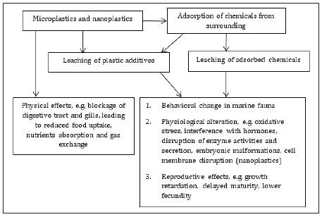
Similar to microplastics, nanoplastics contain chemical additives and provide the surfaces for adsorption of chemicals which upon release into the environment or after ingestion, can interfere with physiological functions and exhibit toxicity on marine fauna (Figure 1). However, due to their smaller sizes, nanoplastics have larger surface areas for chemical leaching and adsorption, making them potentially harder to detect and their effects more complex to characterize than microplastics. Their small sizes also permit them to interact at cellular level resulting in another dimension of toxicological concern. Nanoplastics of polystyrene have been demonstrated to permeate the membrane bilayers and induce alteration of membrane structure due to their solubility in the membrane (Figure 1). This hampers diffusion across membrane and disrupts cell functions [29]. Chemicals adsorbed onto nanoplastics can enter tissues, causing long-term toxicity [14].

Figure 1: Ecotoxicological effects of micro- and nanoplastics.


Latex nanoparticles were already detected in the gills and intestines of the Japanese rice fish (Oryzias latipes), and to lesser extent in their liver, blood and brain, thus indicating the ability of nanoplastics to cross the blood-brain barrier [30]. Exposure to 500nm polystyrene at concentrations of 1.25 and 2.5mg/L was found to lower fecundity of a marine copepod (Tigriopus japonicus) [5]. Blue mussel (Mytilus edulis) produced pseudofeces and experienced reduced filtering activity when exposed to 30nm polystyrene particles at concentrations between 0.1 and 0.3g/L [31]. 50mg/L of 200nm polystyrene stimulated pre-apoptosis among the Mediterranean mussels (Mytilus galloprovincialis) while 90nm polystyrene at less than 3.85mg/L resulted in deformation in the embryos of sea urchin (Paracentrotus lividus) [32,33].

Nonetheless, Booth et al. [34] revealed negligible toxicity of 2 poly(methylmethacrylate)-based Plastic Nanoparticles (PNPs) and fluorescent PNPs at concentrations ranging from 500mg/L to 1000mg/L on Corophium volutator [34]. Baudrimont et al. [35] examined the toxic effect of polyethylene nanoplastics on marine diatoms (Thalassiosira weissiflogii) and found no deleterious effect on their cell growth at concentrations up to 10,000μg/L [35]. Contrarily, polymethylmethacrylate nanoplastics at concentrations exceeding 4.69mg/L caused death of rotifers and the 48h median lethal concentration was estimated at 13.27mg/L [36]. Brine shrimps (Artemia franciscana) exposed to cationic amino-modified polystyrene nanoplastics showed mortality after 14 days and 1μg/L of the nanoplastics increased molting in the shrimp larvae [37]. Similar to microplastics, the toxicity of nanoplastics is type-specific, species-specific and dose dependent.

Conclusion

With increasing use of plastics and entry of plastics into the environment, the presence of micro- and nanoplastics in the marine ecosystems is a persistent problem. In comparison to large plastics, micro- and nanoplastics are harder to detect and it is significantly more complicated to characterize their ecotoxicological effects which are often species-specific, type-specific, size-dependent and dose-dependent. Micro- and nanoplastics can also adsorb chemicals and metals from their surrounding which renders understanding of their ecotoxicological effects even more complex. Current studies focus mainly on exposing various marine fauna to different types of micro- and nanoplastics in laboratory and polystyrene micro- and nanoparticles seem to receive more attention in such studies than other micro- and nanoplastics. These studies revealed the potential of these plastic fragments to cause behavioral change and interfere with physiological processes especially the endocrine and antioxidative systems in marine fauna. They also affect the growth and reproduction of the marine fauna. Furthermore, nanoplastics pose the danger of cellular interactions and membrane disruption. There is a need to also examine the ecotoxicological effects of these plastics at environmental concentrations and their interactions with other environmental contaminants to provide a more realistic picture of how these plastics affect the marine fauna. Research in this area should be expanded to different types of micro and nanoplastics commonly present in the marine environment. With climate change altering the physico-chemical properties of marine environment, it may also be crucial to examine how the ecotoxicological effects of micro- and nanoplastics are influenced [38,39].

References

  1. Desforges JPW, Galbraith M, Dangerfield N, Ross PS (2014) Widespread distribution of microplastics in subsurface seawater in the NE Pacific ocean. Mar Pollut Bull 79(1-2): 94-99.
  2. Rocha ST, Duarte AC (2015) A critical overview of the analytical approaches to the occurrence, the fate and the behavior of microplastics in the environment. TrAC Trends Anal Chem 65: 47-53.
  3. (2011) EU Commission. Plastic Waste: Ecological and Human Health Impact. Europe.
  4. Hartmann NIB, Nolte T, Sørensen MA, Jensen PR, Baun A (2015) Aquatic ecotoxicity testing of nanoplastics: Lessons learned from nanoecotoxicology. Aquatic Sciences Meeting, Denmark.
  5. Costa JP, Santos PSM, Duarte AC, Rocha ST (2016 ) (Nano)plastics in the environment-Sources, fates and effects. Sci Total Environ 566-567: 15-26.
  6. Carr SA, Liu J, Tesoro AG (2016) Transport and fate of microplastic particles in wastewater treatment plants. Water Res 91: 174-182.
  7. Zbyszewski M, Corcoran PL, Hockin A (2014) Comparison of the distribution and degradation of plastic debris along shorelines of the great lakes, North America. J Great Lakes Res 40(2): 288-299.
  8. Shim W, Song YK, Hong SH, Jang M, Han GM, et al. (2014) Producing fragmented micro-and nano-sized expanded polystyrene particles with an accelerated mechanical abrasion experiment. Switzerland, pp. 11-15.
  9. Jiang JQ (2018) Occurrence of microplastics and its pollution in the environment: A review. Sustain Prod Consum 13: 16-23.
  10. Stephens B, Azimi P, Orch Z, Ramos T (2013) Ultrafine particle emissions from desktop 3D printers. Atmos Environ 79: 334-339.
  11. Dai Z, Zhang H, Zhou Q, Yuan T, Tao C, et al. (2018) Occurrence of microplastics in the water column and sediment in an inland sea affected by intensive anthropogenic activities. Environ Pollut 242: 1557-1565.
  12. Obbard RW, Sadri S, Wong YQ, Khitun AA, Baker I, et al. (2014) Global warming releases microplastic legacy frozen in Arctic Sea ice. Earths Futur 2(6): 315-320.
  13. Norén F, Naustvoll LJ (2010) Survey of microscopic anthropogenic particles in Skagerrak. Rep Comm by Climate and Pollution Directorate. Sweden.
  14. Lambert S, Sinclair C, Boxall A (2014) Occurrence, degradation, and effect of polymer-based materials in the environment BT. Reviews of Environmental Contamination and Toxicology 227: 1-53.
  15. Lavers JL, Hodgson JC, Clarke RH (2013) Prevalence and composition of marine debris in Brown Booby (Sula leucogaster) nests at Ashmore Reef. Mar Pollut Bull 77(1-2): 320-324.
  16. Rochman CM, Kurobe T, Flores I, Teh SJ (2014) Early warning signs of endocrine disruption in adult fish from the ingestion of polyethylene with and without sorbed chemical pollutants from the marine environment. Sci Total Environ 493: 656-661.
  17. Besseling E, Wegner A, Foekema EM, Heuvel GMJ, Koelmans AA (2013) Effects of microplastic on fitness and PCB bioaccumulation by the lugworm Arenicola marina (L.). Environ Sci Technol 47(1): 593-600.
  18. Browne MA, Niven SJ, Galloway TS, Rowland SJ, Thompson RC (2013) Microplastic moves pollutants and additives to worms, reducing functions linked to health and biodiversity. Curr Biol 23(23): 2388-2392.
  19. Rochman CM, Hoh E, Kurobe T, Teh SJ (2013) Ingested plastic transfers hazardous chemicals to fish and induces hepatic stress. Sci Rep 3(1): 3263.
  20. Lithner D, Larsson Å, Dave G (2011) Environmental and health hazard ranking and assessment of plastic polymers based on chemical composition. Sci Total Environ 409(18): 3309-3324.
  21. Grün F, Blumberg B (2007) Perturbed nuclear receptor signaling by environmental obesogens as emerging factors in the obesity crisis. Rev Endocr Metab Disord 8(2): 161-171.
  22. Kawahata H, Ohta H, Inoue M, Suzuki A (2004) Endocrine disrupter nonylphenol and bisphenol a contamination in okinawa and Ishigaki Islands, Japan-within coral reefs and adjacent river mouths. Chemosphere 55(11): 1519-1527.
  23. Z YC, YS I, Craig JV, KD J, BG D (2011) Most plastic products release estrogenic chemicals: A potential health problem that can be solved. Environ Health Perspect 119(7): 989-996.
  24. Prokić MD, Radovanović TB, Gavrić JP, Faggio C (2019) Ecotoxicological effects of microplastics: Examination of biomarkers, current state and future perspectives. TrAC Trends Anal Chem 111: 37-46.
  25. Jeong CB, Kang HM, Lee MC, Kim DH, Han J, et al. (2017) Adverse effects of microplastics and oxidative stress-induced MAPK/Nrf2 pathway-mediated defense mechanisms in the marine copepod Paracyclopina nana. Sci Rep 7(1): 41323.
  26. Ribeiro F, Garcia AR, Pereira BP, Fonseca M, Mestre NC, et al. (2017) Microplastics effects in Scrobicularia plana. Mar Pollut Bull 122(1-2): 379-391.
  27. Alomar C, Sureda A, Capó X, Guijarro B, Tejada S, et al. (2017) Microplastic ingestion by Mullus surmuletus Linnaeus, 1758 fish and its potential for causing oxidative stress. Environ Res 159: 135-142.
  28. Paul PI, Lacroix C, González FC, Hégaret H, Lambert C, et al. (2016) Exposure of marine mussels Mytilus spp. to polystyrene microplastics: Toxicity and influence on fluoranthene bioaccumulation. Environ Pollut 216: 724-737.
  29. Rossi G, Barnoud J, Monticelli L (2014) Polystyrene nanoparticles perturb lipid membranes. J Phys Chem Lett 5(1): 241-246.
  30. Almutairi MMA, Gong C, Xu YG, Chang Y, Shi H (2016) Factors controlling permeability of the blood-brain barrier. Cell Mol Life Sci 73(1): 57-77.
  31. Wegner A, Besseling E, Foekema EM, Kamermans P, Koelmans AA (2012) Effects of nano polystyrene on the feeding behavior of the blue mussel (Mytilus edulis L.). Environ Toxicol Chem 31(11): 2490-2497.
  32. Canesi L, Ciacci C, Bergami E, Monopoli MP, Dawson KA, et al. (2015) Evidence for immunomodulation and apoptotic processes induced by cationic polystyrene nanoparticles in the hemocytes of the marine bivalve Mytilus. Mar Environ Res 111: 34-40.
  33. Della TC, Bergami E, Salvati A, Faleri C, Cirino P, et al. (2014) Accumulation and embryotoxicity of polystyrene nanoparticles at early stage of development of sea urchin embryos Paracentrotus lividus. Environ Sci Technol 48(20): 12302-12311.
  34. Booth AM, Hansen BH, Frenzel M, Johnsen H, Altin D (2016) Uptake and toxicity of methyl methacrylate-based nano plastic particles in aquatic organisms. Environ Toxicol Chem 35(7): 1641-1649.
  35. Baudrimont M, Arini A, Guégan C (2019) Ecotoxicity of polyethylene nanoplastics from the North Atlantic oceanic gyre on freshwater and marine organisms (microalgae and filter-feeding bivalves). Environ Sci Pollut Res, pp. 1-10.
  36. Venâncio C, Ferreira I, Martins MA, Soares AMVM, Lopes I, et al. (2019) The effects of nanoplastics on marine plankton: A case study with polymethylmethacrylate. Ecotoxicol Environ Saf 184: 109632.
  37. Bergami E, Pugnalini S, Vannuccini ML, Manfra L, Faleri C, et al. (2017) Long-term toxicity of surface-charged polystyrene nanoplastics to marine planktonic species Dunaliella tertiolecta and Artemia franciscana. Aquat Toxicol 189: 159-169.
  38. Tang KHD (2019) Climate change in Malaysia: Trends, contributors, impacts, mitigation and adaptations. Sci Total Environ 650(pt 2): 1858-1871.
  39. Tang KHD (2019) Are we already in a climate crisis? Glob J Civil Environ Eng 1: 25-32.

Thursday, December 16, 2021

Types of Microorganisms at Nosocomial Infections on the Surface of Laryngoscope Before Intubation at General Anesthesia_Crimson Publishers

 Types of Microorganisms at Nosocomial Infections on the Surface of Laryngoscope Before Intubation at General Anesthesia by Asghar Karbord in Cohesive Journal of Microbiology & Infectious Disease_Infectious disease Journals


Abstract

Introduction: Laryngoscope is using for endotracheal intubations. Inadequate decontamination of laryngoscope can develop nosocomial infections. If the blade isn’t disconnecting from laryngoscope after intubation can be transmit infections to handle. This study considering prevalence and types of bacteria isolated separately from laryngoscope blades and handles.

Method: This cross-sectional study was in the operating rooms of the educational and treatment center of Qazvin University of Medical Science in province Qazvin in Iran at 2018. 40 Laryngoscope blades and 40 laryngoscope handles were sampled after disinfection, with 4 methods disinfection: (1-Water, Povidon Iodine 7.5%, Ethanol 70%), (2-Water, Povidon Iodine 7.5%, Deconex (guaifenesin and phenylephrine 53 plus), (3-Water, Povidon Iodine 7.5%) and (4-Ethanol 70%)”. Samples were cultured on Mueller Hinton 5% sheep blood agar plate, MacConkey agar and Manitol salt agar. Inoculating plates were incubated at 37 ᵒC for 48 hours. Dominant microorganism and other growth bacteria identified comparatively.

Results: Nine various types of microorganisms were isolated and determined that handles were more Contamination than blades. Most negative results derived from blades and handles that were separated from each other and also for blades that were kept on gauze and separated from other instruments.

Conclusion: Performance of a standard disinfection method by software spss22 for both part of laryngoscope (blade and handle) seems to be necessary. Considering a special spot (like a special dish) for laryngoscope separated from other instruments may prevent the development of nosocomial infection

Keywords: Laryngoscope; Nosocomial infection; Intubation

Introduction

One of the most important Anesthesia instruments is laryngoscope. Laryngoscope is a type of endoscope used to endotracheal intubation and composed of a blade and a handle [1]. Laryngoscope blade comes in direct contact with the patient mucous membranes or broken skin and can be contaminated with secretions from the mouth and oropharynx, blood and various species of microorganism. Laryngoscope handle does not usually come in direct contact with patient’s oral mucosa. It can be contaminated indirectly while its routine disinfection is not observed as a standard practice. Laryngoscope blade and handle can transmit a lot of microorganisms to each other when the blade is not removed from handle after use.

When the hand of anesthesia provider touches a contaminated laryngoscope that seems to be clean, microorganisms can transmit to him and to other patients [2]. Also lack of a special keeping spot (for example a special dish) separated from other instruments can cause cross contamination between laryngoscope and other instruments and can spreads nosocomial infections [3-6]. Over two million patients per year develop nosocomial infections, resulting in 90,000 deaths annually and significant added healthcare costs, as well as unanticipated burdens on patients and their families [3]. Laryngoscope blades are placed in the semi critical group of medical instruments classified by Center for Disease Control and Prevention (CDC) and the Association for Professionals in Infection Control and Epidemiology (APIC) and designated by the CDC as “high level” disinfection [5]. In this study the researchers consider the incidence and the type of bacteria isolated from laryngoscope blades and handles which are separately used in an operating room and consider relationship between its contamination and its keeping spot.

Method

This cross-sectional study performed, and Samples were collected from operating rooms laryngoscopes of an educational and treatment center related to Qazvin University of Medical Science in Qazvin in Iran. Out of 80 samples which were earned (random simple sampling) in 4 consecutive days from 9 am to 3pm, 40 samples were taken from laryngoscope blades while other 40 samples were taken from laryngoscope handle. Some information about the laryngoscope that is ready to use include disinfection method, keeping spot, separating blade and handle from each other, and about the patient who was going to be intubated with that laryngoscope. The researchers had no interference in disinfection methods, and it was done within their routine procedures. There were 4 manual disinfection methods: 1- water, Povidon Iodine 7.5%, Ethanol 70% 2-Water, Povidon Iodine 7.5%, Deconex 53 plus 3- Water, Povidon Iodine 7.5% 4-Ethanol 70%. The investigator obtaining the sample wore sterile gloves and held the laryngoscope by its handle to avoid contacting with blade and sampled handle and blade with two sterile swabs separately. Sampling was done in aseptic circumstance. The sterile swab was rubbed to entire surfaces rotatory. After sampling, both divisions of laryngoscope were cleaned with ethanol 70% and prepared for next use. Nutrition broths (Merk) including samples transported to a laboratory with an ice box with a maximum transporting time of 4h. Specimens were inoculated onto a Mueller Hinton 5% sheep blood agar plate (High Media), MacConkey agar and Manitol salt agar (High Media). Inoculated plates were incubated at 37 ᵒC with 5%-10% CO2 for 48 hours. The result of Bacterial culture was accomplished using standard laboratory methods and dominant microorganism and other growth bacteria identified comparatively. Also, number of colonies was reported approximately. Data was entered in Statistical Package for Social Sciences (SPSS version 22) and all the data was analyzed. Data for baseline characteristics type of microorganisms was expressed as statistical relationship by Chi-Square test. In our study all the data was collected on medical checkups. Analysis of the data was done using descriptive statistics like frequencies, percent of microorganisms.

Results

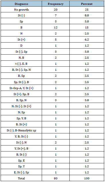
From 40 samples taken from laryngoscope blades 28(70%) were positive and from 40 samples taken from laryngoscope handles 33(82.5%) were positive. Nine different microorganisms were isolated: 1-Staphylococci, 2- Streptococci spp, 3-Bacillus spp, 4-Neisseria spp, 5-Enterobacter spp, 6-Diphtheria spp, and 7- Strep- Grp-A, 8-Yeast, 9- B-hemolytic streptococci. (Table 1) Dominant Organism in blade samples 10(25%) was Gram-Positive Bacillus and in handle samples 18(45%) was Gram-Positive Cocci. From all 80 samples 11(13.7%) staphylococci Coagulase-positive were found that 4(36.3) were for blades and 7(63.6) were for handles. From 40 collected blade samples most, negative cases were related to those were put on gauze separate from other instruments. From 80 samples 60(75%) blade and handle were separated that 19(31.6%) cases were negative and 20(25%) blade and handle were not separated that 1(20%) were negative. From 4 disinfection methods most, negative cases resulted in complex of water, povidon- iodine 7.5% and deconex (Table 2).

Table 1: Frequency of various bacteria from laryngoscope blades.

St= Staphylococci; Sp= Streptococci spp; B= Bacillus spp; N= Neisseria spp; D= Diphtheria spp; E= Enterobacter spp; Y= Yeast spp; (+) =Coagula’s Positive; (-) =Coagulase Negative


Table 2: Frequency of various bacteria from laryngoscope blades by methods disinfection.

St= Staphylococci; Sp= Streptococci spp; B= Bacillus spp; N= Neisseria spp; D= Diphtheria spp; E= Enterobacter spp; Y= Yeast spp; (+) =Coagula’s Positive; (-) =Coagulase Negative


Discussion

In this study the researcher’s different microorganisms on apparently clean laryngoscope that some of them are pathogen and potential for nosocomial infections which indicate that the decontamination procedures are disappointing and are not adopted to international standards. Also, the researchers found a significant relation between disinfection method and contamination quantity (p≤0.05 was significant). And there is a significant relation between keeping blade and handle separated from each other (p≤0.05 was significant) (Table 3). Also, more positive samples on handle laryngoscope indicate inadequate attention to handle disinfection that may cause contamination transmission to clean blade and to another instrument. In 2009 a study in the State University of New York Upstate IRB (Syracuise NY) 60 laryngoscope handles were sampled after low-level disinfection. From 40 samples sent for aerobic bacterial culture 30(75%) was positive and from 20 samples sent for viral testing all was negative [2]. In 2007 an outbreak of Pseudomonas aeruginosa in a neonatal intensive care unit (NICU) was identified that two infants reportedly died. Improper reprocessing of rigid laryngoscopes was identified as the cause of this outbreak [4]. This study supports previous studies and emphasizes laryngoscope disinfection adopted with standard methods. We also suggest considering a special spot or dish for laryngoscope separated from other instruments.

Table 3: Significant relation between disinfection method and place of laryngoscope.


Conclusion

Inadequate attention to decontamination of laryngoscope causes a lot of pathogen microorganism to be remained on this instrument. Teaching of an effective method for decontamination of laryngoscope designed by Center for Disease Control and Prevention (CDC) and supervising its performance in a best way seems to be necessary. Also considering a special spot or special dish for laryngoscope separated from other instruments may prevent the development of nosocomial infection [7].

References

  1. Simmons SA (2000) Laryngoscope handles: A potential for infection. AANA 68(3): 233-236.
  2. Tyler RC, Frederic JA, Scott WR, Deanna LK, Sumena C, et al. (2009) Nasocomial contamination of laryngoscope handles: challenging current guidelines. Anesth Analg 109(2): 479-483.
  3. Laurie A, Roming, David Hudak, Jeff Barnard (2005) Scrub making the case for disposable laryngoscope blades. Emerg Med Serv 34(3): 91-94.
  4. Muscarella LF (2008) Reassessment of the risk of healthcare-acquired infection during rigid laryngoscopy. Journal of Hospital Infection 68(2): 101-107.
  5. Ying HC, Karlokwong, Japing S, Yin ching C, Yichueh, et al. (2006) Use of condoms as blade covers during laryngoscopy, a method to reduce possible cross infection among patients. J Infect 52(2) 118-123.
  6. MJL Bucx, Dankert J, Beenhakker MM, Harrison TEJ (2001) Decontamination of laryngoscopes in the Netherlands. Br J Anaesth 86(1): 99-102.
  7. Junichi O, Kouichiro M, Hiroshi M, Takafumi H, Midori O, et al. (2004) Gargling with povidone-iodine reduces the transport of bacteria during oral intubation. Can J Anaesth 51(9): 932-936.

For more articles in Infectious disease Journals
Please click on below link: https://crimsonpublishers.com/cjmi/

Wednesday, December 15, 2021

Childhood Sexual Abuse: Scars Left for Life_Crimson Publishers

 Childhood Sexual Abuse: Scars Left for Life by Alisha Arora in Psychology and Psychotherapy: Research Study_Journal of Psychology and Psychotherapy


Abstract

The occurrence and prevalence of childhood sexual abuse (CSA) is a growing concern across the globe. The survivors of CSA experience its aftermath for years. The post-trauma consequences were classified broadly as-(i) physical, (ii) psychiatric, and (iii) psychosocial. The present review highlights the extant association of CSA with outcomes ranging from somatic/physical pain to psychiatric disorders to psychosocial dynamics of school dropout, sexual re-victimization, and sexual offending. The scars of CSA are deep rooted and through the review the need was observed to address the problem with extensive programs, interventions and policies, at the earliest stages.

Keywords: Childhood sexual abuse; Trauma; Outcomes; Trauma survivors

Childhood Sexual Abuse: Scars Left for Life

Childhood Sexual Abuse (CSA) is a global concern which is increasing in prevalence worldwide. The World Health Organization [1] defines CSA as “the involvement of a child in sexual activity that he or she does not fully comprehend and is unable to give informed consent to, or for which the child is not developmentally prepared, or else that violate the laws or social taboos of society”. Prevalence rates of CSA are concerning with females being more targeted. In a recent review by Barth et al. [2], it was observed that CSA prevalence rates were 8 to 31% for girls and 3 to 17% for boys. There has been a widespread work on the clinical outcomes and consequences of CSA, depicting the chronic scars it leaves behind. The consequences can broadly be categorized as physical, psychiatric, and psychosocial. CSA survivors are exposed to a varied range of impairment and difficulties in their adulthood and are always more vulnerable than their counterparts. Physical outcomes mostly include gastrointestinal symptoms, obesity, fibromyalgia, fatigue, irritable bowel syndrome, and asthma [3]. Estimates of a history of CSA among a population of women diagnosed with fibromyalgia range from 50 to 67% [4]. Among the ones suffering from non-organic gastrointestinal health issues, CSA survivors account for 53% [5].

Chronic hyper-arousal of the stress response and hyper-vigilance to the environment as a consequence of CSA makes survivors more vulnerable to stress. Stress, in turn leads to complex disease processes. Hence, among survivors, a reduced immune capacity and resultant health consequences are seen for a long time. CSA exposure considerably impacts the metabolic processes, as a result of stress hormones being vulnerable. Obesity hence is found among most of the survivors. Depression, a mental health outcome further reinforces obesity. Psychiatric manifestations depict the severity of mental health outcome. Post-Traumatic Stress Disorder (PTSD) is a commonly associated consequence [6] which can be an etiological cause for poor coping skills and poor problem-solving skills among survivors. Deficits in these life skills pave the way for depression, anxiety, and maladaptive personalities [7]. Along with biochemical factors, psychological mechanisms involving cognitive errors and poor coping skills are primary reasons of mood disorders among survivors. Beliefs such as I am not lovable, People are bad, I cannot deal myself, are errors which persists post-event. Eating disorders are also associated with CSA primarily involving bulimia nervosa and binge eating pattern [8]. Eating and purging can be faulty coping mechanism for stress in victims. Substance abuse, after PTSD is the most commonly studied outcome among CSA survivors [9].

According to Devries et al. [10], CSA is associated with increased odds of suicide attempts. Klonsky and Moyer [11] explain that self-injurious behaviour is a proxy consequence of other psychiatric vulnerabilities which occur as a CSA consequence. Dissociation, alexithymia, and borderline personality disorder has relatively higher link with CSA aftermath and act as moderators for suicidal behaviour/tendency. Psychosocial dynamics are perhaps the most extensive and deep scars CSA leads to. Lloyd et al. [12] explains sexual re-victimization, risky sex behaviour and risk of sexually transmitted diseases (like HIV) prominent among CSA survivors. Poor frustration tolerance and dissatisfaction may validate findings that CSA survivors have poor quality of relationships with partners [13]. Duncan [14] found in his longitudinal study that exposure to CSA leads to school/college dropout (up to 65%) and low scholastic performance. The effects of CSA at the family level may be economic, resulting in loss of income that may lead to change of residency and dropping out of school for the survivor [15]. In addition, sexual offending by survivors, involving offense against adults and/or children has also been indicated [16]. The sexually abused-sexual abuser association can be explained on basis of learning theory as per Burton [17] who mentions it as imitation of the perpetrator’s behavior and reinforcement of associated attitudes, and beliefs.

India is home to a vast youth population. Exposure to trauma, specifically CSA remains a challenge. Less reporting and stigma attached further bleaks the chance of reaching out to victims. The effects are either immediate or long term and lack of help ends most of them in hospitals or jails. Interventions and regulations have led to development of stricter laws. But the destination is still far away. Effects of CSA are varied and not uniform. It affects the victim, their families and their future, for a longer duration. Physical, psychiatric, and psychosocial outcomes have been evidenced in studies across globe. However, non-uniform definition of CSA, non-experimental methodology and reduced report rates of CSA weakens the empirical value. But we cannot ignore the fact that outcomes are there, and they are life changing. The essential message is to extensively work upon the programs, interventions and policies which ensure minimizing the exposure of CSA and provide the children and society, a safer-healthier future.

References

  1. World Health Organization (2003) Child sexual abuse: Guidelines for medico legal care for victims of sexual violence. World Health organization: Child Sexual abuse.
  2. Barth J, Bermetz L, Heim E, Trelle S, Toni T (2013) The current prevalence of child sexual abuse worldwide: A systematic review and meta-analysis. Int J Public Health 58: 469-483.
  3. Danese A, Tan M (2014) Childhood maltreatment and obesity: Systematic review and meta-analysis. Mol Psychiatry 19(5): 544-554.
  4. Wilson DR (2009) Stress management for adult survivors of childhood sexual abuse: A holistic inquiry. Western Journal of Nursing Research 32(1): 103-127.
  5. Hulme PA (2000) Symptomatology and health care utilization of women primary care patients who experienced childhood sexual abuse. Child Abuse Negl 24(11): 1471-1484.
  6. Chen LP, Murad MH, Paras ML, Colbenson KM, Sattler AL, et al. (2010) Sexual abuse and lifetime diagnosis of psychiatric disorders: Systematic review and meta-analysis. Mayo Clin Proc 85(7): 618-629.
  7. Amado BG, Arce R, Herraiz A (2015) Psychological injury in victims of child sexual abuse: A meta-analytic review. Psychosocial Intervention 24(1): 49-62.
  8. Molendijk ML, Hoek HW, Brewerton TD, Elzinga BM (2017) Childhood maltreatment and eating disorder pathology: A systematic review and dose-response meta-analysis. Psychol Med 47(8): 1402-1416.
  9. Halpern SC, Felipe BS, Juliana NS, Anne OS, Mayra P, et al. (2018) Child maltreatment and illicit substance abuse: A Systematic review and Meta‐analysis of longitudinal studies. Child Abuse Review 27(5): 344-360.
  10. Devries KM, Mak JY, Child JC, Falder G, Bacchus LJ, et al. (2014) Childhood sexual abuse and suicidal behavior: A Meta-analysis. Pediatrics 133(5): e1331-e1344.
  11. Klonsky ED, Moyer A (2008) Childhood sexual abuse and non-suicidal self-injury: Meta-analysis. Br J Psychiatry 192(3): 166-170.
  12. Lloyd S, Operario D (2012) HIV risk among men who have sex with men who have experienced childhood sexual abuse: systematic review and meta-analysis. AIDS Educ Prev 24(3): 228-241.
  13. Dennerstein L, Guthrie JR, Alford S (2004) Childhood abuse and its association with mid-aged women’s sexual functioning. J Sex Marital Ther 30(4): 225-234.
  14. Duncan RD (2000) Childhood maltreatment and college drop-out rates: Implications for child abuse researchers. Journal of Interpersonal Violence 15(9): 987-996.
  15. Tavkar P, Hansen DJ (2011) Interventions for families victimized by child sexual abuse: Clinical issues and approaches for child advocacy center-based services. Aggression and Violent Behavior 16(3): 188-199.
  16. Jespersen AF, Lalumière ML, Seto MC (2009) Sexual abuse history among adult sex offenders and non-sex offenders: A meta-analysis. Child Abuse & Neglect 33(3): 179-192.
  17. Burton DL (2003) Male adolescents: Sexual victimization and subsequent sexual abuse. Child and Adolescent Social Work Journal 20(4): 277-296.

Tuesday, December 14, 2021

A Review on Effect of Para-nonylphenol on Male Reproductive System_Crimson Publishers

 A Review on Effect of Para-nonylphenol on Male Reproductive System  by Malmir M in Perceptions in Reproductive Medicine_Journal of Reproductive Health


Abstract

Para-nonylphenol is known as a toxin and an environmental pollutant that has adverse effects on the reproductive system of laboratory animals. In this review, we focus on recent studies on the effect of this pollutant on the reproductive system including testicular tissue, sperm parameters and endocrine system disorders. The reproductive system is one of the most important and extremely sensitive organs of the body that is vulnerable to oxidative stress caused by pollutants. By searching in the scientific databases of PubMed, Google Scholar, Science Direct, Springer and Web of Science related articles were extracted. As a result, all observations have confirmed that Para-nonylphenol can cause multiple damages to the male reproductive system.

Keywords: Para-nonylphenol; Reproductive system; Sperm; Testis

Chemical Structure of Para-nonylphenol

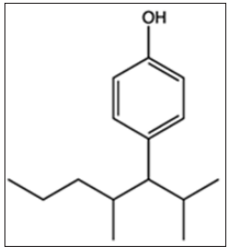
Para-nonylphenol (p-NP) is a term that can be applied to a wide range of isomeric compounds with the general formula C9H12 (OH) C6H4 (Figure-1). p-NP is an organic compound of the alkylphenol group. Alkylphenols are a small group of substances known as Xenostrogen [1]. If the position of the hydrocarbon chain linking to phenol in nonylphenol be para, it is referred to as p-NP or 4-nonyl phenol [1].

Figure 1: Chemical structure of Para-nonylphenol.


Estrogenic Activity of p-NP

p-NP has higher estrogenic activity than other alkylphenols and this effect has been observed in the male reproduction system including mice [1,2]. p-NP has been proposed to act as estrogen mimics by direct action at the estrogen receptor [3]. Estrogen was considered as a female hormone, it is also present in males and is responsible for performing some physiological functions such as maintenance of the skeletal system, normal function of testis and prostate [4]. On the other hand, p-NP can reduce the biosynthesis of testosterone by inhibiting the activity of the 17α-HSD enzymes and the cAMP pathway of Leydig cells [5-15]. Many studies have shown that estrogenic activity disrupts sex hormones such as testosterone [6], estrogen and progesterone [7], which can decrease the chance of fertility (Table-1).

Evaluation of Oxidative Stress and Apoptosis Induced by p-NP

p-NP can induce oxidative stress on germ cells [8] and reduces the level of antioxidant defense system [9] and also increased lipid peroxidation [10] in the testicular tissue [11]. Also, p-NP by increasing Reactive Oxygen Species (ROS) levels that cause increasing active box and the cytochrome exhaust from the mitochondria that leads to activation of the Apaf1/Caspase-9 complex. Activation of this Caspar cascade results in apoptosis [12] of germinal and Sertoli cells [10]. According to the researches presented in Table 1, it can be concluded that this pollutant increases ROS and causes apoptosis in the male reproductive system (Table-2).

Table 1: Evaluation of the adverse effect of p-NP on different species of laboratory animal (male reproductive system).

NIMRI: Naval Medical Research Institute; SD: Sprague-Dawley; NAC: N- acetylcysteine; p-NP: para-nonylphenol; T: testosterone; E: Estrogen; TMDA: Tissue Malondialdehyde; MDA: Malondialdehyde; LH: Luteinizing hormone; FSH: Follicle-stimulating hormone; AEA: Antioxidant; Enzyme Activities; ↑: Increase; ↓: Decrease; +: positive effect on p-NP


Table 2: Evaluation of the adverse effect of p-NP on different species of laboratory animal (testicular tissue).

NIMRI: Naval Medical Research Institute; SD: Sprague-Dawley; NAC: N- acetylcysteine; p-NP: para-nonylphenol; Ap: Apoptosis; CC: Caspase; Cascade; OS: Oxidative Stress; +T: Positive-TUNEL in germinal cells; ↑: Increase; ↓: Decrease; +: positive effect on p-NP.


Evaluation of the Adverse Effect of p-NP on Testicular Tissue (Histological and Stereological Studies)

NP can destroy the linkage of Gap junction by reducing the expression of connexin 43 protein, causing a defect and apoptosis in spermatogenic and Sertoli cells that may be a reason for the reduction in epithelial layer [6,13], as well as disruption of the blood-testicle barrier and the production of tissue edema. On the other hand, NP by stopping the B type spermatogonia in the G1 stage of mitosis because of the product of the XPB1 gene, inhibits the expression of cyclin 1 protein, which is one of the necessary factors for mitosis [5]. These studies listed in Table 2 demonstrates the adverse effect of this pollutant on testicular tissue.

Evaluation of the Adverse Effect of p-NP on Spermatogenesis

p-NP can induce apoptosis in germinal and Leydig cells [6] and decrease testosterone levels [5], as well as, leads to a decrease in the count and production of sperm daily [5,7]. The middle part of the sperm contains a large number of mitochondria that is responsible for movement and ROS reduces the progressive sperm motility by degenerating these mitochondria [14]. ROS by lipid peroxidation causes a decrease in membrane fluidity, damage to proteins and DNA, and eventually, abnormalities occur in sperm morphology [10]. Table 3 shows the studies of the adverse effect of p-NP on spermatogenesis.

Table 3: Evaluation of the adverse effect of p-NP on different species of laboratory animal (Spermatogenesis).

NIMRI: Naval Medical Research Institute; SD: Sprague-Dawley; NAC: N- acetylcysteine; p-NP: para-nonylphenol; Mot: Motility; Abn: Abnormality; Cou: Count; DSP: Daily sperm production; Via: Vaibility; Ant-E: Antioxidant Effect; ↑: Increase; ↓: Decrease; +: positive effect on p-NP.


Conclusion

This study, by collecting various studies using stereological [5], histological [15], biochemical [15-18] and andrological [7] methods, showed that p-NP at different doses and duration of treatment on laboratory animals can induce oxidative stress and apoptosis in germinal cells. This pollutant also reduces the chances of fertility by disrupting the endocrine system [2,7]. Humans are constantly exposed to p-NP through water, soil, food and vegetables. Many studies have shown that the use of antioxidants can prevent the adverse effects of oxidative stress caused by this pollutant in the male reproductive system [5].

References

  1. Routledge EJ, Sumpter JP (1997) Structural features of alkylphenolic chemicals associated with strogenic activity. J Biol Chem 272(6): 3280-3288.
  2. Duan P, Hu C, Butler HJ, Quan C, Chen W, et al. (2017) 4‐Nonylphenol induces disruption of spermatogenesis associated with oxidative stress‐related apoptosis by targeting p53‐Bcl‐2/Bax‐Fas/FasL signaling. Environ Toxicol 32(3): 739-753.
  3. Bindohaish G (2008) Effects of environmental pollution with alkylphenol (4-Nonyl phenol) on reproduction of tilapia, oreochromus spilurs (teleosts). Egyptian Journal of Aquatic research 34: 336-355.
  4. Pettersson K, Gustafsson JA (2001) Role of estrogen receptor beta in estrogen action. Annu Rev Physiol 63: 165-192.
  5. Malmir M, Faraji T, Noreini NS, Mehranjani SM (2018A) Protective antioxidant effects of n-acetylcysteine on testicular tissue and serum testosterone in paranonylphenol-treated mice (a stereological analysis). Reprod Syst Sex Disord 7(2): 2-6.
  6. Malmir M, Soleimani MM, Noreini NS, Faraji T (2018B) Protective antioxidant effects of N‐acetylcysteine against impairment of spermatogenesis caused by paranonylphenol. Andrologia 50(10): e13114.
  7. Momeni HR, Mehranjani SM, Abnosi MH, Mahmoodi M (2009) Effects of vitamin E on sperm parameters and reproductive hormones in developing rats treated with para-nonylphenol. International Journal of Reproductive BioMedicine 7(3): 111-116.
  8. Han XD, Tu ZG, Gong Y, Shen SN, Wang XY, et al. (2004) The toxic effects of nonylphenol on the reproductive system of male rats. Reproductive Toxicology 19(2): 215-221.
  9. Abnosi MH, Masoomi S (2019) Para-nonylphenol toxicity induces oxidative stress and arrests the cell cycle in mesenchymal stem cells of bone marrow. Iranian Journal of Toxicology 13(3): 1-8.
  10. Agarwal A (2005) Role of oxidative stress in male infertility and antioxidant supplementation. US Kidney and Urological Disease p. 122.
  11. Shalaby KA, Saleh EM (2011) Ameliorative effect of honeybee propolis on the nonylphenol induced- reproductive toxicity in male albino rats. Aust J Basic Appl 5: 918-927.
  12. Sayed AEDH, Kotb AM, Oda S, Kashiwada S, Mitani H (2019) Protective effect of p53 knockout on 4-nonylphenol-induced nephrotoxicity in medaka (Oryzias latipes). Chemosphere 236: 124314.
  13. Lu WC, Wang AQ, Chen XL, Yang G, Lin Y, et al. (2014) 90d Exposure to nonylphenol has adverse effects on the spermatogenesis and sperm maturation of adult male rats. Biomed Environ Sci 27: 907-911.
  14. Keikha L (2015) Evaluation of the protective effect of nigella sativa oil on testicular tissue and sperm parameters in adult NMRI mice treated with para-nonylphenol. SSU_Journals 23(2): 1927-1944.
  15. Duan P, Hu C, Butler HJ, Quan C, Chen W, et al. (2016) Effects of 4-nonylphenol on spermatogenesis and induction of testicular apoptosis through oxidative stress-related pathways. Reprod Toxicol 62: 27-38.
  16. Azizi P, Mehranjani MS (2019) The effect of green tea extract on the sperm parameters and histological changes of testis in rats exposed to para-nonylphenol. Int J Reprod Biomed 7(10): 717-726.
  17. Feng M, Chen P, Wei X, Zhang Y, Zhang W, et al. (2011) Effect of 4-nonylphenol on the sperm dynamic parameters, morphology and fertilization rate of Bufo raddei. African Journal of Biotechnology 10(14): 2698-2707.
  18. Doust KS, Noorafshan A, Dehghani F, Panjehshahin MR, Monabati A (2010) Effects of hydroalcoholic extract of Matricaria chamomilla on serum testosterone and estradiol levels, spermatozoon quality, and tail length in rat. Iranian Journal of Medical Sciences 35(2): 122-128.

For more articles in Journal of Reproductive Health
Please click on below link: https://crimsonpublishers.com/prm/

A Close Look at the Application of the Yin-Yang- Based Acupoint Pairs_Crimson Publishers

A Close Look at the Application of the Yin-Yang- Based Acupoint Pairs by Tong Zheng Hong in Advancements in Bioequivalence & Bioavailabi...