Crimson Publishers High Impact Journals

Showing posts with label Journal of Pharmaceutical Sciences Review and Research. Show all posts

Thursday, April 14, 2022

Bilateral Renal Cell Carcinoma and Thyroid Carcinoma with Tertiary Hyperparathyroidism, Clinical Case_Crimson Publishers

Bilateral Renal Cell Carcinoma and Thyroid Carcinoma with Tertiary Hyperparathyroidism, Clinical Case by Chernyshev VA in Developments in Anaesthetics & Pain Management_Journal of Pharmaceutical Sciences Review and Research


Introduction

Multiple primary tumors (MPT) are independent emergence and development of two or more neoplasms in one patient. In the structure of multiple primary cancer of the genitourinary organs in men most frequently there is a combination of two malignant neoplasms of the genitourinary organs (29,2%) with tumors of the gastrointestinal tract (25,0%) and tumors of head, neck and skin (25,5%), respectively [1]. While for men with multiple primary cancer of the genitourinary organs, the development of malignant neoplasms of the prostate glands (33% of cases), the bladder (29,4%) and the kidneys (21,4%) is somewhat less common, for women the kidneys (67,8%) and much less frequently of the bladder (22,6%) [2]. When planning the treatment of primary multiple malignant tumors, it is necessary to evaluate with special objectivity the possibilities of each method of anticancer treatment, the factors limiting its use, and the risk of complications. It is impossible to achieve a high degree of direct impact, suppression of the growth of one or all tumors at any cost without taking into account the prognosis for each of them. In particular, in the practice of oncologists in the treatment of bilateral renal cell carcinoma, the question arises about the possibility of using targeted therapy in patients with end-stage chronic renal failure who are on hemodialysis. However, clinical trials prior to 2017 did not lead to approval of targeted therapy in this group of patients [3,4].

Report

The article presents a case of diagnosis and surgical treatment in a patient who is in the end stage of chronic renal failure with developed tertiary hyperparathyroidism, with bilateral renal cell carcinoma, thyroid carcinoma, with successful use of targeted therapy.

Keywords: Bilateral renal cell carcinoma; Targeted therapy; Thyroid carcinoma; Chronic kidney failure; Tertiary hyperparathyroidism

Material and Methods

In September 2012, a patient of 50 years old turned to the Republican clinical oncology center of the Ministry of health of the Republic of Tatarstan. At the time of treatment, the patient complained of weight loss, weakness, pain in the right side and a painful tumor in the left half of the lower jaw up to 4xcm in size. It is known from the anamnesis that in 1989 the patient was diagnosed with polycystic kidney disease, chronic glomerulonephritis. In 2001, the patient suffered from hemorrhagic fever with renal syndrome with the outcome of terminal chronic renal failure. From then on, patient S. was on long-term hemodialysis 3 times a week. During the examination of the abdominal cavity and retroperitoneal space a multi-chamber cystic solid formation of 116x80 mm was found in the right kidney, as well as foci of destruction in the right scapula up to 7mm, in the medial part of the iliac wing up to 22mm, in the body of the left iliac bone up to 15mm. While running bone scintigraphy, uneven accumulation of radiopharmaceutical in the pelvic bones and in the lower jaw on the left was revealed. After FNA-biopsy of the lower jaw was revealed a cytological picture of osteoblastoclastoma or bone cyst. The level of parathyroid hormone (PTH) at that time was 1390Pg/ml, the level of total blood calcium 2.52mmol/l, and alkaline phosphatase 217u/l. The patient had the following comorbidities: arterial hypertension grade 3 (risk 4), dilated cardiomyopathy, chronic heart failure grade 2a (class 2), mitral valve regurgitation 1-2 degrees, tricuspid valve regurgitation 2 degrees, nephrogenic anemia of the 1st stage, polycystic kidney disease.
In October 2012, the patient underwent an operation in the urology Department of the Republican clinical hospital in Kazan: right-sided nephrectomy. According to the histological examination (No. 41471-80): renal cell carcinoma, mixed-cell variant, was detected (pT3). The patient was diagnosed with cancer of the right kidney pT3N0M0, stage 3, clinical group 2. Taking into account changes in the bones with the formation of foci of parathyroid osteodystrophy and laboratory parameters, a concomitant diagnosis was established: secondary/tertiary hyperparathyroidism, parathyroid osteodystrophy. Ultrasound examination of the neck revealed an increase in the parathyroid glands on both sides from 8 to 12mm, the structure of the thyroid gland was diffusely heterogeneous with nodules up to 8 mm due to concomitant autoimmune thyroiditis, and the cervical lymph nodes were not changed.
In order to improve the standard of living and normalize the phosphorus-calcium metabolism and blood biochemical parameters, the patient was shown surgery to remove all hyperplastic parathyroid glands. In December 2012, the patient underwent an operation: total parathyroidectomy with auto transplantation of part of the lower right parathyroid gland into the muscle fibers of the extensors of the right forearm. During the operation, during the revision of the thyroid gland, the manifestation of thyroiditis were determined, but according to the histological examination (No. 27883/2012), it turned out that in addition to the changes due to nodular hyperplasia of the left lower and right parathyroid glands, the “left upper parathyroid gland” turned out to be a metastasis of papillary thyroid cancer to the paratracheal lymph node. Postoperative laboratory parameters significantly decreased: PTH to 216Pg/ml, total calcium to 2.05 mmol/l and ionized calcium to 1.22mmol/l. After 1 month, the patient was prepared for re-operation on the thyroid gland, while the PTH level increased to 458Pg/ml. In January 2013, the operation was performed: thyroidectomy with central lymph node dissection (level 6), removal of the dystopic left upper parathyroid gland. During the histologic examination the papillary tumor was revealed in the lower pole of the thyroid’s left lobe up to 1cm with extracapsular extension, also there was 2 out of 5 metastatic paratracheal lymph node and parathyroid adenoma. In the postoperative period, the level of PTH decreased to 23.2Pg/ml, ionized calcium to 0.95mmol/l, and total calcium to 1.91mmol/l. A year later, in January 2014, by a dispensary observation and ultrasound of the abdominal cavity and retroperitoneal space revealed a tumor of the left kidney and a tumor formation in the bed of the removed right kidney.
In February 2014, the patient underwent surgery in the oncology center (Kazan): left-side nephrectomy with resection of the left adrenal gland, removal of metastasis of the right kidney bed with the liver resection. During the operation, the revision revealed a tumor in the left kidney up to 4 cm in size with multiple cysts, at the level of the right adrenal gland a tumor up to 2cm with ingrowth into the liver. According to the histological examination (No. 4335/2014): papillary carcinoma of the left kidney, type 2, without extracapsular extension (pT2N0M0), polycystic kidney disease, metastasis of renal cancer to the liver, there were no sign of metastases in lymph nodes. Thus, the patient was given a new diagnosis: papillary carcinoma of the left kidney with metastasis in the right kidney bed, pT1N0M1, condition after right-side nephrectomy for renal cancer pT3N0M0. Papillary carcinoma of the left thyroid lobe pT3N1M0, after surgical treatment. Chronic renal failure (stage 5), at the stage of chronic hemodialysis. Tertiary hyperparathyroidism, condition after total parathyroidectomy. In January 2015, by a dispensary examination according to the CT of the abdominal cavity, metastases of renal cancer were found along the abdominal wall and again in the bed of the removed right kidney. After one month in Republican clinical hospital (Kazan) was performed surgery: laparotomy, removal of metastatic lesions of the abdominal wall and the bed of the right kidney.
According to the results of histological examination, metastases of renal cell carcinoma were found in 8 foci. In August 2015, the next medical examination in oncological center according to results of ultrasound and CT of the abdominal cavity showed a tumor formation in the bed of the right kidney 10mm, in the subhepatic space and in the projection 6 and 7 segments of the liver with sizes up to 27 and 23mm, respectively, tumor in the perinephric space on the right was regarded as metastatic lesions. By the decision of the doctors’ Concilium of may 11, 2015, despite the presence of such a serious concomitant condition as terminal chronic renal failure and long-term hemodialysis, for the first time in the Republic of Tatarstan (probably in Russia), a decision was made to conduct targeted therapy. The drug of choice was Sunitinib at a dosage of 50mg, which had a significantly higher median survival rate in patients with metastatic renal cancer [3,4]. From December 2015 to October 2017, patient S. received targeted therapy with Sunitinib in the amount of 14 courses with the preservation of creatinine levels up to 600 mmol/l and urea up to 20 mmol/l. Determined stabilization process until mid-October, 2017, when during a routine examination according CT was identified a negative dynamics in the increase in the size of metastatic foci and the appearance of a new lesion in a right leg aperture size up to 9 mm. An another doctor’s Concilium was held on October 31, 2017, where it was decided to conduct a second-line targeted therapy with Afinitor (Everolimus) with proven effectiveness and low frequency of side effects as a second-line drug [5], with a decision on further surgical intervention. From January 2018 to may 2018, patient S. received targeted therapy with this drug at a dosage of 10 mg, maintaining acceptable levels of creatinine and blood urea with continued hemodialysis 3 times a week. Then, on May 17, 2018, the patient underwent surgery to remove the metastatic foci of renal cancer with liver resection and cholecystectomy. According to the results of histological examination, 8 foci were identified as metastases of renal cancer. In October 2018, during a routine dispensary examination, according to the CT of the abdominal cavity, metastases of the abdominal wall were found in the amount of number 3, up to 3cm in size. After a year of follow-up with stabilization of the process, the patient died of an ischemic stroke in July 2019.

Conclusion

A case of 7-year life expectancy of a patient with primary multiple tumors (bilateral renal cancer, thyroid cancer) against the background of an end-stage chronic renal failure and hemodialysis with the development of tertiary hyperparathyroidism and multiple surgical interventions for multiple recurrence of renal cancer and long-term targeted therapy with an acceptable quality of life is presented. Analyzing this clinical case, we can conclude how important a personalized approach is in oncology, in particular if we are talking about cancer patients with end-stage chronic kidney failure who require special anticancer treatment: it is necessary to take into account all the features of patient management, timely diagnose the progression of the disease, carefully monitor laboratory biochemical parameters of blood, and most importantly together with doctors of related specialties, ensure a decent standard of living and its maximum duration.

References

  1. Stepanova Yu A, Kalinin DV, Vishnevsky VA (2015) Primary multiple tumors (literature review). Vishnevsky Institute of Surgery of the Ministry of Health of Russia, Medical Visualization, Russia, 6: 94-97
  2. Leonov OV, Dolgikh VT, Kopyltsov EI, Alekseev BY (2010) Primary-multiple malignant neoplasms of the genitourinary organs. Oncourology 6(2): 56-60.
  3. Ravaud A, Robert JM, Pandha SH, George DJ, Allan JP, et al. (2016) Adjuvant sunitinib in high-risk renal-cell carcinoma after nephrectomy. N Engl J Med 375: 2246-2254.
  4. Taneja SS (2012) Efficacy and toxicity of sunitinib in patients with metastatic renal cell carcinoma with severe renal impairment or in haemodialysis. The Journal of Urology 187(4): 1223-1224.
  5. Alekseev B Ya, Kalpinsky AS (2010) Everolimus in the treatment of metastatic kidney cancer. Herzen Moscow State Research Institute. The Urologic Oncology 3: 19-23.

For more articles in Journal of Pharmaceutical Sciences Review and Research
Please click on below link: https://crimsonpublishers.com/dapm/

Thursday, March 31, 2022

Biomedical Waste Management in A Multispecialty Hospital_Crimson Publishers

 Biomedical Waste Management in A Multispecialty Hospital by Vivek Kumar Garg in COJ Reviews & Research_Journal of Pharmaceutical Sciences Review and Research


Abstract

Any waste which is generated during the diagnosis, treatment, or immunization of human beings or animals or in research activities pertaining thereto or in the production or testing of biologicals, is called biomedical waste. The current study is based on BMW in multispecialty hospital at civil hospital, Barnala, India. The material used in the papers regarding BMW management is the Questionnaire, prepared with different topics. Our objective in the study was to evaluate the good points as well as the lacunas in the protocols adopted in the hospital waste management and its disposal, in the multispecialty civil hospital. In the present study, a list of departments of civil hospital, Barnala was made. There are following departments like General Medicine, General Surgery, Gynecology, Pediatrics, Ophthalmology, ENT, Dental, Skin, Orthopedics, Blood bank, OT, Wards, Indoor etc. The results obtained showed lack of awareness of bio medical waste management and its handling even in qualified medical staff. The reason being its complex is system and rules that has not become part of daily curriculum till now. We have studied one month regular protocol followed for bio medical waste collection by the hospital staff. During the study, we have observed that the hospital is properly managing their biomedical waste. Regularly, the hospital segregates the waste according to the specified categories and color coding. Regarding the capabilities and risks of biomedical waste treatment alternatives, it must be emphasized that the only treatment technologies that are usually used to treat pathological waste are the incineration and mechanical/ chemical disinfection systems.

Keywords: Biomedical waste; Healthcare; Multispecialty; Hospitals

Introduction

Any waste which is generated during the diagnosis, treatment, or immunization of human beings or animals or in research activities pertaining thereto or in the production or testing of biologicals, is called biomedical waste [1]. Medical care is indispensable for our life. But the waste generated during medical care needs attention. The nosocomial (hospital acquired) infections are the result of hospital waste. The hazardous and toxic parts of waste from healthcare establishments comprising infectious, biomedical and radioactive material as well as sharps needles, constitute a grave risk. The rag pickers and waste workers are often worst affected. Diseases like cholera, typhoid, plague, tuberculosis, hepatitis, AIDS, Diphtheria, Malaria, etc. pose grave public health risks [2-4]. With a judicious planning and management, the risk of spread of disease can be considerably reduced. The rules framed by the Ministry of Environment and Forests (MoEF), Govt. of India, known as Bio Medical Waste Management and Handling Rules, 1998, provides uniform guidelines and code of practice for the whole nation [5,6]. In Schedule I of the Bio Medical Waste Management rules 1998 (Annexure II), waste has been categorized into 10 points [2,4,7]. There are total 6 schedules in BMW management.

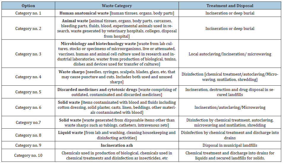
Schedule I: (Table 1)

Table 1: Schedule I.


Schedule II: (Table 1)

Table 2: Schedule II: Colour coding charts and type of container for disposal of BMW is as follows.


Schedule III: Labels for BMW containers/Bags

Schedule IV: Label for transport of BMW containers/Bags

Schedule V: Standards for treatment and disposal of BMW standards for incinerators.

Schedule VI: Schedule for waste treatment facilities like Incinerator/Autoclave/Microwave system.

Materials and Methods

The current study is based on BMW in multispecialty hospital at civil hospital, Barnala, India. The material used in the papers regarding BMW management is the Questionnaire, prepared with different topics.

Methods- The questionnaire was prepared by keeping following points in mind.

  1. Awareness of the importance of BMW management
  2. What is BMW
  3. Types of BMW
  4. BMW management at different levels- means at small clinics to big hospitals to medical and dental colleges.
  5. Classification of BMW
  6. Isolation of BMW
  7. Packaging of BMW and its disposal practices

Selection criteria to distribute questionnaire- The first step is preparation of questionnaire, study sample selected, organized and finalized. The second stage included distribution of questionnaire, analyzing the collected data and determining the response. Selection criteria for the persons are determined- The person must be part of healthcare, graduate or undergraduate, knowledge of English language, medical or paramedical staff, male or female, small clinics, primary health centers, polyclinics, multispecialty hospitals, government civil hospitals.

Aims and Objectives

To evaluate the steps taken in the management of hospital generated bio medical waste. Our objective in the study was to evaluate the good points as well as the lacunas in the protocols adopted in the hospital waste management and its disposal, in the multispecialty civil hospital. In the present study, a list of departments of civil hospital, Barnala was made. There are following departments like General Medicine, General Surgery, Gynecology, Pediatrics, Ophthalmology, ENT, Dental, Skin, Orthopedics, Blood bank, OT, Wards, Indoor etc. It took one month to record bio medical waste generation of all departments separately for Red, Yellow, Blue and White containers/bags. Then we added the waste for Red of all departments into one head, Yellow of all departments into one head, Blue of all departments into one head and White of all departments into one head. This all was infected waste. The non-infected waste e.g. expired medicines were put in Black colored container. But we did not include non-infected waste in our study. Since, our topic is focused on the protocols, that means we have to keep eye on the proper segregation of the waste before putting into their respective color-coded containers in their respective departments.

Result

Red Bag- Solid waste, POP casts, infected cotton & dressing, infected bandages, items containing blood & body fluid like extracted teeth, cysts, granulomas

Yellow Bag- Anatomical waste Microbiology & Biotechnology waste. Wastes from lab cultures or specimen of microbes live or attenuated vaccines, human and animal cell cultures, waste from production of biological, toxins, dishes & devices.

Blue Bag- Disposable plastic glasses, Plastic Tubings, IV sets, syringes, Gloves, Solid waste other than sharp needles/blades.

White Container- Sharp needles/objects that may cause cuts, Needles, Blades, expired injections, cut glasses.

Black Container- All above mentioned color bags contain Infected Waste. But Black color bag contains Non-Infected waste. e.g. cytotoxic drugs, chemical waste, expired drugs tablets & capsule form.

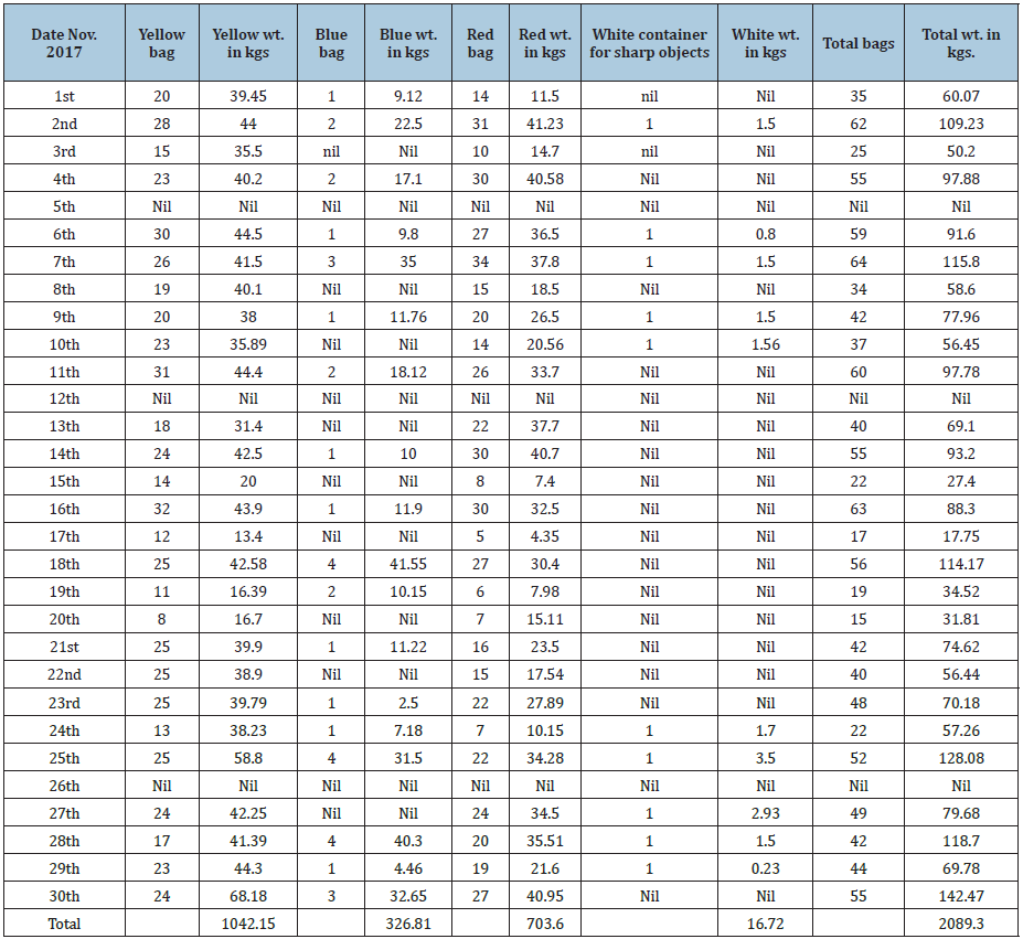
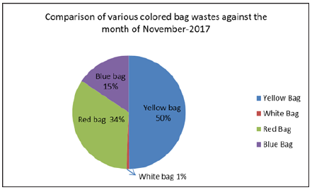
We studied one-month 1st November 2017 to 30th November 2017, BMW collection and disposal and its Protocols followed in the Civil Hospital, Barnala (Table 3) (Figure 1-5).

Table 3: BMW November 2017, civil hospitaal, Barnala.


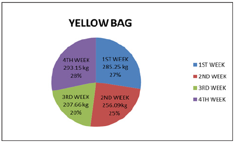
Figure 1: Comparison of average yellow bag waste week wise for the month of November 2017.


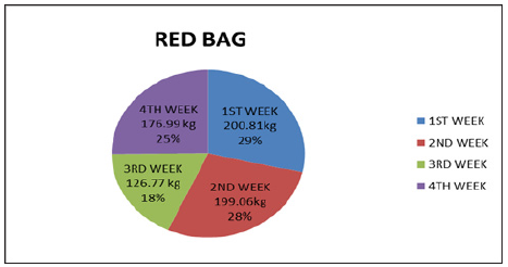
Figure 2: Comparison of average red bag waste week wise for the month of November 2017.


Figure 3: Comparison of average blue bag waste week wise for the month of November 2017.


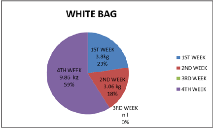
Figure 4: Comparison of average white bag waste week wise for the month of November 2017.


Figure 5: BMW report of November 2017 civil hospital, Barnala.


BMW report of November 2017 civil hospital, Barnala

Yellow container - 1042.15kg, Blue container -- 323.57kg

Red container – 696kg, White bag - 13.12kg, Total waste --2066.64kg

Total deliveries in the Gynae Deptt. --495 in number

Human Anatomical waste from Gynae deliveries inkgs - approx 0.5kg per delivery.

Total deliveries=495

So Total Human anatomical waste from gynae depth was 495 x 0.5=247.500kg

Out of 1034.95kg of waste of yellow container, 1042.15-247.500=794.65kg

Means Yellow container contains 247.500kg Human anatomic waste and 794.65kg is other solid waste.

Liquid waste generated after washing instruments and lab containers, test tubes was 300 liters in one-month November 2017.

Discussion of Diagrams

Yellow bag diagram with human anatomical waste with placentas shows

285.25kg and 27% 1st week of November 2017

256.09kg and 25% 2nd week of November 2017

207.66kg and 20% 3rd week of November 2017

293.15kg and 28% 4th week of November 2017

Blue bag diagram with infected plastics, syringes, gloves, plastic tubing’s shows

93.52kg and 29% 1st week of November 2017

51.78kg and 16% 2nd week of November 2017

65.42kg and 20% 3rd week of November 2017

116.09kg and 35% 4th week of November 2017

Red bag diagram with soiled waste, infected dressings, POP casts shows

200.81kg and 29% 1st week of November 2017

199.06kg and 28% 2nd week of November 2017

126.77kg and 18% 3rd week of November 2017

176.99kg and 25% 4th week of November 2017

White bag diagram with sharp needles and cut glasses shows

3.8kg and 23% 1st week of November 2017

3.06kg and 2nd week of November 2017

Nil 3rd week of November 2017

9.86kg and 59% 4th week of November 2017

The idea, to show one-month study and further breaking it into 4 parts (weeks), is to show regularity in the collection of biomedical waste in the hospital.

Treatment and Disposal of BMW

The agency responsible for picking and carrying the biomedical waste in our area is Medicare Environmental Management Pvt. Ltd., Ludhiana. This agency is responsible for treatment and disposal of biomedical waste generated from hospitals. During the study, it has been noted that the hospital is properly following all the protocols in the management of its biomedical waste. The hospital staff segregates the biomedical waste regularly according to the specified categories and color coding. The hospital staff puts sodium hypochlorite 5% into the white container with sharp waste to disinfect the waste. All other color coded Yellow, Red and Blue are handed over to the agency as such. These instructions are being given by the agency to the hospital.

Waste management problem is a “Serious Public Health Threat” , Niti Aayog’s Draft has prepared a 3 year action agenda of April 2017 , plans to set up 100 waste to energy WTE plants to deal with 170000 tons per day of municipal solid waste for some 7935 urban centers. The Action Agenda states that “Incineration or waste to energy is the best option “Even as Annexure A of the Kyoto Protocol marks out waste incineration as a source of greenhouse gases. This protocol remains relevant because the Paris Agreement on climate crises will come into operation only from 2021.

As to implications for public health, a WHO’s publication of 2016 reported an association between birth outcomes (Preterm birth and spontaneous abortion), chronic or acute respiratory effects in children or adults in relation to increased level of exposure to incinerators. It echoes the findings of a judgment of the Delhi High Court which observed, “Residents living within 10 kms of an incinerator, refinery and waste disposal site “Showed” significant increase in laryngeal cancer in men living with closer proximity to the incinerator and other pollution sources.

Results in the Studies

The results obtained showed lack of awareness of bio medical waste management and its handling even in qualified medical staff. The reason being its complex is system and rules that has not become part of daily curriculum till now.

Conclusion

We have studied one-month regular protocol followed for bio medical waste collection by the hospital staff. During the study, we have observed that the hospital is properly managing their biomedical waste. Regularly, the hospital segregates the waste according to the specified categories and color coding. Regarding the capabilities and risks of biomedical waste treatment alternatives, it must be emphasized that the only treatment technologies that are usually used to treat pathological waste are the incineration and mechanical/chemical disinfection systems.

Recommendations

Some suggestions are recommended to the hospital staff, which is well taken and appreciated. The key challenges need to be resolved for biomedical waste management is as under

  • Enhanced awareness about hazardous waste
  • Improving the segregation of medical waste
  • Identifying appropriate treatment alternatives.

One of the most critical issues regarding biomedical waste management is selecting an appropriate treatment. The availability of permitted landfill space and the demographic and geographic factors need to be considered when selecting the most appropriate management strategy. Safety, reliability and cost of alternative treatment methods also affect selection of treatment alternatives.

Regarding the public and environmental issues, a correct Health Care Waste Management (HCWM) will avoid the negative long-term health effects viz., releasing the toxic substances such as dioxin, mercury and others in the environment.

References

  1. Datta P, Mohi GK, Chander J (2018) Biomedical waste management in India: Critical appraisal. J Lab Physicians 10(1): 6-14.
  2. Yadav M (2001) Hospital waste - A major problem. JK Practitioner 8(4): 276-282.
  3. Sharma S (2010) Awareness about bio-medical waste management among health care personnel of some important medical centers in Agra. Int J Environ Sci Dev 1(3): 251-255.
  4. Bhawan P, Nagar A (2016) Revised Guidelines for Common Bio-medical Waste Treatment Facilities i Revised Guidelines for Common Bio-medical Waste Treatment and Disposal Facilities Central Pollution Control Board (Ministry of Environment, Forest and Climate Change), India.
  5. Radha K, Kalaivani K, Lavanya R (2009) A Case Study of biomedical waste management in hospitals. Glob J Health Sci 1(1): 82-88.
  6. Hospitals P (2015) Study of Biomedical Waste Generation and Management by Government and Journal of pharmaceutical and biomedical sciences Study of Biomedical Waste Generation and Management by Government and Private.
  7. Mattoo K, Singh V, Garg R (2014) Are dental training programs heading towards ecological disaster - results from a survey. J Atmos Pollut 2(1): 17-21.

For more articles in Journal of Pharmaceutical Sciences Review and Research
Please click on below link: https://crimsonpublishers.com/cojrr/

Tuesday, August 10, 2021

Correlation Between Degree of Preoperative Anxiety and Postoperative Pain in Patients Under Elective Surgery_ Crimson Publishers

Correlation Between Degree of Preoperative Anxiety and Postoperative Pain in Patients Under Elective Surgery by Lucía Alvarez Bastidas* in Developments in Anaesthetics & Pain Management_ Journal of Pharmaceutical Sciences Review and Research


Abstract

Introduction: Preoperative anxiety frequently encountered in patients before surgery, contributes and/or predisposes to different undesirable effects in the postoperative period of these patients, highlighting postoperative pain although a close relationship has not been established yet. There are a large number of validated scales that help to assess the degree of anxiety and pain that each patient will experience during their surgical procedure

Objective: To determine the degree of correlation between preoperative anxiety and the level of postoperative pain in patients under elective surgery.

Material and methods: Correlation study carried out in HGR No.1 IMSS Obregón, Sonora from January 2016 to May 2106, in patients electively programmed for orthopedic and orthopedic surgery. Measuring level of anxiety with Amsterdam scale and postoperative pain with visual analogue scale.

Results: There is a positive relationship between the perceived pain of patients with respect to the preoperative anxiety suffered

Conclusion: Preoperative anxiety is directly linked to the postoperative pain of the patient undergoing elective traumatology surgery.

https://crimsonpublishers.com/dapm/fulltext/DAPM.000523.php

Crimson Publishers: https://crimsonpublishers.com/

For more articles in Journal of Pharmaceutical Sciences Review and Research,
Please click on below link: https://crimsonpublishers.com/dapm/

Visual-cognitive Skills and Physical Qualities in Elite Soccer: Practical Considerations for Training and Return-to-Play Protocols: Crimson Publishers

Visual-cognitive Skills and Physical Qualities in Elite Soccer: Practical Considerations for Training and Return-to-Play Protocols by Lukasz...