Triple Noncontiguous Thoracic Spine Fractures
Treated with Hybrid 3D Intraoperative CTNavigated
Minimal Invasive Fixation Technique:
A Case Report by Patrick Misson in Techniques in Neurosurgery & Neurology_Journal of Neurology
Abstract
Introduction: Multilevel noncontiguous thoracic
spine fractures are usually due to high-energy trauma and often
associated with life threatening organ injuries or neurological
deficits. Early posterior percutaneous fixation has shown its efficacy
in non-neurologic fractures and for comorbidity management. However, it
remains a tricky procedure due to spine instability and anatomic
landmark modification. Additional intraoperative CT-assisted navigation
might improve precision in such long and complex spine fixation.
Case Presentation: A 26-year old men was admitted to
ICU after a very high velocity motorcycle accident at 250km/h.
Surprisingly, he had no neurological deficits, but the initial traumatic
assessment revealed triple unstable noncontiguous thoracic spine
fractures of Th4, Th6 and Th8. The patient was treated with a hybrid
minimal invasive approach using an intraoperative 3D CT-scan navigation
system and percutaneous pedicle-screwing method. The postoperative
CT-Scan shown instrumentation from Th2 to Th10 and demonstrated
correction of regional kyphosis. The patient regained his mobility and
started to walk 3 days following the surgery. He was discharged with
temporary additional thoraco-lumbar corset 7 days after surgery without
any postoperative complications.
Discussion: Navigated trans-pedicular screw fixation
has proven its superiority in precision. The use of live intraoperative
CT-scan with spinous process reference marker provides critical
information about spine alignment and pedicular trajectories which
facilitate screw placement in fractured or misplaced vertebrae. The
addition of standard lateral C-arm can be easily implemented.
Conclusion: Noncontiguous triple fractures of the
thoracic spine are an unusual presentation of polytrauma injury. Long
and complex thoracic posterior spine fixation is feasible in a safe way
using an intraoperative CT-navigated minimal invasive technique in
support.
Keywords: Noncontiguous spine fractures; Thoracic spine fixation; Minimal invasive technique; Intraoperative CT-scan navigation
Abbreviations: CT: Computed Tomography; ISS: Injury
Severity Score; ICU: Intensive Care Unit; ASIA: American Spinal Injury
Association; MRI: Magnetic Resonance Imaging; AO-Spine: Arbeit
gemeinschaft für Osteo-synthesefragen Spine.
Introduction
Multilevel noncontiguous thoracic spine burst fractures are rarely
reported in the literature [1-5]. They are usually described as floating
spine injuries. The reported incidence of noncontiguous spine fractures
varies from 1.6 to 19%, with a usual range of 3 to 8%. However, this
incidence has been historically underestimated due to overlooked
injuries or delayed X-rays diagnosis. Recent evidences based on routine
full-spine CT-scan suggest the actual frequency may be as high as 20%
[6]. These injuries are strongly associated with high-energy trauma,
mainly fall from high height or traffic accident [7]. The average age at
the time of accident is 30,9 years. They are frequently associated with
life threatening organ injuries and neurological deficit, resulting in
high Injury Severity Score (ISS) [3,4,6,7]. In such clinical situations,
early surgical fixation has shown its efficacy compared to conservative
treatment. It allows better comorbidity management and leads to better
clinical outcomes in terms of mobilization. Recently, percutaneous
posterior spinal fixation has become the new gold standard compared to a
conventional open approach. It reduces the invasiveness of the
procedure and offers better precision [8]. However, percutaneous
fixation on multilevel traumatic injuries remains a tricky procedure due
to spine instability and anatomic landmark modification. This is
particularly the case at thoracic spine level where anatomical
variability and the restricted size of the pedicles are limiting the
margin of error [8,9]. Additional intraoperative CT-assisted navigation
might improve precision in such long and complex spine fixation
procedure [8-14]. In the light of the above, we report here the case of a
patient with traumatic noncontiguous triple burst fractures of the
thoracic spine. To our knowledge, this is the first report of such a
long thoracic spine fixation using 3D intraoperative CT-navigated
minimal invasive technique for multilevel traumatic fractures. The
purposes of this report are twofold. First, it exposes our technical
report in this specific case. Secondly, we want to discuss the benefits
of intraoperative navigation assistance.
Case Presentation
A 26-year old man was admitted to ICU after a very high velocity
motorcycle accident on a speed track. He has been ejected from his
motorbike at a speed of 250km/h and hit the tire wall directly with the
back. On the hospital admission, he did not show any major abdominal
injury but a respiratory insufficiency due to a bilateral
hemopneumothorax requiring bilateral pleural drain. Initial neurological
evaluation revealed severe back pain but no neurologic deficit.
According to the American Spinal Injury Association (ASIA) impairment
scale, his neurological status has been estimated as ASIA E. A crush
syndrome was also present at the admission but resolved within 24 hours
with optimal hydration and urine alkalization.
Radiologic Findings
The initial whole-body computed tomography revealed three
unstable noncontiguous vertebral body fractures distributed on
Th4, Th6 and Th8. These were complicated by the association of
multiple fractures of pedicles, spinous and transverse processes
at adjacent thoracic spine levels (Figure1). These fractures were
graded according to AO Spine classification as it follows: type
A4 with 32% vertebral compression on Th4; type A3 with 18%
vertebral compression on Th6; type B2 with 75% vertebral
compression on Th8 (Table 1). The regional kyphosis between
Th2 and Th10 was measured with an angle of 38.5°. The computed
tomography also revealed a fracture of the sternal manubrium,
multiple bilateral rib fractures from Th5 to Th9 and a left great
trochanter fracture. Interestingly the whole spine MRI assessment
did not show any spinal cord injury neither epidural lesions. The
ISS was measured at 24.
Figure 1: Preoperative CT-scan. (A) The sagittal and axial views show three unstable noncontiguous vertebral body
fractures (red arrows) distributed on Th4 (B), Th6 (C) and Th8 (D). The regional kyphosis was measured with an
angle of 38.5° between Th2 and Th10 (white lines).
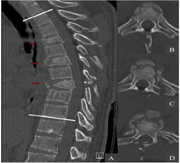
Table 1: Fractures summary according to the AO Spine classification.

Surgical Technique
The patient undergone a surgery based on a hybrid minimal
invasive approach using an intraoperative 3D CT-scan navigation
system and percutaneous pedicle-screwing method. We performed
a posterior thoracic spine fixation from Th2 to Th10. The patient
was placed in a prone position on a mobile, radiolucent, carbon
fiber operating table connected to the Airo ring (Figure 2A).
A fluoroscopy was used to check the spinal alignment and to
determine the vertebral levels to be instrumented. A medial
posterior skin incision was performed from Th1 to Th11 down to
the muscular fascia. At this stage, a navigated reference marker
clamp was firmly placed with minimal muscle dissection on
the spinous process of Th9, which was the closest intact bony
structure adjacent to the fractured levels [10] (Figure 2B). A first
intraoperative CT-scan acquisition was performed, assessing a good
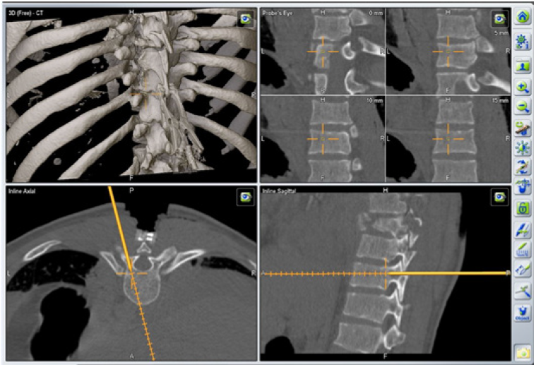
kyphosis correction (Figure 3). The acquired datas were processed
thanks to the intraoperative image-guidance system. A series of
trans-pedicular channel was performed from Th10 to Th7, based
on the 3D navigated CT-scan with a navigated standard Jamshidi
needle (Figure 4) K-wires were inserted into the pedicles before
insertion of cannulated screws. Additional lateral fluoroscopy was
performed at the end of each screw placement in order to verify
the screwing accuracy and the absence of bone displacement [12].
A second series of pedicular screwing was then realized using
the same procedure under the control of a second CT-scan with
a Th4 reference marker clamp. Two 24cm slightly bent titanium
rods were placed in a strictly minimal invasive way to connect the
screws. A vertebral distraction was applied to the vertebral body of
Th8 to restore correct vertebral height and complete the procedure.
Further decompressive laminectomy was unnecessary, as the MRI
did not highlight any spinal cord compression.
Figure 2: Operating room setting with the Airo ring. (A) Midline incision with navigated reference marker fixed on
Th9.
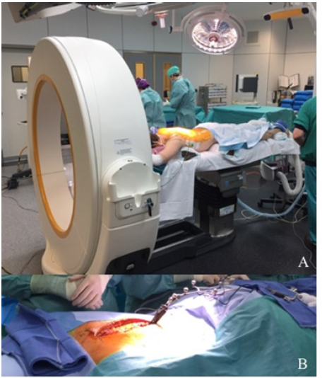
Figure 3: First intraoperative CT-scan acquisition with reference marker fixed on the Th9 spinous process (white
arrow).
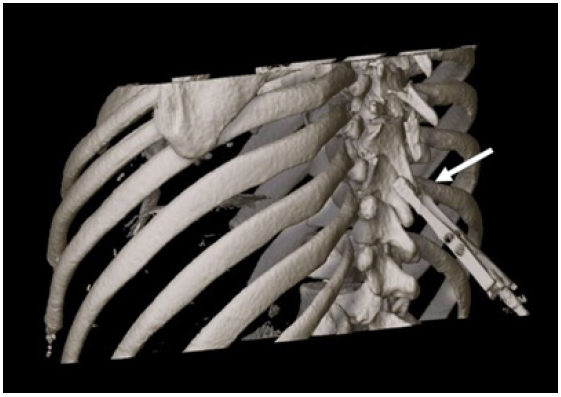
Figure 4: Intraoperative 3D-navigated screw placement trajectory.
Radiologic and Clinical Outcomes
The postoperative CT-scan demonstrated correction of regional
kyphosis with an angle of 32° (Figure 5). Eleven screws were placed
completely within the pedicle unit. Two screws (Th4 and Th7 right)
were considered as “in-out-in” with a moderate penetration of the
external pedicle wall (3-6mm outside the pedicle boundaries)9,
hence there was no indication to reposition these. The patient
regained his mobility and started to walk three days following
the surgery. An additional thoraco-lumbar corset was placed for a
period of 6 weeks. He was discharged 7 days after surgery without
any postoperative complications and resumed his professional
activity a month later.
Figure 5: Postoperative sagittal CT-scan reconstruction of the instrumentation from Th2 to Th10. (A) The regional
kyphosis was measured with an angle of 32° (white lines) after the surgery. (B) Posterior (C) and left posterolateral
(D) 3D images.

Discussion
The case reported here is a unique presentation of triple unstable
noncontiguous thoracic spine burst fractures without any neurologic
lesions. Furthermore, the way he was treated by means of intraoperative
minimal invasive navigation despite the multitude and the non-contiguous
of these fractures has never been reported in the literature. In case
of non-neurologic thoracolumbar fractures, it is more and more commonly
admitted that a rapid surgical fixation allows a better management
compared to conservative treatment. Recent developments of percutaneous
techniques have also optimized the treatment of other comorbidities. It
allows early mobilization, decrease the complication rate and reduce the
duration of hospitalization, as well as it could be appliable to treat
various types of fractures [1,2,4,5,7-9]. Although there are still
debates, our experience shows that this is also the case for
noncontiguous fractures [1-5]. Some factors such as the important
length of fixation, anchoring difficulties on a multi-fractured spine
and repercussion on spine mobility should be considered. It is not
always possible to restore a perfect spine alignment. Reduction as much
as possible of the number of fixed levels is a major concern in order to
optimize the clinical outcome and spine range of motion. Yet, when the
number of intact segments in between two fractured segments that need
surgery is lower than 4, it is recommended to connect both fixations in
one single instrumentation.1 From the very beginning of the medical
care, the objective was to multiply anchor points, especially in between
fractured level and even in fractured vertebrae in order to reduce the
fixation length and maintain spine mobility. This could also increase
the rigidity of the instrumentation and reduce the use of transverse
connector. Percutaneous pedicular thoracic spine screwing is not an easy
procedure for many reasons: the narrowness of the pedicles,
radiological opacification generated by the ribs, anatomical
modifications due to the trauma and instability of fractured fragments.
The use of intraoperative navigation like the Airo system has recently
proved his superiority in terms of precision with a correct placement
rate of 95-99% compared to 89-95% with conventional free hand
percutaneous technique [8,9,11-13]. Regarding these data, there is no
doubt that the 3D-intraoperative navigation technique has the best
accuracy. In this specific case, intraoperative navigation has been
helpful to securely realize pedicular screwing at many segments and in
particular in two fractured vertebrae (Th4 and Th6) 8,9,13. It also
allowed the intraoperative control of screws placement and kyphosis
correction [8,10-13]. When needed it gives you the possibility of
directly replacing misplaced screws and avoid reoperation. However,
intraoperative spine navigation remains a difficult and time-consuming
procedure that requires a learning curve to be comfortable with
[8,9,11-13]. There have been concerns about extended surgical time.
Recent series show that the instrumentation setup for iCT navigation
requires more time, but each screw placement is faster than with
free-hand technique and takes about 2 to 3 minutes. One in the other,
the total time for the whole procedure seems comparable [8,9,11,12].
Another main hazard during minimal invasive surgery is the radiation
exposure that is greater to the surgeon compared with open spine
procedures [9,11]. The use of intraoperative navigation can decrease the
global radiation exposure [9,11]. In case of such a long fixation, the
medical staff irradiation was significantly reduced. However, patient
irradiation is higher with intraoperative CT-scan acquisition than in
usual procedure. On the other hand, regarding radiation hazard, each
intraoperative acquisition remains lower than a standard diagnostic CT
[9,11,13]. Two screws were considered as “in-out-in” (Th4 and Th7 right)
without any consequences for spine stability or neurological damage.
This inaccuracy could be explained by biomechanical instability,
movements of reference marker during trajectory navigation,
instrumentation at a large distance from the tracking device or
inaccuracy of Jamshidi needle setting [8,9,12,13]. From a technical
point of view, some recommendations can be given for the use of
intraoperative navigation in long fixation: The use of an additional
lateral C-arm fluoroscopy can be easily implemented, even at a later
time during surgery, and provide direct radiological information about
screws placement or bone fragment displacement [11,12]. This information
will be correlated by the surgical team with the image-guided
navigation and gives a double check when in doubt or in critical
regions. In order to optimize the precision, a new CT-scan acquisition
should be performed every 4 to 5 segments allowing the repositioning of
the reference marker [10,13]. The setting of the navigated Jamshidi
needle must also be checked frequently, ideally at the start of each
pedicular sight [11]. Due to the number of percutaneous tubes needed in
very long fixation, it is strongly recommended to start the navigation
with the level farthest from the camera in order not to be disturbed by
the interposition of tubes between Jamshidi needle and the camera.
Conclusion
Noncontiguous triple fractures of the thoracic spine are an
unusual presentation of polytrauma injury. Long and complex
thoracic posterior spine fixation is feasible in a safe way using an
intraoperative CT-navigated minimal invasive technique in support
[8-10,12,13]. The use of intraoperative imaging provides critical
information about spine alignment and pedicular trajectories which
facilitate screw placement in fractured or misplaced vertebrae
[13]. It requires optimal surgical preparation and intervention
steps must be followed scrupulously in order to avoid classic traps.
Finally, this technique can be labelled as “hybrid” as it combines the
use of a midline incision with minimal invasive fixation, and also
the use of fluoroscopy in addition to intraoperative navigation.
Acknowledgement
Patrick Misson provided the conception and design of the article,
drafting the article, revised it critically for important intellectual
content, and final approval of the version to be submitted. Didier
Martin and Abdelhammid Mendili were responsible for the article
critically for important intellectual content. Corentin Delhaye
provided radiological interpretation and image management. Jean
Pirson provided the anesthesia. Cristo Chaskis provided the revised
the article critically for important intellectual content and gave final
approval of the version to be submitted. No funding to declare.
Conflict of Interest
The authors declare that there is no conflict of interest.
References
- Cho Y & Kim
YG (2019) Clinical features and treatment outcomes of acute multiple
thoracic and lumbar spinal fractures: a comparison of continuous and
noncontinuous fractures. J Korean Neurosurg Soc 62(6): 700-711.
- Wittenberg RH, Hargus S, Steffen R, Muhr G, Bötel U (2015) Non-contiguous unstable spine fractures. Spine 27(3): 254-257.
- Takami M, Okada
M, Enyo Y, Iwasaki H, Yamada H, et al. (2017) Non-contiguous
double-level unstable spinal injuries. Eur J Orthop Surg Traumatol
27(1): 79-86.
- Seçer M, Alagöz
F, Uçkun O (2015) Multilevel non-contiguous spinal fractures: Surgical
approach towards clinical characteristics. Asian Spine J 9(6): 889-894.
- Kim JK, Moon BJ,
Kim SD, Lee JK (2018) Minimal invasive non-fusion technique for the
treatment of non-contiguous lumbar burst fractures in young age patient:
A case report. Med 97(10): 10-13.
- Nelson DW,
Martin MJ, Martin ND, Beekley A (2013) Evaluation of the risk of
non-contiguous fractures of the spine in blunt trauma. J Trauma Acute
Care Surg 75(1): 135-139.
- Giorgi H,
Blondel B, Adetchessi T, Dufour H, Tropiano P, et al. (2014) Early
percutaneous fixation of spinal thoracolumbar fractures in polytrauma
patients. Orthop Traumatol Surg Res 100(5): 449-454.
- Hubbe U, Kogias
E, Vougioukas (2009) Image guided percutaneous trans-pedicular screw
fixation of the thoracic spine. A clinical evaluation. Acta Neurochir
151(5): 545-549.
- Allam Y,
Silbermann J, Riese F, Greiner PR (2015) Computer tomography assessment
of pedicle screw placement in thoracic spine: Comparison between free
hand and a generic 3D-based navigation techniques. Eur Spine J 22(3):
648-653.
- Citak M, Stubig
T, Kendoff D (2010) Navigated minimally invasive thoracolumbar pedicle
screw placement with flat panel 3-d imaging a feasibility study. Technol
Heal Care 18(2): 101-110.
- Lian X, Navarro
RR, Berlin C (2016) Total 3D Airo® navigation for minimally invasive
transforaminal lumbar interbody fusion. Biomed Res Int 5027340.
- Hecht N,
Kamphuis M, Czabanka M (2016) Accuracy and workflow of navigated spinal
instrumentation with the mobile AIRO® CT scanner. Eur Spine J 25(3):
716-723.
- Scarone P,
Vincenzo G, Distefano D (2018) Use of the AIRO mobile intraoperative CT
system versus the O-arm for transpedicular screw fixation in the
thoracic and lumbar spine: a retrospective cohort study of 263 patients.
J Neurosurg Spine 29(4): 397-406.
- Ushijima T,
Kawaguchi K, Matsumoto T (2018) Double non-contiguous fractures in a
patient with spondylo-epiphyseal dysplasia with spinal ankylosis treated
with open and percutaneous spinal fixation technique: A case report.
BMC Res Notes 11(1): 1-5.
For more articles in Journal of Neurology