Crimson Publishers High Impact Journals

Wednesday, November 10, 2021

Recognizing Early Warning Signs for Patient Safety: Study in Pakistan’s Tertiary Care Hospital _Crimson Publishers

 Recognizing Early Warning Signs for Patient Safety: Study in Pakistan’s Tertiary Care Hospital by Rufina Soomro in COJ Nursing & Healthcare_Nursing & healthcare journal

Abstract

Introduction: It is not uncommon for hospitalized patients to exhibit early warning signs before deteriorating. Clinical deterioration can happen at any point in a patient’s illness, or care process. Nurses are expected to have skills to enable them to assess precisely the severity of deterioration, as nurses are the first to encounter the patients. In keeping views we as team have develop module on Recognition of Early Warning signs for Nurses to assess their knowledge, educate them and reassess their knowledge.

Purpose: We want to assess the knowledge about early warning signs in our staff nurses then educate them and reassess to see their knowledge improvement.

Study design, material & method: A quasi-experimental pre-posttest design was used to achieve the aim of the study. Pre-test for participants were conducted to assess base line knowledge. Interactive lectures were done by experts. Than hands on workshop arranged in which case scenarios were discussed in groups. To assess the effectiveness of educational module same post test were given. In the end qualitative analysis were also done for reflective writing for individual personalized learning.

Results: The overall compared result shows that the average pre and post scores was significant. Pretest mean score was 6.47(3.15) and it improves in Post Test 11.66(2.58). P value is less than 0.01. This shows that the average pre and post scores for education module were significant. The analysis shows that the workshop was effective in improving the knowledge of Nurses.

Conclusion: After this module implementation, we achieved better results in posttests. The key message from this report is continuous education and emphasis on recognizing early warning signs has brought improvement in nurse’s knowledge and self-confidence.

Introduction

Early detection and prevention of patient deterioration is a key aspect of a patient’s multidisciplinary assessment [1]. Nurses are expected to have skills to enable them to assess precisely the severity of deterioration, as nurses are the first to encounter the patients [2]. It is not uncommon for hospitalized patients to exhibit early warning signs before deteriorating [3] Clinical deterioration can happen at any point in a patient’s illness, or care process, but patients are particularly vulnerable after surgery and during recovery from a critical illness. Many patients who suffer cardiopulmonary arrests show signs of deterioration during the 24 hours before the arrest and “it has been estimated that approximately 23,000 in-hospital cardiac arrests in the UK could be avoided each year with better care” by NHS 2012 Royal College of Physicians 2012 [4] The National Patient Safety Agency (NPSA) launched the fifth Patient Safety Observatory report “safer care for the acutely ill patient: learning from serious incidents” in which ‘deterioration’ was identified as a key theme [5]. Claims on improving the knowledge of ward nurses by training them to monitor and analyses various data’s. For example, educational programs like Acute Life-threatening Events-Recognition and Treatment course. In keeping views we as team have develop module on Recognition of Early Warning sign for Nurses to assess their knowledge.

Purpose

We want to assess the knowledge about early warning signs in our staff nurses then educate them and reassess to see their knowledge improvement.

Study Design, Material & Method

A quasi-experimental pre-post test design was used to achieve the aim of the study. Pre-test for participants were conducted to assess base line knowledge. Interactive lectures were done by experts on Introduction to Early Warning Signs & Respiratory Assessment, Hemodynamic Monitoring, Post op Assessment (PACU), Importance of Documentation, Drains & Urine output for early recognition of patient’s deteriorations. 2 specific role plays were also prepared one related to surgery specialty and one related to medicine specialty. Debriefing with discussion done & queries were resolve on spot. Later on small group activity was arranged in which 6 different case scenarios were given to all in a group of 10. For group working, a mentor was assigned for each group. They have worked on scenario and present case study in front of panel of experts [6]. Case based discussion took place and all queries were answered by Panellist.

Recommendations and conclusion were done by team. Participants were encouraged to asked questions rather than assuming things. For key information and take home massage, handout were also prepared and given to all participants. Key messages about patient safety and early detection were pasted on notice board in all wards for continuous reinforcement. To assess the effectiveness same post test were given to all participants. In the end qualitative analysis were also done by reflective writing for individual personalized learning. In reflective writing we have asked to write learning outcome from this course & how will you act differently after this course [7]. Participants have shared their learning and we have analysis it in results part.

Results

Pre-test was conducted to assess base line knowledge of staff. Paper comprises of MCQs-40% on nursing assessment, 30% on drug dose calculation and 30% on nursing documentation. For the purpose of descriptive analysis, mean and standard deviation was calculated for the continuous variables. Frequencies and percentages were reported for the categorical variables. In the inferential analysis, paired t- test was used. The overall compared result shows that the average pre and post scores were significant. Pre-test mean score was 6.47(3.15) and it improves in Post Test 11.66(2.58). P value is less than 0.01. This shows that the average pre and post scores for education module were significant. The analysis shows that the workshop was effective in improving the knowledge of Nurses. Reflective writing was also shows that this training enhances staff knowledge.

In reflective writing Question has been asked from participants to assess the learning that what you learn from this session. Few statements of the participants were as follows. Response shared by staff nurses “I got new information from this session about the importance of vital sign and early warning sign which may cause cardiac arrest, hypoglycemia and other severe complains and I am also learned from this session, no ignorance of complains of patient because the ignorance of little complain of patient becomes very risky for your patient.”, “I learn what to do first in Emergency situation and How to manage emergency condition in words”, “I have learned a lot of new knowledge such as recognizing the Early Warning Sign of deterioration and also the Post Anesthesia care unit and Importance of the Documentation. I learned about the 6th vitals. Speaker shares some knowledge about Urine Output or I/O charting.”

“By the end of session I have learned the Early Warning Sign and Measurement of overall body Assessment and its intervention to increase the survival rate of patient condition and After Demonstration to enhance my skill.” Furthermore, another question which were asked from participant was how you will work differently from now onwards. Responses from participants were “I will assess all early warning sign in my ward and upgraded to my all duty staffs. “This session early warning sign is very important and useful. This session improved my knowledge.” “After this work shop I will try my best to maintain appropriate documentation, take a vital sign and note the deterioration for taken action. By attending this much information lecture and workshop, I will practice all my knowledge and skills and article things to identify early warning signs and prioritize patient need and make quick intervention according to signs to save patient life and prevent him/her from deteriorating and manage patient timely before he goes to worse condition to save a patient’s life and reduce patient stay in or prevent from going ICU or HDU, improve recovery.” Another participant shared that “I am very much proud to be here in this learning session Figure 1. It was very much informative rich in Knowledge and nursing skills. Especially I am not as much able that I can identify early warning signs and can take quick necessary action by doing this. I can save many life and prevent my patient being deteriorated or to save him/her to go in ICU / HDU.”

Figure 1:


Discussion

Six physiological parameters form the basis of the scoring system have highlighted during workshop. Which were Respiratory rate, Oxygen saturations, Temperature, Blood pressure, Pulse rate & urine output. Following strategies were used for awareness. Case scenarios discussions take place in groups of 5 staff nurse. 1 mentor was present for discussion. They have prepare presentation in groups. According to Andrews & Waterman [8] The Early Warning Score leads to successful referral of patients by providing an agreed framework for assessment, increasing confidence in the use of medical language and empowering nurses. This study has also shows improvement in nurse’s knowledge as shared in posttest and reflective writing. We can hope to see good impact on patient care improvement Nurses are expected to have skills to enable them to assess precisely the severity of deterioration, as nurses are the first to encounter the patients [2].

Early identification is important to reduce mortality, avoidable morbidity, and length of stay and associated healthcare costs. By closely monitoring changes in physiological observations, deteriorating patients are more likely to be identified before a serious adverse event occurs. This role should effectively be played by the assign nurse. Andrews & Waterman [5] suggest same “It is also essential that nurses and nursing students are supported in its use and in developing confidence in using medical language by continued emphasis on physiology and pathophysiology in the nursing curriculum.” Nurses are recommended to attend courses on acute life threatening events recognition and treatment and to develop further simulation exercises in both pre and post registration courses, which may foster EWS process and improve patients’ outcome [4].

Conclusion

After this module implementation, we achieved better results in post tests. The key message from this report is continuous education and emphasis on recognizing early warning signs has brought improvement in nurse’s knowledge. As participants were free to ask question from experts at any time, it boost their selfconfidence which improves in communication among health care team. It also plays vital role in enlightening patient’s safety and quality care.

Recommendation

We comprehend that the majority of the nurses have moderate level of knowledge. Furthermore the finding of the study have created a platform for nursing leaders, to start a comprehensive training program in order to improve patient safety and clinical outcomes for acutely ill patients in our hospitals and treatment decisions can be taking timely to improve patient recovery. These type of training session should be a part of ongoing training for nurses and all health care professionals, for team work which, improves communication and build confidence.

References

  1. Mitchell IA, Mckay H, Steen Van LC, Berry R, McCutcheon C, et al. (2010) A prospective controlled trial of the effect of a Multi-faceted intervention on early recognition and intervention in deteriorating hospital patients. Resuscitation 81(6): 658-666.
  2. Beaumont K, Luettel D, Thomson R (2008) Deterioration in hospital patients: early signs and appropriate actions. Nursing Standard 23(1): 43-48.
  3. Bette Case di Leonardi (2014) AMN Healthcare Inc. Patient Deterioration: Early Warning Signs.
  4. Odell M, Victor C, Oliver D (2009) Nurses’ role in detecting deterioration in ward patients: systematic literature review. J Adv Nurs 65(10): 1992- 2006.
  5. Johnston CC, Rattray J, Myers L (2007) Physiological risk factors, early warning scoring systems and organizational changes. Nursing in Critical Care 12(5): 219-224.
  6. Min A Hwanga, Eun Mi Joa, Hyeri Yoona, Dong In Suhba Development of an Early Detection and Monitoring System for Deteriorating Patients Medical information center, Seoul National University Hospital, Korea Department of Pediatrics & Adolescent Medicine, Seoul National University Children’s Hospital, Korea.
  7. Andrews T, Waterman H (2005) Packaging: a grounded theory of how to report physiological deterioration effectively. J Adv Nurs 52(5): 473-481.
  8. Duncan KD, McMullan C, Mills BM (2012) Early warning systems: The next level of rapid response. Nursing 42(2): 38-44.

    Crimson Publishers: https://crimsonpublishers.com/

    For more articles in Nursing & Healthcare Journal
    Please click on below link: https://crimsonpublishers.com/cojnh/


Tuesday, November 9, 2021

Rheumatoid Arthritis in the Cervical Spine_Crimson Publishers

 Rheumatoid Arthritis in the Cervical Spine by Tanaka M in Researches in Arthritis & Bone Study_Journal of Arthritis Bone Disease

Abstract

1. Abstract

Rheumatoid Arthritis (RA) is a chronic, systematic, and progressive autoimmune disorder. More than two-thirds of all RA patients show symptomatic cervical spine lesions. RA in the cervical spine is categorized into three types: Atlantoaxial Subluxation (AAS), Vertical Subluxation (VS), and Sub axial Subluxation (SS). For the diagnosis of RA, plain radiographs are the basis of radiographic evaluation. Computed Tomography (CT) and Magnetic Resonance Imaging (MRI) are important for the further examination. The significantly better results can be achieved with surgical treatment than with conservative treatment. However, the Indications for transoral decompression are limited. For most of the condition, the posterior atlantoaxial fixation can achieve a good result.

2. Keywords: Rheumatoid Arthritis; cervical spine; radiographs; surgical treatment

Introduction

Rheumatoid Arthritis (RA) is a chronic, systematic, and progressive autoimmune disorder, with an incidence of 1-3% [1]. This condition can occur at any age, especially during the fourth or fifth decade of life. The most common sites are the metacarpophalangeal and metatarsophalangeal joints (Figure 1). More than two-thirds of all RA patients show symptomatic cervical spine lesions [2,3], but the lumbar spine is rarely involved. The inflammation in RA causes synovitis, then loosening of the ligaments and capsules, and finally bony erosion, vertebral collapse, and subluxation.

Figure 1:


Clinical presentation

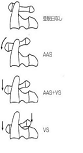
RA in the cervical spine is categorized into three types: atlantoaxial subluxation (AAS), Vertical Subluxation (VS), and Sub axial Subluxation (SS).

Atlantoaxial subluxation (AAS)

Rheumatoid synovitis at the atlantoaxial joints and around the stabilizing transverse, alar and apical ligaments leads to atlantoaxial instability. Disintegration of the transverse ligament causes anterior AAS. The reported prevalence of anterior AAS was 10-55% [4,5]. Compression of the cervical cord is usually from posteriorly, leading to demyelination of the posterior fascicle. Posterior AAS is less frequent (less than 1%) due to odontoid destruction or fracture [6].

Vertical subluxation

Destruction of the lateral atlantoaxial joint leads to joint depression and upward movement of the axis. Pathologically, this destruction spreads to the occipitoaxial joint [7].

Sub axial subluxation

Destruction of the sub axial joints and dysfunction of the posterior ligaments leads to SS. Common sites are C4/5 and C5/6. The prevalence of SAS is 13% among all RA patients [8]. This condition is associated with AAS and onset is usually late.

Plain radiography

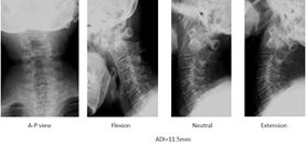
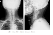
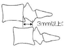
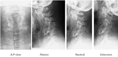
Plain radiographs are the basis of radiographic evaluation for RA in routine screening of the wider population. Anterior AAS may be assessed on a lateral cervical spine radiograph by measuring the Anterior Atlantoaxial Interval (AADI) from the middorsal margin of the anterior ring of C1 to the ventral surface of the odontoid (Figure 2). AADI more than 3mm in an adult or 4mm in a child is considered abnormal [9] (Figure 3). The interval is accentuated on a flexion view if the AAS is present. Posterior AAS may be assessed by measuring the Posterior Atlantoaxial Interval (PADI) from the dorsal aspect of the odontoid to the ventral margin of the C1 lamina.

Figure 2:Atlantoaxial subluxation (AAS).


Figure 3:Radiologic evaluation (Anterior subluxation).


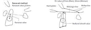
VS can be measured by McRae’s line, Chamberlin’s line or McGregor’s line (Figure 4). McRae’s line connects the front and back of the foramen magnum. If the odontoid process exceeds this line, this condition is regarded as VS. Chamberlin’s line connects the dorsal margin of the hard palate to the dorsal margin of the foramen magnum. If the tip of the odontoid projects more than 6mm, this condition is also regarded as VS. On the other hand, McGregor’s line is easier to use, as it is drawn from the posterior margin of the hard palate to the most caudal point of the occiput. The tip of the odontoid should not project more than 4.5mm.

Figure 4:76 Women, RA, Anterior atlantoaxial subluxation.


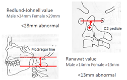
The tip of the odontoid is sometimes difficult to identify because of erosion. In such situations, the Redlund-Johnnell [10] method and Ranawat [11] method are very useful to diagnose VS. The Redlund-Johnnell [10] value is measured as the distance between McGregor’s line and the vertebral body of the axis (Figure 4). A value less than 34mm in men and less than 29mm in women is defined as VS. Ranawat value is measured as the distance between McGregor’s line and the center of the C2 pedicle. A value less than 15mm in men and less than 13mm in women is defined as VS (Figure 5 & 6).

Figure 5:Redlund Johnell method.


Figure 6:Radiologic evaluation (Vertical subluxation).


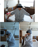
SAS is defined as a slip of more than 3mm in the sub axial vertebra (Figure 7). For RA patients, the lack of osteophyte formation represents a unique feature compared with OA patients (Figure 8). The Space Available for the Cord (SAC) is related to the onset of myelopathy and surgical results [12]. RA patients need to hyper-flex the neck during meals because of ROM (Range of Motion) disturbance of the upper extremities (Figure 9).

Figure 7:79 Women, RA, Vertical subluxation.


Figure 8:79 Women, RA, Vertical subluxation, CT.


Figure 9:Sub axial Subluxation (SAS).


Computed tomography (CT)

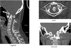
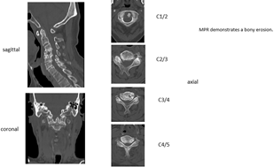
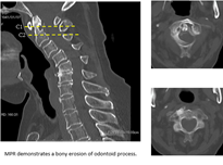
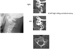
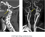
CT is preferred for evaluating bony destruction and ruling out anatomical anomalies. Multiplanar Reconstruction (MPR) provides a three-dimensional evaluation of the cervical spine. Sagittal reconstruction images allow clear visualization of AADI, odontoid erosion, and high-riding vertebral artery (Figures 10-13).

Figure 10:71 Women, RA, Sub axial subluxation.


Figure 11:67 Women, RA, Subaxial subluxation.


Figure 12:76 Women, RA.


Figure 13:74 Women, RA.


Magnetic resonance imaging (MRI)

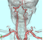
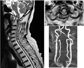
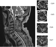
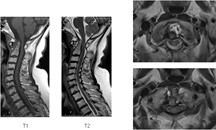
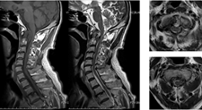
As plain radiograms cannot evaluate soft tissues, MRI is very important to assess pannus, cord compression, myelomalacia, and soft tissue (Figures 14-17). MRI in flexion is used to check compression of the spinal cord in AAAS patients [13]. MRI can be used to evaluate the corticomedullary angle. An angle less than 135° is correlated with neurological compression of the medulla [14]. Magnetic resonance angiography reveals a vertebral artery abnormality in 34% of RA patients [15].

Figure 14:Acetabular


Figure 15:75 Woman, RA.


Figure 16:75 Woman, RA.


Figure 17:75 Woman, RA.


Natural history

The prevalence of cervical subluxation in RA patients is 43-86% [16]. The most common form of subluxation is AAAS. Fujiwara et al. identified AAAS in 31% of RA patients, and reducible AAAS progresses to irreducible AAAS, then to VS (Figure 18) [14]. Approximately 20% of subluxations in RA patients involve VS. Neurological deficits are reported in only 7-58% of RA patients [17,18]. Once paralysis develops, the natural history seems to be one of progressive neurological deterioration. Sudden death occurs in as many as 10% of RA patients due to medullary compression [16].

Figure 18:79 Women, RA.


Symptoms

Local symptoms: Neck pain from RA is very common and is caused by inflammation present in the joint when the disease is active. Pain at rest indicates inflammation, but pain during motion indicates instability of the joint.

3.9.2. Neurological deficit: Neurological examinations may be difficult to interpret in RA patients, as subtle neurological changes may be masked. Manual muscle testing is difficult to perform in patients with multiple joint destruction. Peripheral nerve entrapment syndrome modifies the neurological status. Deep tendon reflex is relatively useful to evaluate myelopathy, even in patients with multiple joint destruction. VS patients may show symptoms of medullary compression or lower cranial nerve symptoms.

Vertebral Artery (VA) insufficiency syndrome: VA insufficiency syndrome may occur in patients with AAAS, especially rotatory subluxation. These patients complain of vertigo, tinnitus, nausea, vomiting, and faintness during neck motion. Approximately 20% of RA patients experience occlusion of a VA [19].

3.9.4. General symptoms: RA patients typically complain of fatigue before other symptoms become obvious. Morning stiffness is often an early sign of RA, followed by joint pain, joint swelling, and loss of joint range of motion. When accompanied by other symptoms, low-grade fever can also be a symptom of RA.
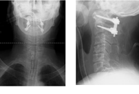
Non-surgical treatment: Marks reported significantly better results can be achieved with surgical treatment than with conservative treatment [20]. Among RA patients with myelopathy who refuse surgical treatment, 76% become bedridden within 3 years, and mean survival is 2.3 years [21]. Mainstay nonsurgical treatments for PA patients are physical therapy, patient education, and medication. Non-steroidal anti-inflammatory drugs, steroids, and new immune-modulatory biologic medications are the cornerstones of pharmacotherapy for RA.

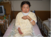
Initial pharmacological therapy typically includes methotrexate at 7.5-15mg/week. Corticosteroids are powerful drugs useful for achieving rapid relief from pain. A soft or hard collar can be used to limit anterior subluxation and improve discomfort.

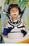
Surgical treatment: Surgery is indicated for RA patients with intractable pain or progressive neurological deficits. The goals of surgery include pain relief, neurological decompression, and prevention of further neurological deterioration. Some patients require awake intubation because of severe cervical cord compression. Preoperative skeletal traction with a halo ring or halo jacket should be considered for patients with severe myelopathy (Figure 19). Somatosensory and motor-evoked potentials are important for monitoring spinal cord function during positioning and surgery.

Figure 19:67 Woman, RA.


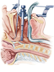
Transoral odontoidectomy: Indications for this technique are limited, because RA patients can experience difficulty opening the mouth completely due to temporomandibular joint disturbances. A distance of at least 25mm between the front teeth is needed for this procedure. As RA patients already have some degree of immunodeficiency, surgical-site infection may be another problem associated with this procedure [22] (Figure 20).

Figure 20:79 Women, RA.


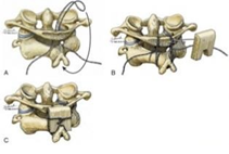
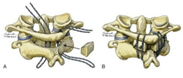
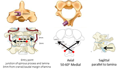
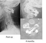
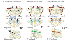
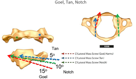
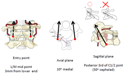
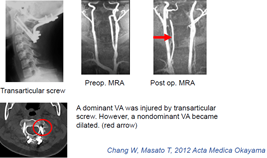
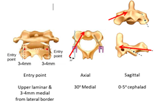
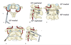
Posterior atlantoaxial fixation: In AAS patients, an AADI of 9mm or more, a SAC of less than 14 mm, or vertebral artery insufficiency are candidates for atlantoaxial arthrodesis surgery [23]. In the historical background, this technique was described Gallie [24] and Brooks & Jenkins [25] (Figures 21 & 22). C2 laminar screw and C1 lateral mass screw fixation, regarded as the Wright technique, is also a safer than Magerl [26] method (Figures 23 & 24). Three famous methods are available to put screws into the C1 lateral mass; Goel, Tan, and Notch methods (Figures 25 & 26). A disadvantage of these techniques is a high rate of nonunion due to reduced stability [27]. Magerl [28] reported trans-articular screw fixation, as the strongest anchor for the upper cervical spine (Figures 27 & 28). However, this procedure has a potential for VA injury in the presence of a high-riding VA [29,30] (Figure 29). Goel reported safer, more rigid fixation using a C1 lateral mass screw and C2 pedicle screw [31] (Figures 30-32). The most important advantage of this technique is that after placement of four screws, AAS can be reduced using screws and rods.

Figure 21:60 Men, RA.


Figure 22:76 Woman, RA.


Figure 23:67 Woman, RA.


Figure 24:


Figure 25:


Figure 26:Transoral adenoidectomy.


Figure 27:Gallie fusion.


Figure 28:Brooks-Jenkins Fusion.


Figure 29: C2 laminar screw (Wright 2004).


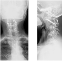
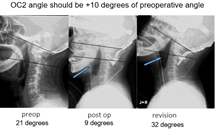
Posterior occiput cervical fusion: This technique is mainly indicated for patients with irreducible AAAS and VS. For VS patients, odontoid migration cranial to McGregor’s line of at least 5mm or an abnormal Redlund-Johnnell [10] or Ranawat [11] value are indications for occipitoaxial (OC2) fusion surgery [32]. A major complication for this procedure is dysphagia (Figures 33-36), so OC2 angle should be considered before surgery. An angle of approximately 20° is ideal for OC2 fusion [33]. The navigation is very useful for identifying the thickest part of the occiput and screwing pedicle screws into the cervical spine [34] (Figures 37-44).

Figure 30:52 Men, RA, AAAS, Wright method.


Figure 31:Three methods of C1 LMS placement.


Figure 32:C1 LMS technique.


Figure 33:C1/2 transarticular screw (Magerl 1987).


Figure 34:62 Woman, RA, AAAS, Magerl fixation.


Figure 35:Iatrogenic VA Injury with Magerl Fixation.


Figure 36:Goel method (C1 LMS & C2 Pedicle screw).


Figure 37:73 Woman, RA, AAAS, Goel fixation.


Figure 38:C2 pedicle screw


Figure 39:C2 pars screw (Judet 1962).


Figure 40:Dysphagia after OC2 Fusion.


Figure 41:75 Woman, RA, AAAS.


Figure 42:75 Woman, RA, AAAS.


Figure 43:75 Woman, RA, AAAS.


Figure 44:75 Woman, RA, AAAS.


Conclusion

The goal in management of RA cervical spine lesion is to prevent severe neurological deficit. The early diagnosis is very important because myelopathy is masked with peripheral joint problems. Among several imaging, the lateral flexion radiogram and MRI are very useful to diagnose cervical subluxation. For the surgery, the posterior fixation with instrumentation is the standard treatment. However, the surgeon should be reminded several severe complications. When the solid bony fusion is obtained, the pain relief and neurological recovery of the RA patient are expected.

References

    1. Helmick CG, Felson DT, Lawrence RC, Gabriel S, Hirsch R et al. (2008) National arthritis data workgroup, Estimates of the prevalence of arthritis and other rheumatic conditions in the United States. Part I Arthritis Rheum 58(1): 15-25.

    2. Papadopoulos SM, Dickman CA, Sonntag VK (1991) Atlantoaxial stabilization in rheumatoid arthritis. J Neurosurg 74(1): 1-7.

    3. Delamarter RB, Bohlman HH (1994) Postmortem osseous and neuropathologic analysis of the rheumatoid cervical spine. Spine (Phila Pa 1976) 19(20): 2267-2274.

    4. Conlon PW, Isdale IC, Rose BS (1966) Rheumatoid arthritis of the cervical spine: an analysis of 333 cases. Ann Rheum Dis 25(2): 120-126.

    5. Park WM, O Brien W (1969) Computer-assisted analysis of radiographic neck lesions in chronic rheumatoid arthritis. Acta Radiologicaa Diagnosis 8(6): 529-34.

    6. Joaquim AF, Appenzeller S (2014) Cervical spine involvement in rheumatoid arthritis-A systematic review. Autoimmun Rev 13(12): 1195-202.

    7. Eulderink F, Meijers KA (1976) Pathology of the cervical spine in rheumatoid arthritis: a controlled study of 44 spines. J Pathol 120(2): 91-108.

    8. Zhang T, Pope J (2015) Cervical spine involvement in rheumatoid arthritis over time: results from a meta-analysis. Arthritis Res Ther 17(1): 148.

    9. Rojas, CA, Bertozzi JC, Martinez CR, Whitlow J (2007) Reassessment of the craniocervical junction: Normal values on CT. American Journal of Neuroradiology. 28(9): 1819-1823.

    10. Redlund-Johnell I, Pettersson H (1984) Vertical dislocation of the C1 and C2 vertebrae in rheumatoid arthritis. Acta Radiol Diagn (Stockh) 25(2): 133-141.

    11. Ranawat CS, O Leary P, Pellicci P, Tsairis P, Marchisello P et al. (1979) Cervical spine fusion in rheumatoid arthritis. J Bone Joint Surg Am 61(7): 1003-1010.

    12. Boden SD, Dodge LD, Bohlman HH, Rechtine GR (1993) Rheumatoid arthritis of the cervical spine. A long-term analysis with predictors of paralysis and recovery. J Bone Joint Surg Am 75(9): 1282-1297.

    13. Reynolds H, Carter SW, Murtagh FR, Silbiger M, Rechtine GR (1987) Cervical rheumatoid arthritis: value of flexion and extension views in imaging. Radiology 164(1): 215-218.

    14. Narváez JA, Narváez J, Serrallonga M, De Lama E, de Albert M, et al. (2008) Cervical spine involvement in rheumatoid arthritis: correlation between neurological manifestations and magnetic resonance imaging findings. Rheumatology (Oxford) 47(12): 1814-1819.

    15. Zenmyo M, Ijiri K, Sasaki H, Sakakima H, Taketomi E, et al. (2010) Magnetic resonance angiography for vertebral artery evaluation in rheumatoid arthritis patients. Neurosurgery 66(6): 1174-1180.

    16. Rasker JJ, Cosh JA (1978) Radiological study of cervical spine and hand in patients with rheumatoid arthritis of 15 years' duration: an assessment of the effects of corticosteroid treatment. Ann Rheum Dis 37(6): 529-535.

    17. Fujiwara K, Owaki H, Fujimoto M, Yonenobu K, Ochi T (2000) A long-term follow-up study of cervical lesions in rheumatoid arthritis. J Spinal Disord 13(6): 519-526.

    18. Conaty JP, Mongan ES (1981) Cervical fusion in rheumatoid arthritis. J Bone Joint Surg Am 63(8): 1218-1227.

    19. Sherk HH (1978) Atlantoaxial instability and acquired basilar invagination in rheumatoid arthritis. Orthop Clin North Am 9(4): 1053-1063.

    20. Marks JS, Sharp J (1981) Rheumatoid cervical myelopathy. Q J Med 50(199): 307-319.

    21. Sunahara N, Matsunaga S, Mori T, Ijiri K, Sakou T (1997) Clinical course of conservatively managed rheumatoid arthritis patients with myelopathy. c Spine (Phila Pa 1976) 22(22): 2603-2607.

    22. Crockard HA, Calder I, Ransford AO (1990) One-stage transoral decompression and posterior fixation in rheumatoid atlanto-axial subluxation. J Bone Joint Surg Br 72(4): 682-685.

    23. Oda T, Yonenobu K, Fujimura Y, Ishii Y, Nakahara S et al. (2009) Diagnostic validity of space available for the spinal cord at C1 level for cervical myelopathy in patients with rheumatoid arthritis. Spine (Phila Pa 1976) 34(13): 1395-1398.

    24. Gallie WE (1939) Fractures and dislocations of cervical spine. Amj Surg 46(3): 495-499.

    25. Brooks AL, Jenkins EB (1978) Atlantoaxial arthrodesis by the wedge compression method. J Bone Joint Surg Am 60(3): 279-284.

    26. Wright NM, Lauryssen C (1998) Techniques of posterior C1-C2 stabilization. Tech in Neurosurg 4: 286-297.

    27. Grob D, Crisco JJ, Panjabi MM, Wang P, Dvorak J (1992) Biomechanical evaluation of four different posterior atlantoaxial fixation techniques Spine 17(5): 480-490.

    28. Grob D, Magerl F (1987) Surgical stabilization of C1 and C2 fractures. Orthopade 16(1): 46-54.

    29. Neo M, Matsushita M, Yasuda T, Sakamoto T, Nakamura T (2002) Use of an aiming device in posterior atlantoaxial transarticular screw fixation. Technical note. J Neurosurg. 97(1 Suppl): 123-127.

    30. Zhang W, Tanaka M, Sugimoto Y, Ikuma H, Nakanishi K et al. (2012) Dominant vertebral artery injury during posterior atlantoaxial transarticular screw fixation in a juvenile rheumatoid arthritis patient with atlantoaxial subluxation. Acta Med Okayama 66(1): 77-81.

    31. Goel A, Laheri V (1994) Plate and screw fixation for atlanto-axial subluxation. Acta Neurochir (Wien) 129(1-2): 47-53.

    32. Riew KD, Hilibrand AS, Palumbo MA, Sethi N, Bohlman HH (2001) Diagnosing basilar invagination in the rheumatoid patient. The reliability of radiographic criteria. J Bone Joint Surg Am 83(2): 194-200.

    33. Izeki M, Neo M, Takemoto M, Fujibayashi S, Ito H et al. (2014) The O-C2 angle established at occipito-cervical fusion dictates the patient's destiny in terms of postoperative dyspnea and/or dysphagia. Eur Spine J 23(2): 328-336.

    34. Krauss WE, Bledsoe JM, Clarke MJ, Nottmeier EW, Pichelmann MA (2010) Rheumatoid arthritis of the craniovertebral junction. Neurosurgery 66(3 Suppl): 83-95.

    https://crimsonpublishers.com/rabs/fulltext/RABS.000522.php

    Crimson Publishers: https://crimsonpublishers.com/

    For more articles in Journal of Arthritis Bone Disease
    Please click on below link: https://crimsonpublishers.com/rabs/    

Monday, November 8, 2021

Fluro Imaging: A Boon to Difficult Subarachnoid Block_Crimson Publishers

 Fluro Imaging: A Boon to Difficult Subarachnoid Block by Anisha Nagaria in Developments in Anaesthetics & Pain Management_Developments in Anaesthetics & Pain Management

Abstract

26-year young female, a known case of long-standing ankylosing spondylitis and suspected case of osteopetrosis having fracture shaft femur had presented to us for lower limb surgery. Difficult spine and limited neck movement due to fusion of vertebras make regional and general anesthesia a challenge for anesthesiologist. After failure of traditional approach to central neuraxial block, fluoroscopic guided block spinal anesthesia was achieved successfully. But with experienced hands, Fluro imaging helps to deal with difficult situation like ankylosing spondylitis safely..

Keywords: Ankylosing spondylitis; Difficult spine; Fluro imaging

Abbreviation: AS: Ankylosing Spondylitis; LS: Lumbosacral; AP: Anteroposterior; SAB: Subarachnoid Block; USG: Ultrasonography; OT: Operation Theater

Introduction

Ankylosing spondylitis (AS) is a chronic inflammatory disease of unknown etiology, having classic “bamboo spine” appearance due to formation of bony bridges between vertebrae. That makes administration of both general and regional anesthesia difficult. Hard lumbosacral (LS) spine with narrow intervertebral spaces, difficult positioning for intubation and spinal anesthesia, and sever pain at the fracture site poses challenge to anesthesiologist. Use of C-arm guided identification of the spine helps in finding severity of LS involvement and identifying widest open intervertebral space is of interest in case of difficult spinal anesthesia. This article reports a successful conduct of orthopedic case, having very difficult spine under C-arm guided subarachnoid block, following failed landmark technique.

Case Report

A 26-year female, a known case of AS, presented with right sided fracture femur after a minor trauma was posted for plating. Patient also had multiple impending fractures in upper and lower limb. At the time of admission, patient was severely anemic (Hb-4.5gm/ dl) which was corrected with multiple blood transfusion (Hb- 9.63gm/dl). MRI of Lumbosacral (LS) spine showed significantly hypertrophied facet joints along with possibility of myelofibrosis/ myeloproliferative disease. These findings suggested possibility of osteopetrosis also, but no other anomalies were found in thorough Pre-anesthesia evaluation. Patient had minimal neck extension and modified mallampati grade 2. After written informed consent patient was electively posted for surgery.

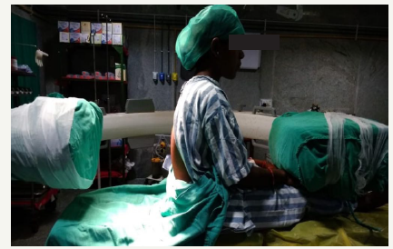
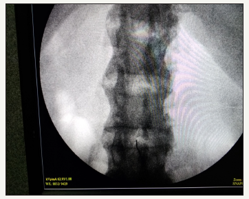
In Operation theater (OT), multipara monitors were attached. A wide bore i.e. line was secured and RL infusion was started. Difficult airway cart was kept ready. Patient was made to sit and under all aseptic precaution, Lumbar punctures were attempted at different spinal levels by median or paramedian approach through landmark technique, but it failed even after multiple attempts. So, it was decided to attempt fluoroscopic guided subarachnoid block under c-arm in same sitting position. An anteroposterior (AP) image of the lumbosacral spine Figure 1&2 showing minimal assessable window at L3-L4 intervertebral space (Figure 3) by variable degrees of lateral and oblique C-arm rotation was obtained. Subarachnoid space was approached in single prick with 25-gauge quincke-Babcock spinal needle using end on view between L3-L4 space. After confirming free flow of CSF, 0.5% Bupivacaine heavy 3.5ml was injected. Patient was immediately made to lie-down in supine position and a level of T10 was achieved. Throughout the surgery hemodynamic vitals were maintained. Surgery was completed uneventfully.

Discussion

Ankylosing spondylitis (AS) is a chronic inflammatory disease of the axial skeleton and peripheral joints, characterized by fusion of adjacent vertebrae, which leads to loss of flexibility of the back and neck. [1] These changes make AS a case of difficult airway and difficult central neuraxial blockade. Cervical spine involvement, with limited movements, makes tracheal intubation difficult which becomes a challenge for anesthesiologist [2]. Prior planning, use of all difficult airway measures and expert hands will ease the situation. Hyperextension of neck should be avoided to prevent cervical fracture. Fiberoptic bronchoscopy can be used successfully to secure the airway [3]. Establishing regional anesthesia in ankylosed spine is also challenging due to reduced articular mobility, obliteration of the inter-spinal spaces and impossibility to position the patient adequately [4].

Figure 1:Position of C-arm and patient in sitting position for AP view of LS spine.


Figure 2:position of c-arm and patient for AP view of LS spine.


Figure 3:AP view from image intensifier showing fused vertebrae and end on view of spinal needle from minimal assessable space between L3-L4 vertebra through which subarachnoid block was approached.


We suspected osteopetrosis also in our patient, because of presence of multiple impending fracture and MRI finding of myelofibrosis, which was another challenge for us, as these patients, add to difficulty in position due to brittle bones. The anaesthetic options are limited and technically difficult with both airway and spine involved in the disease process. The osteoporotic bones are difficult to drill, and surgical time can escalate [5].

Landmark guided subarachnoid block (SAB) becomes difficult and sometime impossible in cases of AS. So, different techniques were reported in various articles for approaching subarachnoid block in AS. The paramedian approach was mentioned in various case reports, this avoids the ossified midline ligaments (interspinous ligament and ligamentum flavum) and has a lower risk of encountering the exiting spinal nerve root [6]. KH Leung et al. [6] had reported a case of sever AS where successful elective mini laminectomy was performed under local anesthesia to approach Subarachnoid space. They had also mentioned about possible complications like inadvertent injury of neural tissues during laminotomy, fracture of the spine and risk of a high spinal anesthetic block with cardio-respiratory impairment. Ultrasonography (USG) of the lumbar spine may facilitate successful central neuraxial blockade in such patients, by indicating the presence and location of a soft tissue window into the vertebral canal [7,8]. Rakesh Goyal and colleagues have presented a case of bilateral THR, who was previously operated under general anesthesia due difficult spine. Later in his next surgery electively planned USG guided SAB was achieved successfully [9].

Neuraxial anesthesia is technically difficult in patients with AS but fluoroscopic guided imaging could successfully be performed. It has proved to be useful alternative option in difficult spinal anesthesia but requires expertise and associated with biological hazards to the patient and staff present in the OT. Reducing the exposure time, distance from radiation source, proper shielding, use of low dose and pulse mode helps in reducing radiation exposure while fluoroscopic guided approach to SAB [6]. SM Channabasappa and his colleagues have reported a case of fluoroscopic guided SAB in patient posted for uretroscopic removal of stone. Fluoroscopic guided transforaminal approach to epidural space was attempted in prone position after failed classical approach [2].

In most of the articles, fluoroscopic guided SAB was performed in prone and in lateral position. We had preferred sitting position for patient as our patient had multiple fracture and putting her in lateral or prone position would had been painful and to reposition patient in supine position would become difficult and time taking. This could have led to inadequate level of anesthesia because we used hyperbaric drug. In the sitting position c-arm was placed in anteroposterior (AP) direction and SAB was achieved.

Conclusion

Ankylosing spondylitis although is a challenge for anesthetist for both managing airway and regional anesthesia but should not be considered unachievable in any case. Understanding the situation and proper planning could be key to success. Fluoroscopic guided neuraxial block is a big boon to difficult spinal anesthesia in expert hand and can be preferred as choice in anticipated difficult spine.

References

  1. Woodward LJ, PCA Kam (2009) Ankylosing spondylitis: Recent developments and anesthetic implications. Journal of Association of Anesthesia of Great Britain and Ireland 64(5): 540-548.
  2. Channabasappa SM, Dharmappa S, Pandurangi R (2016) Fluoroscopy guided transforaminal epidural anesthesia in ankylosing spondylitis. Saudi J Anaesth 10(1): 101-103.
  3. Dave N, Sharma RK (2004) Temporomandibular joint ankylosis in a case of ankylosing spondylitis anaesthetic management. Indian J Anaesth 48: 54-56.
  4. Preeti R, Geeta AP, Bharati AT (2016) Management of a case of ankylosing spondylitis for percutaneous nephrolithotomy in prone position under combined spinal epidural anesthesia. Medical Journal of 9(6): 765-767.
  5. Ashima S, Poojitha R, Sreedhar R, Ramchandran G (2015) Marble bone disease and the anesthesiologist. Indian Journal of Anesthesia 59(6): 390-391.
  6. Leung KH, Chiu KY, Wong YW, Lawmin JC (2010) Case report: Spinal anesthesia by mini-laminotomy for a patient with ankylosing spondylitis who was difficult to anesthetize. Clin Orthop Relat Res 468(12): 3415- 3418.
  7. Chin KJ, Macfarlane AJ, Chan V, Brull R (2009) A case report: The use of ultrasound to facilitate spinal anesthesia in a patient with previous lumbar laminectomy and fusion. J Clin Ultrasound 37(8): 482-485.
  8. Chin KJ, Vincent C (2010) Ultrasonography as a preoperative assessment tool: Predicting the feasibility of central neuraxial blockade. Anesthesia & Analgesia 110(1): 252-253.
  9. Goyal R, Singh S, Shukla RN, Singhal A (2013) Management of a case of ankylosing spondylitis for total hip replacement surgery with the use of ultrasound-assisted central neuraxial blockade. Indian J Anaesth 57(1): 69-71.

The Efficacy of Sap Obtained from Commiphora gileadensis Against Cadmium-Induced Liver Toxicity in Rats: Crimson Publishers

The Efficacy of Sap Obtained from Commiphora gileadensis Against Cadmium-Induced Liver Toxicity in Rats by Amal S Soliman in Research in Med...