Crimson Publishers High Impact Journals

Monday, August 29, 2022

A Close Look at the Application of the Yin-Yang- Based Acupoint Pairs_Crimson Publishers

A Close Look at the Application of the Yin-Yang- Based Acupoint Pairs by Tong Zheng Hong in Advancements in Bioequivalence & Bioavailability_Bioequivalence And Bioavailability International Journal


Abstract

Yi-Yang is the unique concept that serves as the foundations for the developments of acupuncture and traditional Chinese medicine theories, such as the Five Elements, and Qi and Blood. Aimed at the balance of Yin-Yang, acupoint pairs consisting of acupoints on the Yin-Yang meridians, the locations classified Yin or Yang, and the Five-Shu acupoints are presented and discussed. However, it deserves more research to understand whether or not acupoint pairs can outperform the other protocols.

Keywords: Yin-yang; Five elements theory; Five-shu acupoints

Introduction

The safety and effectiveness of acupuncture have been verified with scientific evidence and acupuncture is highly recommended by the WHO for the pain management, which has been recognized in the Western healthcare systems, though it is at present viewed and classified as the complementary or alternative medicine [1]. Acupuncture, as part of the traditional Chinese medicine (TCM), has not only been viewed as one of the major healthcare systems in the Chinese communities, such as Hong Kong, Macau, and Taiwan, for more than two thousand years, but also has gained acceptance and popular in the Asian countries at the present time in Japan, Korea, Singapore, and Thailand [1].

The philosophical and abstract concepts like Yin-Yang, the Five Elements theory, Qi, Blood, Wei-Qi-Yin-Blood sequence, and pattern identification based on the observation of the nature have been used as the foundations to establish the whole systems of acupuncture and the TCM, even though they are difficult to understand for clinical practice. The ancient literature of acupoint pairs developed by the acupuncture masters represents the clinical application of Yin-Yang and the Five Elements theory, which highlights the essence of acupuncture theories. This study aims to explore some acupoint pairs for understanding the application of the rules of Yin-Yang and the Five Elements theory in the acupuncture and TCM theories.

Typical examples

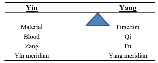
Yin-Yang and the Five Elements are unique and the keys of the TCM theories for many centuries, which absolutely distinguishes the TCM from the Western medicine. Based on the concept of Yin-Yang, a disease is understood to be the imbalance of Yin and Yang which bears the characteristics shown in Figure 1 [2,3].

Figure 1:Characteristics of Yin-Yang.


It is believed and required that an experienced acupuncturist must count on the four skills to get the information for identifying Patterns based on Ying-Yang balance for diagnosis, which is critical to the successful treatment outcomes. The typical acupoint pair in the acupuncture literature showing the application of Ying-Yang is the Four Gates that consists of LV3 and LI4 in Table 1.

Table 1:Yin and Yang of the four gates.


As shown in Table 1, either of the acupoints in this pair has the characteristic of Yin or Yang. It shows in the literature that this acupoint pair has been for long commonly used to promote the circulation of Qi (Yang) and to hold Blood (Yin) throughout the body. On the other hand, it is proved to be effective for treating subhealth [4], verifying the effectiveness of Yin-Yang balance. Both LV3 and LI4 are the Yuan-source acupoints classified in the Five-shu acupoints, to which the quotation “Treating the Zang organ with Yuan-source acupoint” in the Huang-Di-Nei-Jing (Yellow Emperor’s Inner Classic) can apply. It deserves attention that the Five Elements can only apply to the Five-shu acupoints among the acupoints on the twelve regular meridians, the Four command acupoints, the Hui meeting acupoints, and the Four Seas acupoints. The Five Shu acupoint system represents the clinical application of Yin-Yang and the Five Elements theory. Each of the five specific acupoints is categorized as Jing-well, Ying-spring, Shu-stream, Jing-river, and He-sea, to which the Five Elements correspond. It is noted that the five Zang organs have five Shu acupoints and the six Fu organs have six Shu acupoints of the twelve regular meridians. A Shu acupoint on either the Yin meridian or the Yang meridian can collaborate with a Shu acupoint on the either the Yin meridian or the Yang meridian, which is the typical representation of Yin-Yang as in Table 2.

Table 2:A Shu acupoint on Yin meridian combines with a Shu acupoint on Yang meridian.


In order to treat cough through clearing Heat in the Lung, both LU11 and LI1 are Well acupoints used at the same time. In this pair, LU11 classified as Yin acts to clear Heat to benefit the throat, while LI1 is used to reduce swelling and alleviate pain caused by Heat. The most critical notion that “Feng (Wind), the beginner of the illness” is also presented in Huang-Di-Nei-Jing, which suggests that Wind is the major cause of illnesses with its pernicious influence. Blood mobilization and Qi regulation need to be considered at a time because Blood stasis and impeded Qi can result in Blood Xu (deficiency of Blood) that can finally generate Wind.

The pair for Heart Blood deficiency consisting of LV3 and ST36 in Table 3 also represents the application of Yin-Yang. Blood classified as Yin is produced with the stimulation at ST36 while Yin is reinforced by the promotion of Qi belonging to Yang in this pair [4]. Different from the pair in Table 2, Yin and Yang are distinguished with the acupoint locations. ST36 is located on the leg, which is viewed as Yang, and one fingerbreadth lateral to the anterior crest of the tibia. Contrast to ST36, LV3 is classified as Yin because it is on the dorsum of the foot and in the hollow distal to the junction of the first and second metatarsal bones.

Table 3:Features of LV3 and ST36.


Discussion

Acupoint pairs are collected in the Zhen-Jiu-Da-Cheng (The Great Compendium of Acupuncture and Moxibustion) which provides the comprehensive understanding of acupuncture. Yin- Yang is the top principle in the TCM and acupuncture, which serves as the foundations of the Five Elements theory. In addition, both Yin-Yang and the Five Elements are the cores of the acupuncture and the TCM theories and seen as the guides for the diagnosis and the treatment. It is suggested that the understanding and accurate interpretation of the acupuncture and TCM theories are required for the best clinical outcomes [5].

Through the examples discussed above, it is clear that acupoint pairs can consist of either meridians or acupoints classified as Yin or Yang. However, it should be noted with the pair presented in Table 3 in clinical practice that the relationship between Yin and Yang is unfixed. On the other hand, the pair in Table 3 distinguishes itself from the general characteristics of Yin-Yang in Figure with the locations of the two acupoints and highlights the unfixed characteristic of Yin-Yang. Blood classified as Yin can nourish Qi while Qi can move Blood. The concept that Blood is the mother of Qi and Qi is the commander of Blood indicates that one is inconceivable without the other because the circulation of Qi and Blood in the body should be constant and cannot be disrupted [6].

Furthermore, the examples presented above show Blood and Qi are vital and each acupoint pair is aimed to balance Qi and Blood.

Conclusion

In spite of the ancient usage as a therapeutic method to treat illnesses and acupuncture has been proved in some scientific research to be effective for pain relief, inflammatory conditions, etc., acupuncture is facing the evidence-based challenge at the present time. Acupoint pairs may be the optimal approach in terms of cost and time in clinical practice to balance Ying-Yang. However, whether or not there could be more effective options based on the Yin-Yang theory for the better outcomes deserves more evidencebased research.

References

  1. Hong TZ (2018) Challenges in learning and understanding traditional Chinese medicine and acupuncture. Open Acc J Comp & Alt Med 1(1).
  2. Hong TZ (2018) Reminders for clinical application of extra acupoints. Curr Trends Biomedical Eng & Biosci 16(3): 555939.
  3. Hong TZ (2018) Notes for clinical use of extra acupoints. J Complement Med Alt Healthcare 8(1): 555728.
  4. Hong, TZ (2017) Principles of protocol to treat heart blood deficiency. J Complement Med Alt Healthcare 4(4): 555644.
  5. Hong TZ (2018) Cautions for learning traditional chinese medicine and acupuncture. Advancements Bioequiv Availab 2(2).
  6. Hong TZ (2018) A Review for integrating western and chinese medicines in primary dysmenorrhea. Advancements Bioequiv Availab 2(1).
For more articles in Bioequivalence And Bioavailability International Journal
Please click on below link: 

Friday, August 26, 2022

Providing Fair Payment for Prescription Medications in the United States_Crimson Publishers

Providing Fair Payment for Prescription Medications in the United States by Michael M Costello in Examines in Physical Medicine and Rehabilitation: Open Access_physical rehabilitation medicine journal


Opinion

With US health care costs now approaching $3 trillion per year, strenuous efforts to restrain cost growth continue to focus the efforts of policymakers at the federal and state levels. With pharmaceuticals contributing about 20% of total domestic health expenditures, pharmaceuticals are a natural target of cost control efforts. A primary means of attempting to control pharmaceutical costs in the use of Pharmacy Benefit Managers (PBMs), intermediary firms which function as go-betweens in the payer-provider relationship. Medicare, most state Medicaid programs, and many private managed care organizations use PBMs to manage pharmaceutical use for their beneficiaries.

However, a recent review of regulatory efforts combined with the complaints of many retail community pharmacies raises questions as to whether PBMs always act in the best interest of patients, and whether their benefit management practices are harming independent community retail pharmacies. Arnold [1] indicates that PBMs generate their revenues from three primary sources: fees generated from the supply chain, rebates provided by manufacturers and “spreads,” the difference between what the insurer pays the PBM and what the PBM pays the pharmacy which dispenses the medication.

A noted lack of transparency in PBM operations arises from two of those revenues streams. Critics have argued that payers are not benefitting fully from the rebates which PBMs receive from pharmaceutical manufacturers. In addition, pharmacies argue that PBMs are not adequately reimbursing them and are retaining an undue share of the revenues derived from the payers. The market dynamic of PBMs is further complicated by the fact that three large PBMs control 76% of the market of PBM services, and each of the three is owned by a major health insurance company which stands to benefit financially from decisions made by its affiliated PBM Morgan [2].

Smaller independent retail pharmacies complain that they do not have the ability to negotiate favorable acquisition costs and that PBMs reimburse them at less than their acquisition costs for certain prescriptions and charge the pharmacies back for certain patient management discrepancies. Such practices put the pharmacies in a tenuous financial position, leading some to believe they may close. Several large regional grocery store chains that maintained pharmacy counters in their stores are closing or selling those services Terlep [3].

These concerns have been brought to the attention of the US Senate Finance Committee which has written to the US Department of Health and Human Services requesting that it reform the Direct and Indirect Remuneration System by which PBMs remit funds to Medicare Morgan [4]. State legislators are expected to address PBM issues, including rebates to state Medicaid programs and private health insurance companies.

Some suggested areas for consideration in reforming PBM practices are:

Require that all pharmaceutical company rebates be paid to the insurance carrier

Medicare currently requires that all manufacturer rebates be paid back to the program in order to help reduce Part D prescription drug benefits. Such a requirement should be put in place for all payers using PBMs.

Devise a standardized administrative fee protocol

Rather than tying administrative fees together with manufacturer rebates as the Medicare DIR mechanism does, separate the two so as to clarify fees from rebates. Fees can then be regulated as a percentage of rebate payments.

Regulate the “spread” between PBM payments to pharmacies for prescriptions and the acquisition costs which the pharmacies incur

Smaller independent retail pharmacies should not be penalized for the acquisition costs they pay. They should be assured of a fair reimbursement tied to their acquisition costs.

Investigate PBM-insurer relationships for anticompetitive practices

Since the three largest PBMs are owned by large national insurance companies, they have the ability to favor their insurance companies to the detriment of the pharmacies with which they deal. Concerns lie not only with reimbursement rates paid to pharmacies but with the control PBMs exert over provider network configuration.

References

  1. Arnold J (2018) Are pharmacy benefit managers the good guys or bad guys of drug pricing.
  2. Morgan BT (2020) What’s the cost. The Scranton Sunday Times. p. A14.
  3. Telep S, Kang J (2020) Groceries exit from pharmacy business. Wall Street Journal. p. B1.
  4. Morgan BT (2020) Independent pharmacies stuck in a difficult spot. The Scranton Sunday Times. p. A13.
For more articles in physical rehabilitation medicine journal
Please click on below link: https://crimsonpublishers.com/epmr/

Tuesday, August 23, 2022

Exposure to Secondhand Smoke Exacerbated Natural Aging Cardiac Hypertrophy_Crimson Publishers

Exposure to Secondhand Smoke Exacerbated Natural Aging Cardiac Hypertrophy by Jia-Ping Wu in Open Journal of Cardiology & Heart Diseases_American Journal of Cardiology


Abstract

Secondhand Smoke (SHS) exposure is associated with an increased risk of coronary artery disease. This study’s aim was to investigate the relationship of SHS exposure in old rats’ left ventricle impaired and fibrosis. To explore the mechanism of cardiac remodeling of exposure to SHS exposure whether it was exacerbated cardiac impaired, especially in the older left ventricle. The animals were placed in a transparent exposure chamber, connected to smoking device and exposed to 15 cigarettes, smoke for 30 min, twice a day, 6 days/week, for 1 month. Histopathologic of left ventricular sections were stained with hematoxylin-eosin staining (H&E) and Masson’s trichrome. Left Ventricular (LV) morphological variables assessed using H&E stained and Mass weight changes. The cardiac structures were measured by echocardiographic analysis.

LV remodeling and fibrosis-related proteins were detected by gelatin zymography and western blotting analysis. Inflammatory and hypertrophy related proteins were also detected. Results showed in old rats’ group and old rats in the secondhand smoke exposure group (Old SHS Exp) were observed LV wall and mass increased, collagen accumulation and fibrosis, and extracellular space increased. From echocardiographic results, we found LV functions were apparently decreased, LV interventricular septum at systolic and diastolic diameters increased in the Old SHS Exp group. Cardiomyocyte width was increased in old rats, but the length was increased in Old SHS Exp group. Reduced MMP 2 proteins expression and TIMPs increased were induced fibrosis in the Old SHS Exp group. JNK1, p38, IL-6, TNFα were increased by western blotting and immunohistochemistry antibody-positive expression was observed in the Old SHS Exp group.

Keywords: Secondhand smoke exposure; Left ventricle; Cardiac impaired; Left ventricular hypertrophy; Echocardiographic; Cardiomyocyte

Introduction

Secondhand smoke (SHS) exposure is a serious health hazard causing the risk of coronary heart diseases. It causes a wide range of damaging health effects in children and older. SHS exposure affected on the cardiovascular system, including atherosclerosis, arterial stiffness and coronary cardiac disease [1]. In the previous studies reported, SHS exposure is the combination of smoke given off by the burred end of a tobacco or cigarette product to exposure to environment and the smoke exhaled by the smoker [2]. However, low-level chronic cigarette smoke exposure harmful older adults are still unclear. In this study, we use the older rats who underwent 15 cigarette exposure 30 minutes to determined left ventricular remodeling and function. Hypertrophy is an initial adaptive response. Long-term exposure to secondhand smoke increases the risk of developing cancer in younger and elderly people [3].
Low levels of fine particulate exposure from secondhand cigarette smoke are sufficient to induce increase the risk of cardiovascular disease mortality. The SHS exposure-response relationship between cardiovascular disease mortality and fine particulate matter is relatively steep at low levels of SHS exposure and flattens at higher exposures [4]. There are many compensatory mechanisms to increase cardiac workload and stimulation of left ventricular sustains. However, aging is a progressive disease which is a typic natural course whose worsening of the disease until death occurs. Slowly progressive age-related diseases are also chronic diseases; many are also degenerative diseases [5].
The aging mature organism that occurs normally the gradual changes in the structure over time and increases the probability of death. This growing process is unavoidable.

These physiologic changes of old cardiac include left ventricular hypertrophy, increased cardiac fibrosis, and valvular degeneration. Cardiovascular disease is a major risk factor for the aging cause of death [6]. Aging changes in the elderly heart are associated with physiological Left Ventricular Hypertrophy (LVH). However, SHS exposure is associated with pathological LVH [7]. SHS exposure in the elderly maybe leads to cardiovascular diseases such as heart failure and atherosclerosis. SHS exposure in the old heart is still unclear. Heart failure is a related change in cardiac morphology, including decreased in myocyte number, increased in myocytes size decreased in matrix connective tissue, increased in left ventricular wall thickness increased in conduction fiber density and decreased in sinus node cell number [8-10]. SHS exposure in the elderly may stimuli first induce a phase of cardiac hypertrophy, especially in left ventricles individual. Health aging changes may produce clinical heart disease and may mimic heart diseases, such as cardiomyopathy, aortic valve calcium and mitral valve annular calcium [11,12]. Therefore, we detected the molecular mechanisms behind the aging in SHS exposure treatment to identify pathological of cardiac disease disorder and elusive.

Materials and Methods

Animals

We purchased SD rats of 6 weeks years-old age from National Science Council Animal Center and used according to the guidelines of the Helsinki Declaration. One group of rats of 6-weeks-old rats as our young, another group of rats of 18-months-old as our older age groups. Rats were housed in cages in an environmentally controlled animal room. Use committee approved animal care and experiments. Animal room temperature is maintained at 25°C, and relative humidity was approximately 40%.

Secondhand Smoke (SHS) exposure experimental

The elderly SD rats placed in a whole-body transparent exposure chamber with a volume of approximately 95x85x85cm, connected to a smoking device. Filtered air is introduced into the chamber at a low rate. Puffs of SHS exposure were collected in the smoking chamber, is then thrown into the chamber for 30 minutes. The smoke is released at a rate of 15 cigarettes, twice a day in the morning and twice in the afternoon with 30 minutes rest intervals, until the end of 4 weeks.

Echocardiography

After 4 weeks of exposure treatment, all the rats underwent echocardiographic study according to the previously described method. Rats used anesthetized with ketamine hydrochloride (50mg/kg) and xylazine hydrochloride (1mg/kg). Transthoracic echocardiography was performed at 4 weeks after secondhand smoke (SHS) exposure using a Hewlett-Packard Sonos 5500 ultrasound machine with a 7.5-15 MHz linear-array transducer, as described previously. In the short-and long- axis parasternal view, we could obtain a transverse left ventricular one-dimensional image, the ultrasound beam right below the mitral valve plane between the papillary muscles by using the 2D image as a guide for positioning. The M-mode image was recorded and analyzed offline.

Hematoxylin-eosin (H&E) and massons trichrome (mt) stained

Left ventricular cross-sections were cut 10μm thick and placed on slides. Slides deparaffinization and dehydration were performed. They were passed through a series of graded alcohols from 100% to 90% to 70%, 5min each. Hematoxylin-eosin and Masson trichome stained were prepared, incubated for 5min at room temperature. After rinsing with Phosphate-Buffered Saline (PBS), each slide was then soaked with 85% alcohol, 100% alcohol for 5min. After rinsing with water, each slide was then soaked with 85% alcohol, 100% alcohol for 15 min. Stained sections were then rinsed with PBS and air-dried before mounting.

Gelatin zymography

Proteins were separated by 8% non-reducing SDS-PAGE copolymerized with 1mg/ml gelatin. The PAGE was washed at room temperature twice 10 minutes with 2.5% Triton-X 100 and subsequently incubated overnight at 37℃ for maximum sensitivity in Zymogram Developing Buffer mixture (50mmol/L Tris-HCl, pH 7.4 containing 5mmol/L CaCl2 and 1μmol/L ZnCl2). Gels were stained with Coomassie brilliant blue G250 (Methanol, Acetic acid, and water mix) and then destained. The amounts of proenzyme and active metalloproteinase were analyzed by densitometry scanning of the gel.

Western blot

We prepared the tissue extract samples as described above. SDS-PAGE was carried out with polyacrylamide gels. The samples were electrophoresed at 100V for 1hr. Electrophoresed proteins were transferred to PVDF membranes at 150mA for 2hr. We incubated PVDF membranes in blocking buffer (5% non-fat milk in PBS-Tween) for 1hr at room temperature. Polyclonal antibodies against JNK1/2, p38α, IL-6, TNFα, MMP2, MMP9, TIMP-1, TIMP-2, TIMP-3 and TIMP-4 (Santa Cruz, Dallas, Texas, U.S.A.) were diluted 1:200 in antibody buffer (TBS). Incubations were performed at room temperature for 3.5hr. We washed the immunoblots three times in 5ml PBS-Tween for 10min and then immersed in the second antibody solution containing alkaline phosphatase goat anti-rabbit IgG for 1hr and diluted 1,000-fold in binding buffer. Color development was presented in ECL chemiluminescence.

Ethical Statement

Animals guidelines for the animal experimental use of Taipei Medical University Animal Care and Use Committee (IACUC) (LAC- 2019-0264) and ARRIVE Guidelines were followed. The Taiwan Council approved the animal care and experiment. All procedures followed have been performed in accordance with the ethical standards laid down in the 1964 Declaration of Helsinki and its later amendments.

Statistical Analysis

Quantitation was carried out by scanning and analyzing the intensity of the hybridization signals using the FUJIFILM Imagine program for western blot analysis. Statistical analysis of the data was performed using Sigma Stat software. Results were expressed as mean ± SEM. Statistical analysis was performed using the analysis of variance. When assessing multiple groups, one-way ANOVA was utilized with the students t-test was used when indicated.

Result

Histopathologic of a left ventricular cross-sectional analysis assessed cardiac changes in old rats in cigarette smoke exposure by H&E stained and masson’s stained. To investigate the effects of secondhand smoke (SHS) exposure on cardiac functions and structural changes were determined in rats model recommended for gerontological. Heart cross-sections were stained with Massonss trichrome or hematoxylin/eosin staining for visualization of morphology and identification of the location. A cross-sectional analysis assessed left ventricular changes in old rats and old rats in the SHS exposure group. As shown in Figure 1A. left ventricular chamber becomes narrowed in old rats in the SHS exposure group (Old SHS Exp).

Figure 1:Representative histopathological analysis of left ventricular cross-sections with hematoxylin & eosin (H&E) and Masson’s trichrome staining in young, old rats and old rats in the SHS exposure groups (Old SHS Exp).
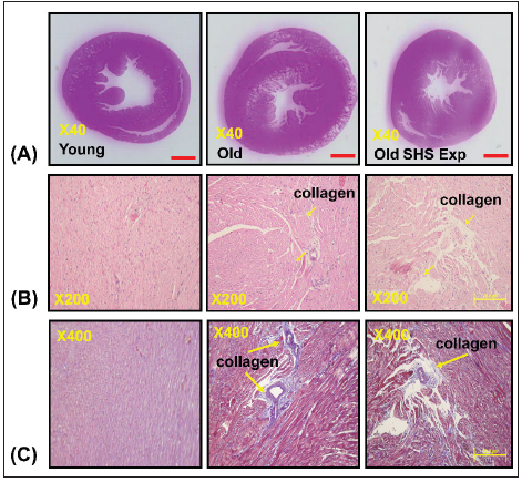
(A) Representative by hematoxylin & eosin staining of left ventricular sections in young, old and old SHS Exp groups
(B) Representative collagen accumulation in the left ventricle by hematoxylin & eosin staining in young, old and old SHS Exp groups. Scale bars 20μm. The images of left ventricular architectures were magnified 200x. Yellow arrows express
(C) Representative collagen accumulation in the left ventricle by Masson’s trichrome staining in young, old and old SHS Exp groups. Scale bars 20μm. The images of left ventricular architectures were magnified 400x. Yellow arrows express.


At the same time, SHS exposure resulted in old rats’ left ventricular papillary muscle deteriorated which led to left ventricular dysfunction. Left ventricular muscle fibers interstitial and extracellular space was broad. Muscle fibers’ rearrangement is disordered. In old rats and old rats in the SHS exposure group, we also could observe ECM degradation resulted in collagen release in cardiomyocytes interstitial (Figure 1B) and collagen accumulation induced fibrosis. Indeed, from Masson’s trichrome stained results, we could observe blue color staining in cross-sections (Figure 1C).

Changes in structures development of heart in old rats and old rats in SHS exposure group

Figure 2 presents heart and Left Ventricular (LV) characteristics in young, old and old rats in the SHS exposure (Old SHS Exp). The whole heart weights of old rats and old rats in the SHS exposure were heavier than young rats. Aging and SHS exposure rats were also enhanced left ventricular weights (*p<0.05 vs. young rats; #p<0.05 vs. old rats) (Figure 2A). The HW-to-body weight and LVto- body weight ratios were significantly increased compared with younger age group. Compared with old rats, the HW-to-body weight and LV-to-body weight ratios also had significantly increased (Figure 2B) in Old SHS Exp groups (*p<0.05 vs. young rats; #p<0.05 vs. old rats). However, body weight is easily caused by the gradual increase in a consequence of aging. Sometimes affected by alcohol, smoke or toxic material effects. The use of tibia length has been evidenced as more reliable than body weight. Because of in which body weight differences may occur condition errors.

Figure 2: Left ventricular hypertrophy takes place in old rats and old rats in the SHS exposure group (Old SHS Exp).
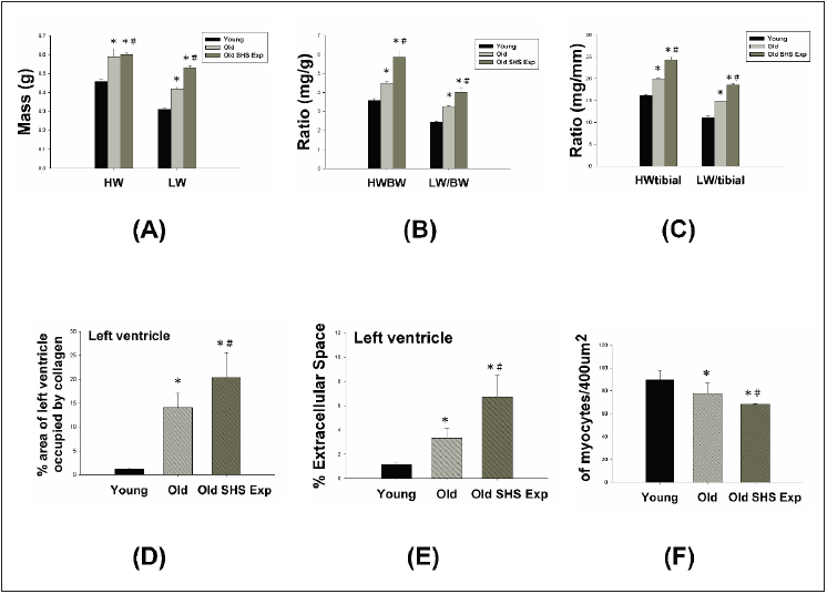
(A) Quantification of heart weight and left ventricle weight statistical analysis. All data are represented as the means ± SEM. *p<0.05 compared with young rats. #p<0.05 compared with old rats
(B) Quantification of the heart weight to body ratio and the ratio of left ventricular weight to body weight statistical analysis. All data are represented as the means ± SEM. *p<0.05 compared with young rats. #p<0.05 compared with old rats
(C) Quantification of heart weight to tibial ratio and the ratio of left ventricular weight to tibial statistical analysis. All data are represented as the means ±SEM. *p<0.05 compared with young rats. #p<0.05 compared with old rats
(D) Quantification of percent of area of left ventricle occupied by collage. All data are represented as the means ± SEM. *p<0.05 compared with young rats. #p<0.05 compared with old rats
(E) Quantification of percent (%) left ventricle extramyocyte connective tissue space (area). All data are represented as the means ±SEM. *p<0.05 compared with young rats. #p<0.05 compared with old rats (F). Quantification of average number of myocytes per 400μm. Values were calculated from myocardial regions. All data are represented as the means ± SEM. *p<0.05 compared with young rats. #p<0.05 compared with old rats.


Furthermore, we detected and measured HW-to-tibial and LVto- tibial ratios to determine heart or left ventricular hypertrophy in old rats and to exposed to SHS exposure (Old SHS Exp). The result showed both of HW-to-tibial and LV-to-tibial ratios were increased significantly as age increased, although exposure to SHS exposure (Figure 3C) (*p<0.05 vs. young rats; #p<0.05 vs. old rats). Therefore, we obtained HW-to-tibial and LV-to-tibial ratios were increased compared with old rats. To detect whether SHS exposure led left ventricular fibrosis exacerbated, we independently calculated the percentage of the per cross-sectional area from hematoxylinstained sections. Quantification of the percentage of the area of left ventricle occupied by collagen (%), collagen area was measured in old rats and old rats in SHS exposure group (Old SHS Exp). The percentage of tissue attributed to collagen distribution increased more rapidly in old rats in the SHS exposure group than in old or young rats (*p<0.05 vs. young rats; #p<0.05 vs. old rats) (Figure 3). To detect whether SHS exposure led left ventricular hypertrophy exacerbated, we independently calculated the percentage of monocytes per cross-sectional area from hematoxylin-stained sections.
Quantification of left ventricular muscle fibers interstitial width of extracellular space, we found left ventricular muscle fibers interstitial become broad resulted in the percentage of extracellular space (%) increased (*p<0.05 vs. young rats; #p<0.05 vs. old rats) (Figure 3). During old age or exposure to SHS exposure, left ventricles from old rats and old rats in the SHS Exp group exhibited fewer percentage per unit area than young rats (*p<0.05 vs. young rats; #p<0.05 vs. old rats) (Figure 3). But cardiomyocytes density is increased per unit area than young rats (result does not show in this article). Indeed, according to tissue architecture using H&E staining analysis, we determined cell size measurement. We observed left ventricular cell size increased width in old rats.

Left ventricular function and structures development of left ventricle on echocardiographic analysis

The echocardiographic analysis is a primary imaging method in the assessment of cardiac impaired and function declined (Figure 3). Parasternal long-axis and short-axis echocardiographic views in young, old and old rats in the SHS exposure (Old SHS Exp) groups showing severe left ventricular hypertrophy. We found left ventricular wall thickness increased. However, M-mode echocardiograms result taken proximal to the papillary muscle deterioration in old rats and old rats in the SHS exposure group (Figure 3A). On the other hand, we found left ventricular wall thickness increases in old rats and old rats in the SHS exposure at systolic and diastolic. Quantification of myocardial hypertrophy for old rats and old rats in the SHS exposure groups are displayed in Figure 3B. Echocardiography of interventricular septal in systolic (IVSs), interventricular septal in diastolic (IVSd), left ventricular internal dimension at end-systolic (LVIDs), left ventricular internal dimension at end-diastolic (LVIDd), left ventricular posterior wall thickness in systolic (LVPWs) and left ventricular posterior wall thickness in diastolic (LVPWd) were presented in young, old and old rats in the SHS exposure group.

Figure 3: Histopathologic of left ventricular hypertrophy using M-mode echocardiograms in young, old rats and old rats in the SHS exposure group (Old SHS Exp)

(A) Representative M-mode echocardiograms taken proximal from young, old and old SHS Exp group. These images were obtained from short-and long-axis imaging at the mid-papillary level. Parasternal short-axis echocardiography views (up-panel), parasternal long-axis echocardiography views (down-panel)
(B) Quantification of left ventricular posterior wall thickness at diastolic and systolic, interventricular septal in systolic (IVSs), interventricular septal in diastolic (IVSd), left ventricular internal dimension at end systolic (LVIDs), left ventricular internal dimension at end diastolic (LVIDd), left ventricular posterior wall thickness in systolic (LVPWs) and left ventricular posterior wall thickness in diastolic (LVPWd) shown in right panel. All data are represented as the means ± SEM. *p<0.05 compared with young rats. #p<0.05 compared with old rats
(C) Quantification of the percentage of fractional shorting and ejection fraction at diastolic and systolic. All data are represented as the means ± SEM. *p<0.05 compared with young rats. #p<0.05 compared with old rats.


The morphological variables obtained from the echocardiographic study are shown in Figure 3. The old rats in SHS exposure had greater IVSs, IVSd, LVIDs, LVIDd, LVPWs and LVPWd dimension compared with young rats (*p<0.05 vs. young rats; #p<0.05 vs. old rats). After exposure to SHS, the old rats had statistically greater dimensions than nonsmoking old rats did. This variable change was used to confirm the efficacy of the exposure of old rats to secondhand smoke (SHS). In addition, considering the left ventricular variables, the ejection and shortening fractions were significantly declined. As Figure 3C shown, shortening (FS%) and ejection fraction (EF%) displayed a progressive impairment in old rats and old rats in the SHS exposure group (Old SHS Exp) (*p<0.05 vs. young rats; #p<0.05 vs. old rats).

Cardiomyocytes width and length of left ventricular hypertrophy in old rats and old rats in the SHS exposure group

The phase of left ventricular hypertrophy during adaptive stress or overload is individual left ventricular myocyte grown in length and/or width as compensated or dilation hypertrophy (Figure 4A). According to the left ventricular cell width size, old rats in the SHS exposure compared with old rats were increased (*p<0.05 vs. young rats). According to compared with left ventricular cell length size in old rats and old rats in the SHS exposure group was increased (Figure 4B). Compared with old rats, left ventricular cell length size in old rats in the SHS exposure group was significantly increased (*p<0.05 vs. young rats; #p<0.05 vs. old rats).

Figure 4: Longer cardiomyocytes occur left ventricular hypertrophy in the SHS exposure group by hematoxylin & eosin stained.
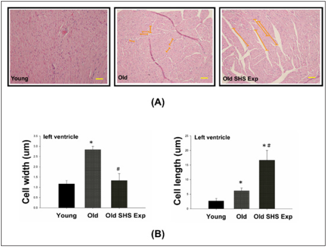
(A) Representative long cardiomyocytes occur left ventricular hypertrophy using histological sections by H&E stained to determine cell width (μm) and cell length (μm) in young, old rats and old rats in the SHS exposure group (Old SHS Exp)
(B) Quantification of cell width (μm) and cell length (μm) in young, old rats and old rats in the SHS exposure group (Old SHS Exp). Values are represented as the means ± SEM. *p<0.05 compared with young rats. #p<0.05 compared with old rats.


Changes to MMPs’ protein expression can explain agerelated heart failure disease

Fibrosis occurs from changes in the balance between synthesis and degradation of extracellular matrix components. Therefore, we sought to determine whether aging and SHS exposure-related collagen accumulation and fibrosis could be related to changes in the regulation of MMP2 and MMP9. Gelatin zymography detected 2 major gelatinolytic bands, MMP-2 and MMP-9, in the left ventricular extracts. MMP-9 gelatinolytic bands did not observe. As Figure 5A shown. Because its regulation was the sum of pro-MMP-9 and TIMP- 1, it may be MMP-9 complexed with TIMP-1. The down regulation of MMP-2 in old rats and old rats in the SHS exposure group was statistically significant compared with young rats. The extent of changes in MMP-2 gelatinolytic activity was lower than old rats (*p<0.05 vs. young rats; #p<0.05 vs. old rats). Consistent with the results of gelatin zymography, MMP-2 and MMP-9 protein content as measured by western blotting was also significantly reduced in old rats and old rats in the SHS exposure group (Old SHS Exp) (Figure 5B).

These results suggest that MMP2/MMP9 contributed to the remodeling of the extracellular matrix in left ventricular fibrosis. While aging, we found MMP2 and MMP9 protein expression decreased. Once exposure to SHS exposure, MMP2, and MMP9 protein expression were significantly lower than old rats (*p<0.05 vs. young rats; #p<0.05 vs. old rats). The dysregulation of MMP2/ MMP9 was now believed to contribute to fibrosis in old and SHS exposure. Elevated TIMPs expression induced fibrosis is present in old rats and old rats in SHS exposure. MMPs catalyze ECM degradation, TIMPs is physiological inhibitors which controlled MMPs activity.

Figure 5: Molecular mechanisms of the imbalance of MMPs and TIMPs induced ECM remodeling.
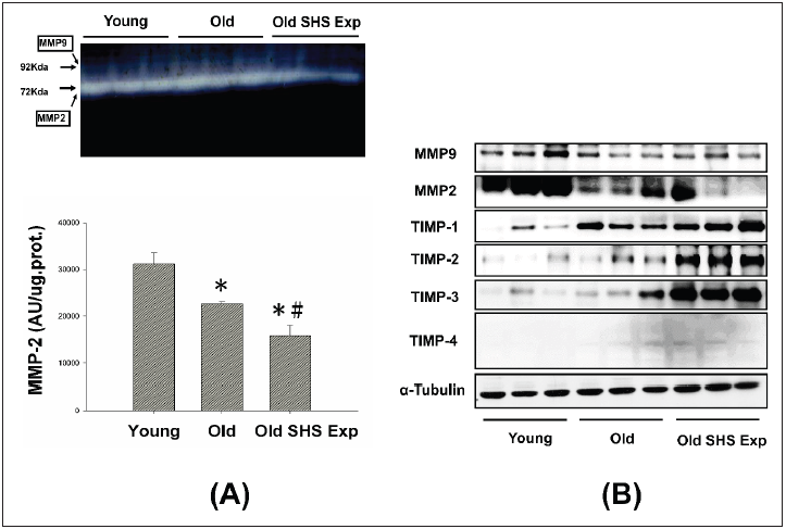
(A) Representative zymographical analysis from young, old rats and old rats in the SHS exposure group (Old SHS Exp). Up-panel shows a gelatin zymography graphic representative of MMP-2 and MMP-9 activity. Down-panel shows MMP-2 activity quantified by densitometry and expressed as mean pixel density. Values are represented as the means ±SEM. *p<0.05 compared with young rats. #p<0.05 compared with old rats
(B) Protein expression activity dysregulation of MMP-2, MMP-9 and protein expression elevated of TIMP-1, TIMP- 2, TIMP-3 and TIMP-4 in old rats and old rats in the SHS exposure group (Old SHS Exp) by western blotting.


To explore whether there are regulation changes in the expression of TIMPs, we assessed the protein expression of TIMPs (TIMP-1, -2, -3 and -4) by Western blot. An up regulation of TIMPs is associated with heart failure and fibrosis. To determine whether aging or SHS exposure modulates cardiac matrix remodeling. As Figure 5B shown, we found TIMP-1, TIMP-2, TIMP-3 and TIMP- 4 induced left ventricular fibrosis, protein expression levels by western blotting analysis in old rats and old rats in the SHS exposure group were significantly higher than young rats. Thus, this apparent cause of fibrosis and heart failure can be explained by differential regulation between MMPs and TIMPs.

SHS exposure results in higher sensitivity to inflammation and heart failure in old rats

SHS exposure is an environmental stressor induced pathological LVH. In this study, we determine whether low concentration SHS exposure to old rats also will be induced pathological LVH. Results showed p-MEK5/MEK5 and p-ERK5/ERK5 increased in old rats in the SHS exposure group (Old SHS Exp), but not in the old hearts (Figure 6A). To further determine the potential of inflammation in the old hearts, we examined JNK1/2, p38α, IL-6, and TNFα protein expression levels by western blotting and immunohistochemistry. As Figure 6B and Figure 6C shown, JNK1/2, p38α, IL-6, and TNFα protein expression levels were increased in old rats and old rats in the SHS exposure group (Old SHS Exp). Immunohistochemical study showed that densities of both old rats and old rats in the SHS exposure hearts were higher in young rats (Figure 6C). IL-6 and TNFα play an important role in promoting LVH and inflammation. With greater age comes, we found that MAPK (JNK1/2 and p38) protein expression were increased in old rats and rats in the SHS exposure group. SHS exposure may enhance proinflammatory cytokines (IL-6 and TNFα) and MAPK cascade expression in old rat hearts. Thus, cytokines in left ventricular hypertrophy tissues increased markedly in keeping with a denser inflammatory cell infiltration.

Discussion

Secondhand Smoke (SHS) exposure has been linked to a number of harmful health outcomes and is an important cause of morbidity and mortality. SHS exposure is an important cause of morbidity and mortality. There is a lot of evidence indicated that SHS exposure a formidable health hazard. However, there is no evidence indicated that SHS exposure presents a challenging health hazard [13]. It is also well understood that toxic air contamination causes lung cancer and cardiovascular diseases. In this study, we investigated the effects of SHS exposure associated with the elderly age, specifically in the left ventricles of male rats. As is well known, old age in humans always accompanies an increased incidence of atherosclerotic vascular disease and cardiovascular disease [14]. In contrast, aging is a physiological process due to increasing injuries and vulnerability, which reduces the ability of organisms to survive. Aging affects various aspects of left ventricular morphology and function and has recently been considered to be a major risk factor for cardiovascular disease and to have effects on various aspects of left vascular morphology and function. Aging can refer to a time-related process, however, it is commonly used for postmaturational processes. The main characteristics associated with aging is a progressive decrease in physiological capacities [15].

Figure 6: The effects of SHS exposure on the activation of molecular mechanisms of inflammation and left ventricular hypertrophy during aging.
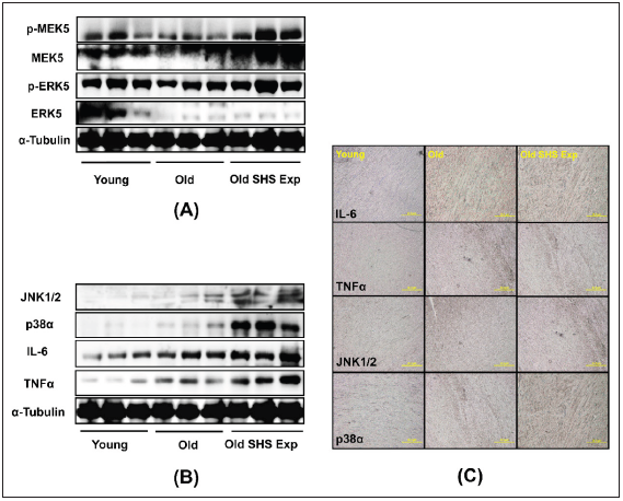
(A) Upregulation of left ventricular hypertrophy proteins, p-MEK5/MEK5 and p-ERK5/ERK5 in old rats in the SHS exposure group by western blot
(B) Upregulation of the expression of inflammatory mediators, cytokines (IL-6 and TNFα) and MAPKs (JNK1/2 and p38), which lead to inflammation response
(C) Immunohistochemistry of IL-6, TNFα, JNK and p38 in young, old rats and old rats in SHS exposure groups.


The overall effect is highly debatable aging and disease. As age increases, whether there will occur diseases itself. However, the most obvious evidence of changes in the aging heart and liver. Biological phenomena appear related to the aging process [16]. Aging exacerbates cardiac damage, leading to cardiac hypertrophy, fibrosis [17] and dysfunction, developed compensatory concentric hypertrophy [18] and fibrosis in response to induced cardiomyocytes hypertrophy in a similar manner [19]. In the aging heart, demonstrated severe left ventricular chamber dilation, wall thinning and
fibrosis, leading to congestive heart failure. In this study, we want to know low-level chronic cigarette smoke exposure whether is harmful to left ventricular function in old rats and to explore the related mechanisms. We found that changes associated with SHS exposure lead to cardiovascular pathological outcomes resulted in age-related disease exacerbated. We observed left ventricular chamber narrowing and rupture and increased left ventricular wall thickness. These results demonstrated left ventricular hypertrophy in old rats and old rats in the SHS exposed group (Old SHS Exp). On the other hand, we could from echocardiography results to determine left ventricular dimension, posterior wall thickness, interventricular septal at end-systole and end-diastole were increased, and left ventricular function declined. Stiffening of these fibers cause left ventricular fibrosis and could also affect the efficient functioning. SHS exposure is linked to a number of harmful health outcomes.
As is well known, SHS exposure is a key risk factor for pathological hypertrophy associated with various cardiovascular disease risk factors [20], however, the old annual human always accompanies with atherosclerotic vascular disease and cardiovascular disease. As the heart reaches senescence, it undergoes a modest degree of Heart failure [21]. It is now determined the differences several signaling molecules play unique role in regulation of old rats in the SHS exposure group. We discuss molecular signaling mechanisms associated with old rats in the SHS exposure, including MMPs, TIMPs, JNK1/2, p38α, IL-6 and TNFα signaling. We suggested that upregulation of pro-inflammatory related protein expression of JNK1/2, p38α, IL-6 and TNFα enhanced left ventricular pathological hypertrophy. Down-regulation of MMP2 and MMP9 in old rats in the SHS exposure accelerated TIMPs-induced cardiac fibrosis. Despite the evidences that chronic exposure to SHS exposure resulted in cardiac changes [22,23], the exact mechanisms involved in lowlevel concentration process remain to be elucidated. These explore knowledge may influence therapeutic strategies for the treatment of cardiovascular disease in old age.

Acknowledgment

The study was approved by National Taipei University of Nursing and Health Sciences. No extra-institutional funding must be reported for this article.

References

  1. Spychala MS, Venna VR, Jandzinski M, Doran SJ, Durgan DJ, et al. (2018) Age-related changes in the gut microbiota influence systemic inflammation and stroke outcome. Ann Neurol 84(1): 23-36.
  2. Stout MB, Justice JN, Nicklas BJ, Kirkland JL (2017) Physiological aging: Links among adipose tissue dysfunction, diabetes, and frailty. Physiology 32(1): 9-19.
  3. Liu P, Xu B, Cavalieri TA, Hock CE (2002) Age-related difference in myocardial function and inflammation in a rat model of myocardial ischemia-reperfusion. Cardiovasc Res 56(3): 443-453.
  4. Zhu J, Rebecchi MJ, Glass PS, Brink PR, Liu L, et al. (2011) Cardio protection of the aged rat heart by GSK-3beta inhibitor is attenuated: Age-related changes in mitochondrial permeability transition pore modulation. Am J Physiol Heart Circ Physiol 300(3): H922-H930.
  5. Jiang Z, Rebecchi MJ, Qiang W, Glass PSA (2013) Chronic tempol treatment restores pharmacological preconditioning in the senescent rat heart. Am J Physiol Heart Circ Physiol 304(5): H649-H659.
  6. Dinas PC, Koutedakis Y, Flouris AD (2013) Effects of active and passive tobacco cigarette smoking on heart rate variability. International Journal of Cardiology 163(2): 109-115.
  7. Flouris AD, Vardavas CI, Metsios GS, Tsatsakis AM, Koutedakis Y, et al. (2010) Biological evidence for the acute health effects of secondhand smoke exposure. Am J Physiol Lung Cell Mol Physiol 298(1): L3-L12.
  8. Brink TC, Demetrius L, Lehrach H, Adjaye J (2009) Age-related transcriptional changes in gene expression in different organs of mice support the metabolic stability theory of aging. Biogerontology 10(5): 549-564.
  9. Garvin AM, Jackson MA, Korzick DH (2018) Inhibition of programmed necrosis limits infarct size through altered mitochondrial and immune responses in the aged female rat heart. Am J Physiol Heart Circ Physiol 315(5): H1434-H1442.
  10. Liu P, Xu B, Cavalieri TA, Hock CE (2002) Age-related difference in myocardial function and inflammation in a rat model of myocardial ischemia-reperfusion. Cardiovasc Res 56(3): 443-453.
  11. Juonala M, Magnussen CG, Venn A, Gall S, Kähönen M, et al. (2012) Parental smoking in childhood and brachial artery flow-mediated dilatation in young adults: The cardiovascular risk in young finns study and the childhood determinants of adult health study. Arterioscler Thromb Vasc Biol 32(4): 1024-1031.
  12. Kizaki K, Momozaki M, Akatsuka K, Fujimori Y, Uchide T, et al. (2004) Impaired gene expression of beta 1-adrenergic receptor, but not stimulatory G-protein Gs alpha, in rat ventricular myocardium treated with isoproterenol. Biol Pharm Bull 27(7): 1130-1132.
  13. Kelly G (2010) A review of the sirtuin system, its clinical implications, and the potential role of dietary activators like resveratrol: Part 1. Altern Med Rev 15(3): 245-263.
  14. Bard RL, Dvonch JT, Kaciroti N, Lustig SA, Brook RD, et al. (2010) Is acute high-dose secondhand smoke exposure always harmful to microvascular function in healthy adults? Prev Cardiol 13(4): 175-189.
  15. Zhu L, Di PY, Wu R, Pinkerton KE, Chen Y, et al. (2015) Repression of CC16 by cigarette smoke (CS) exposure. PLoS One 10(1): e0116159.
  16. Koutros S, Silverman DT, Alavanja MCR, Andreotti G, Lerro CC, et al. (2016) Occupational exposure to pesticides and bladder cancer risk. Int J Epidemiol 45(3): 792-805.
  17. Yang Z, Ming XF (2012) mTOR signalling: The molecular interface connecting metabolic stress, aging and cardiovascular diseases. Obesity Reviews 13: 58-68.
  18. de Almeida AJPO, Ribeiro TP, de Medeiros IA (2017) Aging: Molecular pathways and implications on the cardiovascular system. Oxid Med Cell Longev p. 7941563.
  19. Venn A, Britton J (2007) Exposure to secondhand smoke and biomarkers of cardiovascular disease risk in never-smoking adults. Circulation 115(8): 990-995.
  20. Sheydina A, Riordon DR, Boheler KR (2011) Molecular mechanisms of cardiomyocyte aging. Clin Sci 121(8): 315-329.
  21. Ungvari Z, Csiszar A (2012) The emerging role of IGF-1 deficiency in cardiovascular aging: Recent advances. Gerontol A Biol Sci Med Sci 67(6): 599-610.
  22. Gielen S, Sandri M, Kozarez I, Kratzsch J, Teupser D, et al. (2012) Exercise training attenuates MuRF-1 expression in the skeletal muscle of patients with chronic heart failure independent of age: The randomized leipzig exercise intervention in chronic heart failure and aging catabolism study. Circulation 125(22): 2716-2727.
  23. Alagiyawanna AMAAP, Veerasingam EQ, Townsend N (2018) Prevalence and correlates of exposure to secondhand smoke (SHS) among 14 to 15-year-old schoolchildren in a medical officer of health area in Sri Lanka. BMC Public Health 18(1): 1240.
For more articles in American Journal of Cardiology
Please click on below link:  https://crimsonpublishers.com/ojchd/

Monday, August 22, 2022

Calcified Chronic Subdural Hematoma_Crimson Publishers

 Calcified Chronic Subdural Hematoma by Gómez Fortty María delos Angeles and González Echeverría Kléber Eduardo in Techniques in Neurosurgery & Neurology_Journal of Neurology


Medical Image

Simple skull tomography

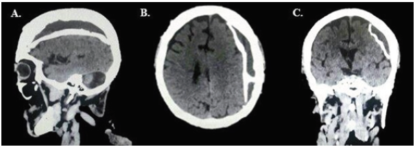
An isodensal image with a wide area of perimeter calcification of left frontoparieccipital extraxial location is observed in a 68-year-old male patient entering with generalized tonic-clonic seizure box, hemiplegia on the right side due to a reduction in brain tissue of up to 200g, with an increase in extra-brain space by 6 to 11% allowing parenchyma to adapt to the hematoma and that it is stood (Figure 1). The clinical presentation of this pathology is often insidious, with symptoms of decreased level of consciousness, problems in gait due to changes in balance, cognitive dysfunction, memory loss, motor deficit (hemiparesias), headache, or aphasia [1,2]. With the presumptive diagnosis, a left parietal craniotomy was performed, through which an encapsulated and calcified lesion was exposed in its entire parietal and visceral face, and that contained a granular substance of dark red color, totally avascular. Radical resection was performed without surgical complications (Figure 2). Final result, successful patient surgical ablation without seizures, regained strength 5/5 in right hemibody, oriented and conscious. It is concluded that chronic subdural hematomas calcification is a rare form of imaging presentation today, known as armoured brain or matryoska brain [3]. Since pseudomembrans are calcified, the chances of brain re-explosion are virtually non- existent [4]; Finally, the decision of the surgery conforms to the clinical or radiological evidence of mass effect. When there is evidence and the need for a craniotomy approach it could be a better option than trepanation in the management of these entities [4,5].

Figure 1:


Figure 2: Craniotomy plus capsulotomy for evacuation of chronic subdural hematoma calcified in 68-yearold patient. (A) Dural opening, (B)Chronic calcified subdural hematoma, (C)Release of chronic calcified subdural hematoma of the cerebral parenchyma, (D) Surgical bed hemostasis, (E)fragments of retired calcified subdural hematoma, (F)Dural closure, (G) Placement of bone flap fixed with microplates, (H) Surgical boarding closure.

References

  1. Ortega SO, Gil Alfonso M, Bacallao GL, Hechevarría AJ, García DM, et al. (2019) Diagnóstico del hematoma subdural: unproceso de clínica e imágenes diná Rev Med Electrónabril 41(2).
  2. Balser D, Farooq S, Mehmood T (2015) Traumatismo craneoencefálico en el adulto mayor. J Neurosurg 1(7).
  3. Arán Echabe E, Fieiro Dantes C, Prieto Gonzales Á (2014) Hematoma subdural crónico calcificado: cerebro blindado. Rev Neurolenero 58(9).
  4. Santarias T, Kolias A, Hutchinson P (2012) Surgical management of chronic subdural hematoma in adults. Operative Neurosurgical Techniques pp. 1573-1578.
  5. García Pallero M, Pulido Rivas P, Pascual Garvi J, G Sola R (2014) Hematomas subdurales cronicos. La arquitectura interna del hematoma como predictor de recurrencia. Rev Neurol octubre 59(7).
For more articles in Journal of Neurology
Please click on below link:  https://crimsonpublishers.com/tnn/

Friday, August 19, 2022

Advances in Syngas Production Technologies: Catalysis and Engineering_Crimson Publishers

Advances in Syngas Production Technologies: Catalysis and Engineering by Makarand R Gogate in Progress in Petrochemical Science_Journal of Petrochemical Science


Opinion

Methane is the principal constituent of natural gas and constitutes over 90% (v/v) by volume, regardless of the source. Coal, oil, and natural gas have traditionally been the 3 fossil fuels of choice for further conversion and upgrading to fuels and fuel additives, chemicals, and petrochemicals, and for generation of electric power. The advent of “Fracking”, a technology first commercialized in the United States around 2008, made it possible to harvest and recover huge quantities of shale gas and associated gas liquids, trapped within tight pore spaces of shale rock deposits or coal bed methane, considered to be 2 unconventional sources of methane [1-3], other than gas hydrates. The production of domestic natural gas saw a hugh spike in about 2008 (about 1Tnm3) and is expected to grow by up to 44% by 2035. The U.S. is now the world’s largest producer of natural gas, and the cost of natural gas is at about the lowest it has been in over 2 decades, at $1.85/MM BTU. The historical trends in the production and price of domestic natural gas, 1900 onwards to 2020, is shown in Figure 1. Instructively, the hugh spike in the production capacity of natural gas is clearly seen around 2005-2010, which coincides with the advent of new “Fracking” capacity in the United States [4].

Figure 1: Historical trends in price of domestic natural gas, 1900 onwards to 2020 [4].


Not surprisingly, natural gas surpassed coal (in 2007) for the largest installed electricity generation capacity in the United States. In addition, the E.I.A. estimates that the unconventional resource base (primarily shale and coal bed methane) of natural gas is around 65Tnm3, out of which about 5Tnm3 are considered to be “proven” reserves, i.e., recoverable under the current economic and environmental conditions [4]. Natural gas, a versatile fuel feedstock, with a high calorific value - one with the lowest C footprint on account of its highest H:C ratio – is primarily used for electricity generation, and for home heating/cooking applications. More than 90% of U.S. domestic production is burned to create energy, for heating, cooking, and transportation purposes, or for generation of electric power (for residential and commercial use). The use of natural gas as a chemical feedstock for further conversion into fuels/fuel additives, chemicals, and petrochemicals, is still very limited. The reason for this is primarily economic in nature. Most natural gas wellhead locations/deposits are found in remote, inaccessible locations. Natural gas, a vapor under ambient conditions, has a very low mass and volumetric energy density, and is difficult and uneconomical to transport over long distances, using gas pipelines, or even LNG tanker trailers. Unfortunately, thus, apart from conversion to synthesis gas, hydrogen cyanide, acetylene, and chlorinated hydrocarbons, methane conversion pathways are not yet cost-competitive to oil-based fuels and chemicals/petrochemicals. However, natural-gas based indirect liquefaction technologies, based on syngas, offer a critical potential avenue (to reduce our dependence on oil and to reduce the C footprint), for further high-volume growth and market share in syngas-based bulk chemicals. The top 3 chemical products, based on natural gas-based syngas, are, ammonia (worldwide capacity 175MMtpa, 11MMtpa U.S.), methanol (110MMtpa, 4.5MMtpa U.S.), and F.-T.-based synfuels products (over 220,000bpd). While the U.S. capacity of the top-3 chemicals above is still a very small fraction of the worldwide capacity, more than a dozen methanol mega projects are currently in various stages of planning, design, and construction, all along the U.S. gulf coast. Natural-gas based syngas is an ideal feedstock for production of above 3 chemicals, as it affords a stoichiometric inlet H2:CO ratio of 2-2.5, directly possible with both conventional steam reforming and autothermal reforming (ATR), without need for any additional shift conversion [5-8].

In this article, we offer an insightful analysis of the current status of syngas production technologies and assess future projections and forecast for the industry. Steam reforming of natural gas is a conventional and now mature technology, for synthesis of “syngas”, a mixture of CO, CO2, and H2 [9-12]. In steam reforming, CH4 reacts with steam to reform CH4 into a mixture of CO, CO2, and H2, as given below:

The synthesis reactions are highly endothermic and thus limited by chemical equilibrium; heat is supplied to the reformer tubes (in a vertical, parallel arrangement), by combustion of natural gas inside a firebox. The product gases leave the reformer unit at 855 oC and 2MPa. In industrial practice, heat is recovered from this gas stream by a series of heat exchange operations.

As noted above, only 2 of the 3 reactions above are independent; the H2:CO ratio for the overall product gas is between 2-2.5. The kinetic studies on a commercial Ni/γ-Al2O3 or a ceramic support indicate that it is the reforming of CH4 to CO and the water gas shift reaction that take place under industrial conditions. The dry reforming reaction can also be postulated to occur in the overall series of reactions, as follows [13]:

As discussed above, steam reforming of natural gas is a mature process technology, and discussed extensively in several recent reviews [6,7,9-12].

Steam reforming is catalyzed by Group VIII transition metals, including Ru, Rh, Ir, and Ni. While extensive experimental and theoretical studies (DFT calculations, scaling relationships, and microkinetic models) show that Ru and Rh are the most active transition group metals for steam reforming, Ni-based catalysts are almost exclusively used in the chemical industry due to high price of Ru and Rh metals. Ir is also an excellent choice as a catalyst, with identical activity and selectivity profiles.

In industry, Ni catalysts are highly susceptible to severe deactivation, by,

  1. sintering and particle growth,
  2. selective poisoning by trace impurities in feed gas, including As, Pb, S, and P, and
  3. C deposition and pore blockage to active sites, by Boudouard reaction, and methane cracking/decomposition, at the high temperatures (>823-1073 K) encountered in steam reforming [14].

Of the three causes mentioned above, sintering and particle growth occur by atom-by-atom translation and migration from one particle to other, a mechanism termed as Ostwald ripening. It has been proposed that Ni surface atoms are quite labile at the high process temperatures and migrate over surfaces by formation of Ni-H and Ni-OH entities. However, promoters like Au and K that selectively bind to active sites (both terrace and step sites) and form surface alloys are shown to impart both sintering and coking resistance and extend catalyst lifetimes in industrial practice. For further reading on causes and consequences of catalyst deactivation, the reader is referred to some recent reviews on this topic [9,12,15-17].

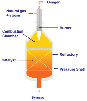
Compared to conventional steam reforming, autothermal reforming (ATR) is a transformational advance in syngas technology, and consists of a combination of a combustion chamber at the reactor inlet in series with a packed bed further away from the inlet, to improve the overall reactor efficiency and operational flexibility [5,12,18]. Natural gas and steam, fed to the combustion zone in presence of O2, at temperatures of ~1300 oC (and higher in the flame core, right at burner entrance mounted ahead of the combustion chamber), undergo homogeneous gas phase reactions, including steam reforming and water gas shift (among other radical reactions). The final conversion takes place in the catalyst bed, and the product gases leave the reformer, at chemical equilibrium, between 850-1100 oC. The ATR operation is very flexible, uses low steam/CH4 and O2/CH4 ratios (sub-stoichiometric), and produces syngas with a wide range of H2:CO ratios, of both CO-rich and H2-rich type. In addition, the ATR operation is typically soot-free and particulate-free. The operational concept of the ATR system is illustrated in Figure 2. Like conventional reforming, Ni-based catalysts are used, but with refractory or spinel support which have high thermal and mechanical strength/stability, to withstand the very high temperatures of operation [9]. Both α-Al2O3 and MgAl2O4 spinel oxide are industrially used as supports.

Figure 2: Illustration of the ATR reactor operation [9].


In catalytic partial oxidation (CPO), still an exploratory technology in nature, the hydrocarbon feed and oxidant (O2) are mixed in the inlet zone of the CPO reactor, and passed over the packed bed of catalyst. Distinct with the ATR operation, the CPO reactor design does not incorporate a burner ahead the catalyst zone. The steam reforming and water gas shift reactions take place over a noble metal-based catalyst, and the product gases typically leave the reactor exit at chemical equilibrium, at temperatures typically >1100 oC. Noteworthy research advances in this area include the development of ms-time scale contact reactors, which incorporate design allowances that circumvent needs for pre-heating, and steam addition [19,20]. Regardless, CPO technology is unlikely to have commercial merit, mainly due to safety concerns, associated with handling of the hydrocarbon-O2 flammable mixture at the inlet. The feed inlet is also designed to be outside the auto-ignition temperature of the mixture, which inherently leads to a higher consumption of O2.

References

  1. Bruijnincx PC, Weckhuysen BM (2013) Shale gas revolution: an opportunity for bio-based chemicals. Angewandte Chemie International Edition 52(46): 11980-11987.
  2. Wang Q, Chen X, Jha AN, Rogers H (2014) Natural gas from shale formation - the evolution, evidences, and challenges of shale gas revolution in the United States. Renew Sustain Energy Rev 30(C): 1-28.
  3. Makogon YF, Holditch SA, Makogon TY (2007) Natural gas-hydrates - a potential energy source for the 21st J Petro Sci Eng 56(1-3): 14-31.
  4. Energy Information Administration (2012).
  5. Rostrup Nielsen JR, Christiansen LJ (2011) Concepts in syngas manufacture, Imperial College Press, London, UK.
  6. Lu K, Song C, Subramani V (2010) Syngas and hydrogen production and purification technologies, Wiley, Hoboken, New Jersey, USA.
  7. Rostrup Nielsen JR (2008) Handbook of heterogeneous catalysis. In: Ertl G, Knözinger H, Schüth F, Weitkamp J (Eds.), Wiley-VCH, Weinheim, Germany.
  8. Tillerson R (2005) Leading the way fundamentals of gas-to-liquids. In: 2nd (edn), Petroleum Economist, London, UK.
  9. Petersen AK, Dybkjaer I, Ovesen CV, Schjodt NC, Sehested J, et al. (2011) Natural gas to syngas: Catalysts and catalytic processes. J Nat Gas Sci Eng 3(2): 423-459.
  10. International energy agency (2012) Golden rules for a golden age of gas.
  11. Wood DA, Nwaoha, C, Towler BF (2012) Gas-to-liquids: a review of an industry offering several routes for monetising natural gas. J Nat Gas Sci Eng 9: 196-208.
  12. Rostrup JR (1993) Production of synthesis gas. Catal Today 18(4): 305-324.
  13. Bradford MJ, Vannice MA (1993) CO2 reforming of CH4. Catal Rev Sci Eng 41(1): 1-42.
  14. Horn R, Schlögl R (2015) Methane activation by heterogeneous catalysis. Catal Lett 145: 23-39.
  15. Ratnasamy C, Wagner JP (2009) Water-gas shift catalysis. Catal Sci Rev Eng 51(3): 325-340.
  16. Rasmussen FB, Sehested J, Teumssen HT, Molenbroek AM, Clausen BS (2004) Sintering of Ni/Al2O3 catalysts studied by anomalous small angle X-ray scattering. Appl Catal A 267(1-2): 165-173.
  17. Sehested J, Carlsson A, Janssens TW, Hansen PL, Datye A (2001) Sintering of Nickel steam reforming catalysts on MgAl2O4 J Catal 197(1): 200-209.
  18. Reyes SC, Sinfelt JH, Feeley JS (2003) Evolution of processes for syngas production: Recent developments in an old technology. Ind Eng Chem Res 42(8): 1588-1597.
  19. Schmidt LD (2000) Millisecond chemical reactions and reactors. Stud Surf Sci Catal 130: 61-81.
  20. Specchia S, Vella L, Montini T, Fornasiero P (2011) Syngas production by short contact-time catalytic partial oxidation of methane, Hydrogen production: Prospectives and processes. In: Honnery DR, Moriarty D (Eds.), (1st edn), Nova Science Publishers Inc., Hauppauge, New York, USA, pp. 95-139.
For more articles in Journal of Petrochemical Science
Please click on below link:  https://crimsonpublishers.com/pps/

Thursday, August 18, 2022

Intestinal Intussusception in an Adult Affected by Vanek Tumor: A Case Report_Crimson Publishers

Intestinal Intussusception in an Adult Affected by Vanek Tumor: A Case Report by Morazán AD in Surgical Medicine Open Access Journal_journal of Surgical Medicine


Abstract

Inflammatory fibroid polyp (IFP) is a rare benign lesion, originating from the submucosa in the gastrointestinal tract. It generally appears as an isolated benign lesion, rarely located at the level of the ileum. Its origin is controversial. Clinical presentation varies depending on its location; invagination and obstruction are the most common indicative symptoms when the polyp is located at the level of the small intestine. We report the case of a 60-year old patient with abdominal pain, nausea and vomiting and a personal history of intermittent constipation. Radiological imaging objectified ileo-ileal invagination completely obstructing the ileum light. Segmental resection of the obstructed ileal segment and terminalterminal anastomosis were performed. The final diagnosis of IFP was established using histological examination.

Keywords: Inflammatory fibroid polyp; Ileum; Invagination

Introduction

Adult intussusception is relatively rare, constituting only 1% of patients with bowel obstructions. Adult intussusception is mostly caused by tumors and 80% of the tumors associated with small bowel intussusception are benign. Vanek tumor (inflammatory fibroid polyp) is a rare pseudo-tumorous lesion of the gastrointestinal tract first described by Vanek in 1949 [1]. It occurs most frequently in the gastric antrum of adults, inflammatory fibroid polyp (IFP) rarely cause ileal intussusception [2]. The IFP is a benign lesion that arises from the submucosa of the gastrointestinal (GI) tract, most commonly in the antrum (70%) and ileum (20%) and, only occasionally, in the duodenum and jejunum. Its frequency from 1% to 4% of diagnoses among benign lesions, and usually occurs between the fifth and the seventh decade of life [3]. The underlying cause of IFPs is still unknown. Many have suggested etiologies possibly related to chemical, physical, or metabolic triggers. Genetic study of IFP showed mutations in platelet derived growth factor alpha (PDGFRA) in some cases. The frequency of mutations among case series ranges from 21.7% to 69.6%. Activating PDGFRA mutations occur in exons 12, 14 and 18. It also shows for the presence of androgen receptor-positive cells whose location corresponds with the distribution of Ki67- positive cells. When localized in the small bowel, the presenting symptoms are colicky abdominal pain and obstruction. Intussusception due to IFPs is uncommon; moreover, jejunojejunal and ileoileal intussusception has only rarely been reported [4-14]. We present the case of a 60-year-old male presenting with symptoms preceding an intestinal obstruction caused by an ileoileal intussusception with an inflammatory fibroid polyp acting as the lead point.

Clinical Case

A 60-year-old male patient, married, farmer, from Tegucigalpa, with a history of a month and a half of evolution, insidiously presenting moderately intense abdominal cramps, diffuse location with predominance in the right iliac fossa and mesogastrium, short duration, accompanied by nausea, vomiting twice a week, which is attenuated after the intake of analgesics and antispasmodics. Evaluated by general practitioner and managed with antispasmodics, changes in lifestyle and diet. The patient continued with insidious, sporadic, and self-limited episodes of pain until one day prior to admission presented an exacerbation with increased intensity without attenuating the intake of analgesics and antispasmodics, so he went to the emergency surgery assessment general. Pathological personal history of systemic arterial hypertension managed with irbersartan 300mg orally every day, benign prostatic hyperplasia treated with alfuzosin once daily, grade I obesity. On physical examination, the patient presented complaining with pain facies, hemodynamically stable with vital signs of BP 130/90mmhg, FC 96x, FR 16x, temperature of 36.5 ℃ without data of respiratory distress, oxygen saturation of 98% with ambient air , chest with well-ventilated lung fields with vesicular murmur, globular abdomen at the expense of adipose tissue, hypoactive intestinal noises 2 per minute, without distension, percussion without alterations, soft, depressible, moderate pain on deep palpation in the iliac fossa and right flank, no involuntary muscular resistance, no data of peritoneal irritation, no palpable tumors or visceromegalies. Genitals and inguinal region without alterations. No stool to the rectal touch. Laboratory tests were requested with the following results: Hb 12.1g/dL, H to 38.5mL/dL, VCM 91.9μm3, WBC. 10,800/mm3, neutrophils 72.2%, platelets 474,000/mm3; Biochemical: Glucose 100mg/dL, Na 140mEq/L, K 4.4mEq/L, creatinine 1.0mg/dL, BUN 13.8mg/dL, urea 32.5mg/dL, Alb 5.1g/dL, TGO 23U/L, TGP 36U/L.

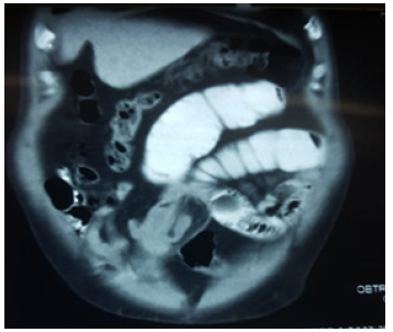
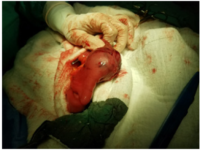
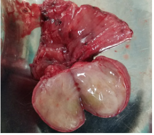
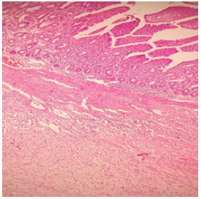
Subsequently, his study was complemented by requesting abdominal tomography. In which a proximal ileal loop was observed with the presence of distal cystic tumor that measures approximately 3.5cm with proximal dilation of this intestinal loop with a hydro-air level, related to intestinal invagination. No data of intestinal obstruction, free fluid, or pneumoperitoneum were observed (Figure 1). He was admitted for surgical treatment with a diagnosis of intestinal invagination. A diagnostic laparoscopy was performed and turned in open laparotomy with the following findings, a firm consistency tumor in the small intestine at the level of the ileum 7cm long and 3.5cm in diameter, occupying the entire lumen of the intestine, with an ileo-ileal invagination segment of approximately 15cm (Figure 2). The tumor was evaluated small intestine segment which measures 7cm long by 3.5cm in diameter with a light brown, smooth, shiny serosa, wall 4cm thick, trabeculated mucosa preserved in the intestinal wall, a nodular mass measuring 4 by 3.5cm, gray, cream attached directly to the wall, clean surgical edges of soft consistency and gelatinous appearance, well defined, not encapsulated with moderate amount of non-foul, non-hot inflammatory fluid (Figure 3). It was decided to perform resection and ileo-ileum anastomosis, the patient evolved satisfactorily and began diet 24 hours post-operative and discharged 4 days later without complications. Histological reported myxoid-like lesion made up of elongated fibroblasts, also with an inflammatory infiltrate polyp accompanied by eosinophils. It grows in the submucosa, pushes and ulcerates the submucosa, with an acute inflammatory reaction. well-defined tumor without cell atypia or mitosis, normal surgical edges (Figure 4).

Figure 1: Proximal ileal loop with the presence of distal cystic tumor that measures approximately 3.5cm with proximal dilation of this intestinal loop with a hydro-air level, related to intestinal invagination.


Figure 2: Firm consistency tumor in the ileum 7cm long and 3.5cm in diameter, occupying the entire lumen of the intestine, with an ileo-ileal invagination.


Figure 3: The tumor with a light brown, smooth, shiny serosa, trabeculated mucosa preserved in the intestinal wall, a nodular mass, cream attached directly to the wall, clean surgical edges of soft consistency and gelatinous appearance, well defined, not encapsulated.


Figure 4: Histological reported myxoid-like lesion made up of elongated fibroblasts, also with an inflammatory infiltrate polyp accompanied by eosinophils.


References

  1. Daum O, Hes O, Vanecek T, Benes Z, Sima R, et al. (2005) Vanek’s Tumor (Inflammatory Fibroid Polyp). Report of 18 cases and comparison with three cases of original Vanek’s series. Annals of Diagnostic Pathology 7(6): 337-347.
  2. Akbulut S, Mahsuni M, Cakabay Bahri, Sule Bakir, Ayhan Senol (2009) Giant inflammatory fibroid polyp of ileum causing intussusception: a case report. Cases J 2: 8616.
  3. Fazzio C, Madeira E (2015) Inflammatory fibroid polyp (Vanek’s polyp): a case report and literature review. J Bras Patol Med Lab 51(2): 117-120.
  4. Bays D, Anagnostopoulos G, Katsaounos E, Filis P, Missas (2004) Inflammatory fibroid polyp of the small intestine causing intussusception: A report of two cases. Dig Dis Sci 49(10): 1677-1680.
  5. Jukic Z, Ferencic Z, Radulovic P, Mijic A (2014) Estrogen and androgen receptors in inflammatory fibroid polyp (Vanek’s Tumor): Case report. Anticancer Res 34(12): 7203-7206.
  6. Lee C, Yuen MK (2014) Inflammatory fibroid polyps causing intussusception in adult patients: Two case reports and review of literature focusing on radiological features. Hong Kong J Radiol 17: 271-276.
  7. Rossi P, Montuori M, Balassone V, Ricciardi E, Anemona L, et al. (2012) Inflammatory fibroid polyp. A case report and review of the literature. Ann Ital Chir 83(4): 347-351.
  8. Ahtil R, Bensghir M, Meziane M, Houba A, Jaafari A, et al. (2017) Rare etiology of subocclusive syndrome: inflammatory fibroid polyp of the ileum, about a clinical case. Pan Afr Med J 26: 146.
  9. Madriz DeHaan P, Martínez Y, Gutiérrez F (2014) Intestinal intussusception due to an inflammatory fibroid polyp (Vanek's tumor). Legal Medicine of Costa Rica-Virtual Edition 31(1): 1409-1415.
  10. Paikos D, Moschos J, Tzilves D, Koulaouzidis A, Kouklakis G, et al. (2007) Inflammatory fibroid polyp or Vanek’s tumour. Dig Surg 24: 231-233.
  11. Yakan S, Calıskan C, Makay O, Denecli AG, Korkut MA (2009) Intussusception in adults: Clinical characteristics, diagnosis and operative strategies. World J Gastroenterol 15(16): 1985-1989.
  12. Gara N, Falzarano J, Limm W, Namiki T (2009) Ileal inflammatory fibroid polyp causing chronic ileocolic intussusception and mimicking cecal carcinoma. World J Gastrointest Oncol 1(1): 89-92.
  13. Bassam Abboud (2015) Vanek’s tumor of the small bowel in adults. World J Gastroenterol 21(16): 4802-4808.
  14. Zakaria A, Daradkeh S (2012) Jejunojejunal intussusception induced by a gastrointestinal stromal tumor. Case Rep Surg 2012: 173-680.
For more articles in journal of Surgical Medicine
Please click on below link:  https://crimsonpublishers.com/smoaj/

A Close Look at the Application of the Yin-Yang- Based Acupoint Pairs_Crimson Publishers

A Close Look at the Application of the Yin-Yang- Based Acupoint Pairs by Tong Zheng Hong in Advancements in Bioequivalence & Bioavailabi...