Crimson Publishers High Impact Journals

Tuesday, August 16, 2022

Clinical Features of Pathological Processes of the Cervix Associated with Human Papillomavirus Infection_Crimson Publishers

Clinical Features of Pathological Processes of the Cervix Associated with Human Papillomavirus Infection by Unanyan A in Investigations in Gynecology Research & Womens Health_Journal of Obstetrics and Gynecology


Abstract

Background: Cervical Diseases associated with Papillomavirus infection are one of the most pressing problems modern gynecology due to the high frequency of occurrence (in the structure gynecological morbidity - from 20 to 45%) and the ability to steady progression of the pathological process. A significant place in the structure of CM pathology is occupied by cervical intraepithelial neoplasia (CIN), which are often malignancy. Of particular importance are the issues of oncological transformation of the cervix in patients of reproductive age, taking into account the upcoming implementation of reproductive function. In Russia over the past 15 years, there has been an increase in the proportion of neglected cases of cervical cancer (cervical cancer), which ranks second (after breast cancer) in the structure of malignant tumors in reproductive age in women. Unfortunately, to date, the data of only an anamnesis, general clinical laboratory and instrumental methods of research are not productive enough to assess the risk of developing cervical onychopathology.

Objective: To identify the most significant clinical and anamnestic risk factors for the progression of pathological processes associated with human papillomavirus infection and the development of malignant transformation of the cervix in women of reproductive age, as they are important, taking into account statistical analysis.

Material and methods: The study conducted prospective and retrospective analyzes of 185 patients of reproductive age with cervical diseases; the age of the women surveyed ranged from 25-44 years.

Results: We have revealed a moderate relationship (using Spearman's correlation analysis) between the degree of progression of cervical disease and the presence of aggravated heredity (cancer of the reproductive system) (R = 0.5679, p1.0, which allowed us to attribute these factors of hereditary burden to cancer with statistically significant risk factors for malignant transformation and progression of the pathological process of cervix.

Conclusion: According to our study the most clinically and statistically significant risk factors are high virus HPV an oncogenic risk of more than 105 genomic equivalents; HPV persistence of high oncogenic risk; previously morphologically verified CIN; sexually transmitted mixed infections (3 or more).

Keywords: HPV; CIN; Cervical cancer; Gynecological oncology

Introduction

Cervical diseases associated with human papillomavirus (HPV) are an urgent problem of modern obstetrics and gynecology, primarily due to the high incidence and possibility of development of precancerous processes and cervical cancer [1]. Cervical intraepithelial neoplasia (CIN), which often leads to the development of cervical cancer, occupies a significant place in the structure of cervical diseases associated with HPV [2]. The current management and treatment tactics for detecting CIN are different: from observation using HPV testing and cytological control to excision, which in some cases is repeated, and even hysterectomy [1]. At the same time, the reason for such different tactics of management is not always unambiguous and needs a pathogenetic justification in order to identify the most effective approaches in the treatment of women of reproductive age. A factor requiring special attention in the management of women with CIN is the possibility of developing residual/recurrent disease [3]. Residual/recurrent disease is detected, as a rule, in patients with a neoplastic process at the edges of the resected tissue.

Currently, existing methods for predicting the progression of the residual neoplastic process, up to its malignant transformation, are not effective enough, and therefore the principles of the formation of high cancer risk groups require further development. Recently, an important role in the process of carcinogenesis has been played by epigenetic changes [4]. One of the main epigenetic events necessary for tumor development is acetylation of histones and methylation of tumor suppressor genes, which are the earliest events in the process of carcinogenesis [5]. However, the identification of their clinical significance and features in the genesis of the progression of pathological processes of CM require further research due to the insufficient number of works in this direction.

Materials and Methods

The study was conducted on the clinical bases of the Department of Obstetrics and Gynecology No. 1 (head of the department-MD, professor Ishchenko A.I.) First Moscow State Medical University named after I.M. Sechenov and LLC "Invitro". The study conducted prospective and retrospective analyzes of 185 patients of reproductive age with cervical diseases; the age of the women surveyed ranged from 25-44 years. In retrospect, all patients were divided into 5 groups depending on the morphological type of the pathological process of CM: group 1-patients with benign processes associated with HPV (n=32); Group 2-women with CIN I (n=43); Group 3-patients with CIN II (n=46); Group 4-women with CIN III (n=42); Group 5-patients with invasive squamous cervical cancer (stage I; n=22). Patients with a morphologically verified diagnosis of CIN III or invasive squamous cervical cancer for further treatment were referred to an oncologist. Patients with CM adenocarcinoma were not found in the study.

Inclusion criteria-reproductive age, morphologically verified cases of pathological processes of cervix associated with HPV, HPV-detected HPV (high oncogenic risk), the presence of paraffin blocks in women with previous biopsy or excision of CM. Exclusion criteria: pregnancy and lactation, the use of HPV vaccine, severe somatic burden of patients (renal, hepatic, cardiovascular insufficiency), morphologically verified malignant formations (except squamous cervical cancer). In the process of determining urogenital infections, the diagnostic systems DNA-Sorb-AM, Amply Sense HPV Raman Screen-Titer-FL, Сobas 4800 (Roche Molecular Systems, USA) were used. Conducted traditional and liquid methods of cytological examination (classification of the Bethesda system), colposcopy (Sensitec OC-100 and SLC-2000). An aim biopsy and LLETZ, or conical excision, were performed on an EHVCh-140-02 Fotek device (Russia) and Surgitron DF 120 4.0 MHz (USA). An ultrasound scan was performed on an Acuson 128 XP 10 device (USA), Dornier AI 5200 "(Germany)," Logiq C5 "(USA). Destructive methods of treatment were performed using the PlasmaJeT apparatus (Great Britain). The latest WHO classification [6] distinguishes between LSIL, whose synonym is CIN I, and HSIL, whose synonyms are CIN II and CIN III.

However, it should be noted that the classification that divides CIN into CIN I, CIN II, and CIN III [7] reflects in more detail the phased development of the neoplastic cervical process, which was the basis for the use of this particular classification in our own research work. All quantitative data obtained were processed by the method of variation statistics using spreadsheets "Microsoft Excel", as well as the application package "Statistica for Windows" v. 7.0, Stat Soft Inc. (USA). The probability of the occurrence of an event for some case was calculated by the formula, p=1/(1+e^(-z) ) ,where z = b1*X1+b2xX2+...+bnxXn+... X1 are the values of independent variables, b1 are the coefficients, calculation which is the task of binary logistic regression, e is the natural logarithm and is equal to 3.714. In the resulting binary logistic regression equation, the prognosis (low risk–p<0.5) of the progression of the pathological process of cervix associated with HPV was calculated.

Research Results and Discussion

We carried out a detailed comparative analysis of clinical, medical and diagnostic parameters in the formed groups of patients. The average age of the examined patients was 38.4±1.4 years. Using Pearson's correlation analysis, we found a direct moderate relationship between age and the degree of progression of the pathological process of cervix (r=0.3485, p1.0. It should be noted that, according to WHO 2013 (Guidelines for screening and treatment of precancerous lesions for cervical cancer prevention), the highest risk of cervical cancer occurs between the ages of 30 and 49. The age of women as a risk factor for the occurrence of pathological processes of cervical cancer has also been noted by other researchers [8].

We have revealed a moderate relationship (using Spearman's correlation analysis) between the degree of progression of cervical disease and the presence of aggravated heredity (cancer of the reproductive system) (R=0.5679, p1.0, which allowed us to attribute these factors of hereditary burden to cancer with statistically significant risk factors for malignant transformation and progression of the pathological process of cervix. Hereditary factor, as a rule, is realized through the mechanism of predisposition to the disease, when and some inherited adaptive-protective mechanisms are not able to counteract exogenous carcinogenic effects [9]. Several researchers have noted a high degree of hereditary burden of patients in the development of tumor diseases of the genital organs, which, in their opinion, is an important factor predisposing to the development of cervical disease [10].

When studying somatic diseases, it was found that the differences in the frequency of diseases of the respiratory, urinary, nervous systems, gastrointestinal tract in the comparison groups were statistically insignificant (p>0.05). In the study of reproductive history, it is noteworthy that the number of births and abortions did not statistically significantly differ between the groups, however, a significant predominance of post-abortion and postpartum traumatic cervical injuries was revealed in women with CIN III (83.3%) and invasive cervical cancer (90, 9%) compared with patients in the first three groups (9.4%, 11.6% and 43.5%, respectively); (p<0.05). Traumatic injuries of cervix (postpartum, post-abortion, etc.) in the progression of the pathological process of cervix were identified as a significant risk factor based on the calculation of RR. According to Prilepskaya et al. [10] traumatic injuries of cervix with its further deformation lead to a violation of the physiological barrier, which is accompanied by a decrease in local immunity and easier penetration of the infection. Data from other researchers indicate that precancer and cervical cancer are often formed against the background of post abortion and postpartum cicatricial deformity of cervical cancer and ectropion [11].

Using Pearson's correlation analysis, we revealed a direct moderate dependence between the duration of smoking and the degree of progression of the pathological process of cervix (r=0.4176, p<0.0001), which indicates a tendency to increase the incidence of cervical diseases with long-term smoking. Tobacco smoking for more than 7 years was chosen as a factor in the progression of the neoplastic cervical process in connection with the highest possible RR compared to other similar indicators, and also taking into account the fact that the lower limit of 95% CI is higher than 1, which indicates the statistical significance of this factor. According to numerous studies, smoking women compared with non-smokers are 2-3 times higher risk of developing cervical cancer [12].

Noteworthy data on infections in the examined patients. The most significant increase in the frequency of mixed infections (combination of 3 or more) in the comparison groups from 1 (3.1%) to 5 (72.7%) in the examined patients was revealed; (p <0.05), as well as recurrent bacterial vaginosis in the same comparison groups in 1 (9.4%) and 5 (86.4%); (p <0.05). Mixed infection factors (a combination of 3 or more) and recurrent bacterial vaginosis due to the highest RR and a CI above 1 were evaluated as statistically significant. The findings of other studies indicate that the presence of mixed infections can contribute to the development of dystrophic changes in stratified squamous epithelium, disruption of intercellular contacts, leukocyte and lymphocytic infiltration of the stroma [13]. A systematic review by Murta et al. [14] confirms the significant association between bacterial vaginosis and CIN [14].

In our own study, a high frequency of chronic cervicitis was found in women with CIN II (58.7%), CIN III (59.5%) and squamous cervical cancer (86,4%); (p<0.05) relative to this condition in the comparison groups, which, along with the indicators of RR and CI, made it possible to identify chronic cervicitis as a risk factor for the progression of cervical pathology. According to Lebedeva et al. [15], in 47.1% of patients with chronic cervicitis there is a high frequency of infection with HPV-BP. According to several other authors, chronic cervicitis is one of the main exogenous factors contributing to the development of dysplasia and cervical cancer [16].

When analyzing the characteristics of sexual life and contraception in the examined women, statistically significant indicators of the early onset of sexual activity (up to 18 years of age, without the use of barrier contraception) and the number of sexual partners more than 7 (from the moment of the onset of sexual activity) in women 4 (38, 1%) and 5 (36.4%) groups compared with similar indicators in patients of groups 1, 2 and 3 (9.4%, 16.3% and 21.7%, respectively); (p<0.05). The calculation of the RR factor of the number of sexual partners of more than 7 (since the onset of sexual activity) and the onset of sexual activity up to 18 years (without the use of barrier contraception) made it possible to single out this factor as a statistically significant criterion in the progression of cervical pathology. The relationship between HPV infection and promiscuous sexual life has been noted in other studies [1].

The analysis of the clinical picture in patients with pathological processes of cervix shows that the clinical symptoms in patients with cervical diseases are usually due to the presence of a combined uterine pathology (endo- and myometrium). The combination of uterine fibroids with adenomyosis and/or endometrial hyperplastic process in patients with CIN III (47.6%) and cervical cancer (59.1%) was much more frequent compared with the incidence of combined uterine pathology in women of comparison groups; (p<0.05). This fact, apparently, is due to the mutually stimulating effect of pathological processes of endo- and myometrium on the state of cervix, which is realized through the mechanisms of intercellular interactions of growth factors. According to Sidorova et al. [16], there are significant differences in the incidence of CIN in women with combined uterine pathology compared with isolated uterus pathology (p<0.001), where the relative risk of CIN in women with combined uterine diseases (uterine fibroids, adenomyosis, endometrial hyperplasia) is 62 times higher than women with any isolated uterine disease [16].

Our study revealed the persistence of HPV-BP in 80 women (43.2%), the viral load of HPV-BP more than 105 genomic equivalents in 83 women (44.9%), as well as the simultaneous combination of two or more types of HPV-VR infection in 96 women (51.9%). An analysis of HPV infection and the calculation of RR allowed us to determine the persistence of HPVR, the viral load of HPV-HR more than 105 genomic equivalents, and the simultaneous combination of two or more types of HPV-HR as significant factors in the progression of the neoplastic process. Literature data indicate that one-year or two-year persistence, especially HPV type 16, is a high-risk factor for the development of CIN 3 or 3+ [1]. In the study Rogovskaya et al. [1] combined HPV infection is more common in patients with HSIL (58%) than in patients with latent papilloma-virus infection (PVI) and LSIL (46%) [1]. Data from other literature sources also indicate that viral load is associated with a degree of CIN but indicating that viral load as the only marker for determining the risk of neoplastic transformation is not sufficiently informative [1,16].

An assessment of the cases of a morphologically verified CIN in the anamnesis (this fact was recorded when requesting paraffin blocks and reviewing the “glasses” of the preparation) revealed its statistical significance with respect to the possible progression of the pathological process of CM in 4 (28.6%) and 5 (31.8%) groups compared with the other three groups (3.1%, 9.3% and 15.2%, respectively); (p <0.05).

Thus, the results of clinical and medical history and diagnostic features in the studied groups of patients allowed us to determine statistically significant risk factors for the development of malignant transformation of CM. To further, improve the approach to the formation of high cancer risk groups. We carried out the next stage of the study, aimed at identifying pathogenic risk factors for the development of cervical cancer, by a detailed analysis of the molecular biological characteristics of acetylation and methylation with the above clinical risk factors in pathological processes of cervical cancer associated with HPV.

Conclusion

The most clinically and statistically significant risk factors (in terms of significance) of the progression of the pathological process associated with HPV and the risk of developing malignant transformation of the cervix uteri are (with the highest possible RR>1 and with a lower limit of 95% CI>1): high virus HPV an oncogenic risk of more than 105 genomic equivalents; HPV persistence of high oncogenic risk; previously morphologically verified CIN; sexually transmitted mixed infections (3 or more); recurrent bacterial vaginosis; traumatic injuries of the cervix (postpartum, post abortion); age more than 40 years; early onset of regular sexual activity (up to 18 years) without the use of barrier methods of contraception; more than 7 sexual partners (since the sexual debut); the simultaneous combination of two or more types of HPV high oncogenic risk; chronic cervicitis; hereditary burden of cancer of the organs of the reproductive system; hereditary burden of common oncological diseases; a combination of uterine fibroids with adenomyosis and/or endometrial hyperplastic process; smoking.

References

  1. Rogovskaya SI, Lipova EV (2014) Cervix vagina, vulva. A Guide for Doctors, p. 830.
  2. Korolenkova LI (2014) Clinical and morphological conception of the occurrence and course of severe CIN. Clinical Laboratory Diagnostics 6: 18-22.
  3. Ulrich D, Tamussino K, Petru E, Haas J, Reich O (2012) Conization of the uterine cervix: does the level of gynecologist's training predict margin status? Int J Gynecol Pathol 31(4): 382-386.
  4. BarrowTM, Michels KB (2014) Epigenetic epidemiology of cancer. Biochem Biophys Res Commun 455(1-2): 70-83.
  5. Kiselev VI, Drukh VM, Kuznetsov IN, Muizhnek EL, Apolikhina IA, et al. (2014) Evaluation of the effectiveness of drug therapy of cervical intraepithelial neoplasia of mild to moderate severity with Cervicon-DIM suppositories (interim results of a clinical trial). Gynecology 6: 58-62.
  6. WHO (2014) Classification of tumors of female reproductive organs is the sixth volume in the Fourth Edition of the WHO series on histological and genetic typing of human tumors? In: Robert JK, Maria LC, Herrington s, et al. (Eds.), Young (4th edn), Switzerland, p. 316.
  7. WHO (2003) Pathology and genetics of tumors of the breast and female genital organs (IARC who classification of tumors)? Lyon, France.
  8. Saslow D, Solomon D, Lawson HW, Killackey M, Kulasingam SL, et al. (2012) American cancer society, American society for colposcopy and cervical pathology, and American society for clinical pathology screening guidelines for the prevention and early detection of cervical cancer. Am J Clin Pathol 137(4): 516-542.
  9. Nazarova NM, Prilepskaya VN, Sulamanidze LA, Mzarelua GM, Bestaeva NV (2013) Papillomavirus infection: prevalence, diagnosis and treatment. Attending Physician 11: 15-18.
  10. Prilepskaya VN, Bebneva TN (2011) Primary prevention of cervical cancer: Achievements and prospects. Farmateka 13: 42-46.
  11. Radzinsky VE (2013) Problems of diagnosis and treatment of diseases of the cervix, vagina and vulva VE Radzinsky. Post-release and materials of the scientific program of the International Interdisciplinary Forum “Cervix and Vulvovaginal Diseases”. M: Editorial Office of the Journal Status Praises, p. 3-4.
  12. Mirabello L, Schiffman M, Ghosh A, Rodriguez AC, Vasiljevic N, et al. (2013) Elevated methylation of HPV16 DNA is associated with the development of high grade cervical intraepithelial neoplasia. Int J Cancer 132(6): 1412-1422.
  13. Strizhakov AN, Belotserkovtseva LD, Budanov PV (2014) A systematic approach to the selection of a clinical solution for vulvovaginal infections. Gynecology, Obstetrics and Perinatology 1: 60-66.
  14. Murta EF, Souza MA, Araújo E, Adad SJ (2000) Incidence of Gardnerella vaginalis, Candida SP and human papilloma virus in cytological smears. Sao Paulo Med J 118(4): 105-108.
  15. Lebedeva LM (2009) Papillomavirus infection in inflammatory diseases of the cervix. Pathogenetic Approaches to Treatment, p. 22.
  16. Sidorova IS, Unanyan AL, Zaletaev AV, Sha S, Zholobova MN, et al. (2012) The role of microsatellite instability of the genome in diseases of the cervix uteri in women with uterine myoma, adenomyosis, endometrial hyperplasia and their combination. Russian Journal of Obstetrician Gynecologist 4: 13-16.
For more articles in Journal of Obstetrics and Gynecology
Please click on below link:  https://crimsonpublishers.com/igrwh/

Monday, August 15, 2022

Seasonal Abundance and Species Composition of Cetacean Caught as By-Catch by Artisanal Driftnet Off the Niger Delta Nigeria_Crimson Publishers

Seasonal Abundance and Species Composition of Cetacean Caught as By-Catch by Artisanal Driftnet Off the Niger Delta Nigeria by Ana Rita Onodera Palmeira Nunes in Examines in Marine Biology & Oceanography_ocean modelling journals


Abstract

A total of 315 dolphins were captured as by-catch, during the 575 fishing trips between 2017 and 2018, in two-landing sites (Imbikiri, Bayelsa State and Finima River State) offshore the Niger Delta Nigeria. The catches which consisted of 6 different species of cetacean , the short-beaked common dolphin (Delphinus delphis), the Risso’s dolphin (Grampus griseus), the Atlantic bottlenose dolphin (Tursiops truncatus), the Atlantic spotted dolphin (Stenella frontalis), Fraser’s dolphin (Lagenodelphis hosei). and the Long beak dolphin (Delphinus Capensis). The rainy season (April to August) recorded higher Cetacean catches than during the dry season (January, February, March, October, November and December). The short beaked dolphin (Delphinus delphis), was the most abundant (over 56%) during dry and wet seasons while the least abundant was Risso dolphin (Grampus griseus) about 6% during the dry and wet seasons. The variable seasonal abundance and species composition of cetacean observed in this study could provide benchmarks for the sustainability of cetacean species abundance of variability in the Niger Delta and more so in the Gulf of Guinea.

Keywords: Cetacean; Drift net; Catch composition; Artisanal fisheries

Introduction

The Niger Delta is the second largest delta region in the world with a coastline of about 450 kilometers and area of about 70,000km2 and makes up 7.5% of Nigeria's land. The delta has been described as the largest wetland in Africa and among the three largest in the world Whiteman [1]. Their major occupation is fishing (Figure 1). The Niger Delta is a rich fishing ground in Nigeria. Fisheries studies especially dealing with Cetaceans studies in the Niger Delta have been scarce in recent times. This is primarily because most information on cetaceans have been obtained from fishermen, who are known to catch dolphin species through bycatch. Dolphins in the Niger Delta reportedly prefer shallow coastal and estuarine waters of less than 20m depth or more, especially during the bonga season (their prey) from February to June. Dolphins captured around Brass Island in the Niger Delta where they are known to feed on adult bonga, mackerels, and ‘jellyfish’ occur Michael [2].

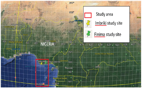
Figure 1: Map of Nigeria showing the study area within the Niger Delta.


Artisanal fisheries target primarily small coastal pelagic, such as the herring species called bonga (Ethmalosa fimbriata) and Sardinella. Most artisanal canoes range in size from 7-12m and are powered by 9.5 to 40 horsepower outboard engine. The gears used include gill nets, drift net, beach seines, large meshed shark drift nets, hooks and longlines Nedelec [3] & Nadreev [4]. The artisanal fisheries unintentionally catch marine mammals like killer whales, dolphins, and sperm whales. Rutherford [5].

Study Methods

The study was carried out for 2 years (January 2018 to December 2019) during the 575 fishing trips between 2017 and 2018, in two-landing sites (Imbikiri Bayelsa State and Finima River State) offshore the Niger Delta Nigeria. Migrant Ghanaians (8 fisher men) and 2 observers were employed to assist in sample collection. Fishing was done two to four times in a month with two or four operational net casts made per trip using drift gill nets. Cetacean by catch from catches were sorted and recorded afterwards. By catch parameters like length were determined by electronic digital caliper, measuring tape and digital thickness. Fishing depth ranges from 11-2460m.

Results

Cetacean abundance

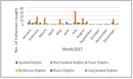
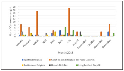
A total of 315 dolphins were captured by the drift net during the 575 fishing trips within the study period of 2017 and 2018, in the two-landing sites (Imbikiri in Bayelsa State and Finima int River state Nigeria). Figure 2 & 3 shows the monthly catches of the cetacean caught during the study period. The rainy season (April to August) recorded higher, than Cetacean catches during the dry season (January, February, March, October, November and December). Cetacean total catch peaked during the months of July and August (peak of wet season) in both 2017 and 2018. September and October (onset of dry season) no catch. While the months of November showed the least catches. A slight increase in total abundant catch was observed in the months of December to March (the real dry season).

Figure 2: Number of cetaceans caught throughout the period 2017.


Figure 3: Number of cetaceans caught throughout the period 2018.


Cetacean species abundance

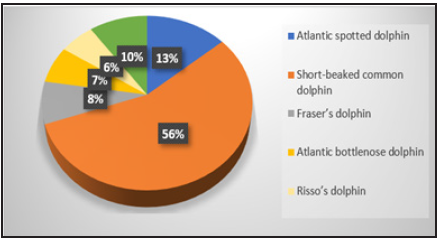
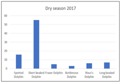
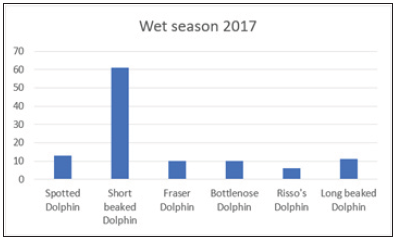
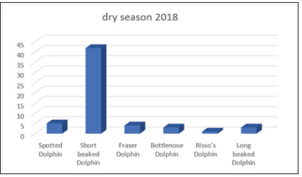
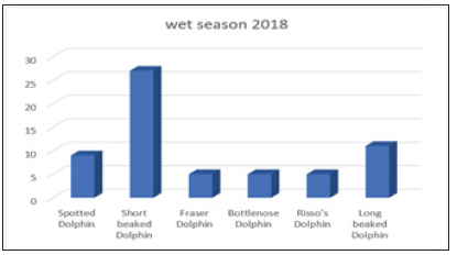
Throughout the study period from 2017/2018, the most abundance (56%) is short beaked dolphin, followed by spotted dolphin (13%), long beak dolphin was 10%, Freser dolphin 8%, bottle nose was 7% and the least abundance was Risso dolphin 6% (Figure 4). During the dry seasons of 2017 and 2018 (Figures 5-8), the short beaked dolphin showed the highest percentage caught while the least were the Bottle nose and Risso’s dolphin. The wet season of 2017 and 2018 also showed the short beaked dolphin specie as the highest while the least was the Risso dolphin.

Figure 4: The percentages of the six different species in the period of the two years study (2017/2018).


Figure 5: The percentages of the six different species in the period of the two years study (2017/2018).


Figure 6: Seasonal Cetacean species abundance of wet season in the month of April, May, June, July, August and September 2017.


Figure 7: Seasonal Cetacean species abundance of dry season in the month of January, February, March, October, November and December 2018.


Figure 8: Seasonal species abundance of cetacean during the wet season in the month of April, May, June, July, August and September 2018.


Discussion

Every year, at least 7.3 million tons of marine mammals are caught incidentally by drift net, purse seine (Rutherford-Fortunati, 2014). The seasonal abundance observed in this study could be due to a variety of reasons. This could be due to the availability of preys for the dolphins, the physicochemical parameters of the water, abundance of nutrients etc. The wet season when Cetaceans are most abundant could be attributed to higher primary productivity characteristics like increased dissolved oxygen, transparency, total dissolved solid, increased pH values and increased zooplankton and phytoplankton species Ndebele [6]. This conclusion however needs further studies to show probable links between species abundance and seasonal variability of physicochemical parameters and nutrients in the waters in the Niger Delta.

Conclusion

This study shows the abundance and species variability of cetacean bycatch in the Niger Delta fishing ground. It also provides information that may highlight the dangers inherent in fishing methods that could eventually lead to the mortality of some cetacean species. Further studies will however be required to assist in measures necessary for the biodiversity conservation of Cetaceans.

Acknowledgement

Our appreciation goes to Professor Larry Awosika for his critical review. We are also grateful to the management and staff of the Nigeria Institute for Oceanography and Marine Research for their assistance during the study. Lastly, we appreciate the local fishermen and others who provided local logistics in the study area.

References

  1. Whiteman A (1982) Nigeria: Its petroleum geology, resources and potential. 1: 238.
  2. Michael U, Koen V (2010) Initial evidence of dolphin takes in the Niger Delta Region and review of Nigerian Cetaceans. Wetland International Nigeria, Conservation and Research of West Africa Aquatic Mammals, Ghana, p. 1883.
  3. Nedelec C (1990) Definition and classification of fishing gear categories. Rome, p. 92.
  4. Nadreev N (1996) Handbook of fishing gear and it is rigging. Jerusalem Programm for Scientific Translations, p. 454.
  5. Rutherford F (2014) Commercial fishing: Methods behind the madness. Retrieved from Be fair to Be Vegan.
  6. Ndebele MR, Musil CF, Kaitt L (2012) Phytoplankton biomass and primary production dynamics in lake Kariba. In Lake and Reservoirs Research and Management 17(4): 274-289.
For more articles in ocean modelling journals
Please click on below link:  https://crimsonpublishers.com/eimbo/

Thursday, August 11, 2022

Modeling the Transmission Dynamics of LASSA Outbreaks in Nigeria States_Crimson Publishers

Modeling the Transmission Dynamics of LASSA Outbreaks in Nigeria States by Samuel Oluwafemi OYAMAKIN in Cohesive Journal of Microbiology & Infectious Disease_Infectious disease Journals


Abstract

A nonlinear deterministic model was considered to study the dynamics transmission and control of LASSA fever virus. The total population was divided into seven mutually exclusive classes between human and rodents as susceptible human, exposed humans, infected human, removed human, susceptible rodents, exposed rodents and infected rodents. Existence and uniqueness of the solution of the model were determined, the basic reproduction number is derived the model threshold parameter was examined using next-generation operator method. The existence of disease-free equilibrium point and endemic equilibrium point was carried out. The model result shows that diseases free equilibrium is local asymptotically stable at Ro< 1 and unstable at Ro> 1, the model is globally asymptotically stable. Sensitivity analysis of the model parameters was carried out in order to identify the most sensitive parameters on the disease transmission using data from the Nigeria States.

Keywords: Nigeria; Infectious diseases; LASSA Virus; Transmission dynamics; Disease free equilibrium

Introduction

This paper aimed at investigating LASSA through the dynamics of various factors that contribute in every stage of its transmission using a mathematical compartmental model based on data from the Nigerian states. LASSA Fever (LF) is an acute and sometimes severe viral hemorrhagic illness endemic in West Africa. The earliest cases of LASSA fever were thought to have occurred between 1920 and 1950, in Nigeria, Sierra Leone and Central African Republic and perhaps in other West African countries. However, the disease was first recognized in Nigeria in 1969, Ms. Lily (Penny) Pinneo (not real names), who was the first documented case of LF and from whom the first LASSA Fever Virus (LASV) was isolated in 1969. LASV is a single stranded RNA virus belonging to the Arenaviridae family of viruses. The virus is often named hemorrhagic fever virus because of the tendency to cause bleeding from body orifices. Sequencing of the small segment of the RNA of LASV has revealed the presence of four major lineages in West Africa with three in Nigeria (lineages I, II, and III) and one in the area comprising Ivory Coast, Sierra Leone, Liberia, and Guinea (lineage IV). Various viral strains have been associated with these major lineages with differences in their genome, serologic, and pathogenic characteristics. In Nigeria, mastomys natalensis have been identified by various names in some local languages including Eeku Asin (Yoruba), Jagba (Hausa), Nkapia or Nkakwu- (Igbo), and Isun (Kolokuma Ijaw) [1-10].

Infectious disease modelling

The central idea about transmission models, as opposed to statistical models, is a mechanistic description of the transmission of infection between two individuals. This mechanistic description makes it possible to describe the time evolution of an epidemic in mathematical terms and in this way connect the individual level process of transmission with a population level description of incidence and prevalence of an infectious disease. The rigorous mathematical way of formulating these dependencies leads to the necessity of analyzing all dynamic processes that contribute to disease transmission in much detail. Therefore, developing a mathematical model helps to focus thoughts on the essential processes involved in shaping the epidemiology of an infectious disease and to reveal the parameters that are most influential and amenable for control. Mathematical modeling is then also integrative in combining knowledge from very different disciplines like microbiology, social sciences, and clinical sciences. For many infections-such as influenza and smallpox-individuals can be categorized as either “susceptible,” “infected” or “recovered and immune.” The susceptible that are affected by an epidemic move through these stages of infection.

Compartmental models in epidemiology

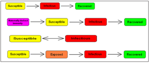
Compartmental models are a technique used to simplify the mathematical modelling of infectious disease. The population is divided into compartments, with the assumption that every individual in the same compartment has the same characteristics. Its origin is in the early 20th century, with an important early work being that of Kermack and McKendrick in 1927. The models are usually investigated through ordinary differential equations (which are deterministic), but can also be viewed in a stochastic framework, which is more realistic but also more complicated to analyze. Compartmental models may be used to predict properties of how a disease spreads, for example the prevalence (total number of infected) or the duration of an epidemic. Also, the model allows for understanding how different situations may affect the outcome of the epidemic, e.g., what the most efficient technique is for issuing a limited number of vaccines in a given population (Figure 1).

Figure 1: Common compartmental models in infectious disease modeling.


Framework for modeling for LASSA transmission dynamics

LASSA fever model presented in this study incorporates a model for the reservoir, which is Susceptible, exposed and infectious. This is done based on the assumption that susceptible rodent becomes exposed and then progress to infectious stage when they share unprotected storage of garbage, food stuff and water with infectious rodents or from inhalation of aerosols from urine and feces. These infected rats eventually tend to infect humans by direct contact or by inhalation of air follicles from dead infected rodents. It is also important to note that the existence of the exposed compartment is because of the incubation time that is the time it takes for an infected human to become infectious, which is twenty-one days for LASSA fever virus (Figure 2).

Figure 2: The SEIR model dynamics.


Methodology

LASSA fever model presented in this study incorporates diagnostic factor, γ(ai)α1(ai)Eh(t,ai) (since early diagnostic enables treatment, that is 6 days after infection) and vaccination parameter (νh)which are critical for prediction and the need of extensions to enhance their predictive power for decision support; also a subdivision of rodent compartment called susceptible, exposed and infectious rodent with discrete age structure denoted by Sr(t,ej), Er(t,ej) and Ir(t,ej) respectively. This is done based on the assumption that susceptible rodent becomes exposed and then progress to infectious stage when they share unprotected storage of garbage, food stuff and water with infectious rodents or from inhalation of aerosols from urine and faeces [11-18].

Parameters

  1. Recruitment term of humans Λh(ai)
  2. Effective transmission rate in susceptible humans by infected rodents ρ(ai)
  3. Effective transmission rate in susceptible humans by infected humans η(ai)
  4. Treatment rate of exposed humans α1(ai)
  5. Treatment rate of infected humans α2(ai)
  6. Rate of inoculation κ(ai)
  7. Progression rate of humans from the exposed state to the infectious state εh(ai)
  8. Diagnostic factor of exposed humans γ(ai)
  9. proportion of effective treatment of infected humans ψ(ai)
  10. Natural death rate of humans µh(ai)
  11. Progression rate of reservoirs from the exposed state to infectious state εr(ai)
  12. Effective transmission rate in susceptible reservoirs by infected reservoirs β(ej)
  13. Disease induced death rate of humans δh(ej)
  14. Mortality of reservoirs due to hunting δr(ej)
  15. Natural death rate reservoirs µr(ej)
  16. Recruitment term of reservoirs Λr(ej)
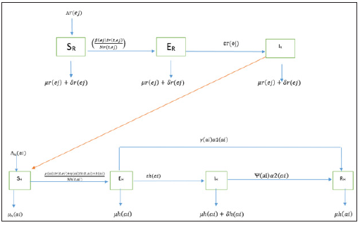
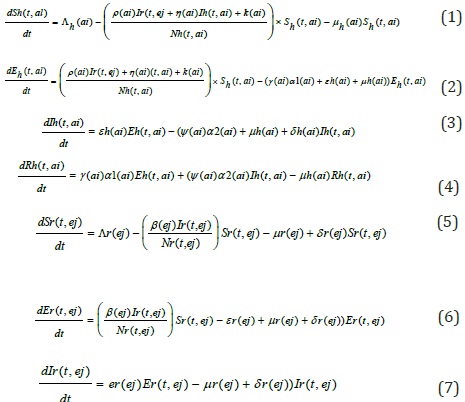
We assume that the population is closed i.e. it is assumed that individuals who recovered from LASSA fever will never go back to susceptible class again (they remain recovered for life). The assumptions above suggest that LASV can only be transmitted from: (i) human to human (ii) rodent to human (iii) rodent to rodent, and as a result, we have the following schematic diagram and system of nonlinear ordinary differential equation (Figure 3).

Figure 3: Schematic diagram of transmission dynamics of Lassa fever


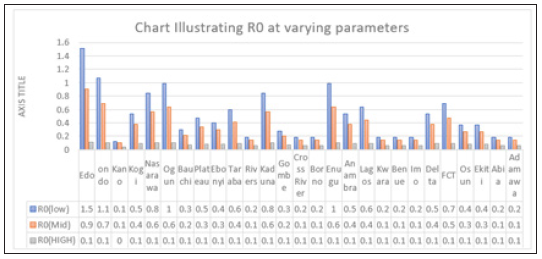
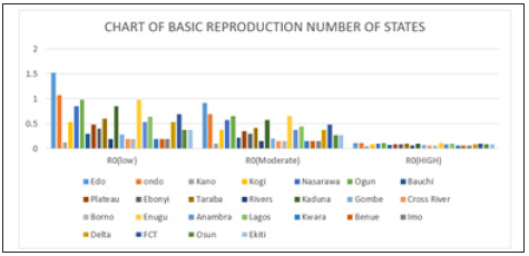
Figure 4: Multiple bar chart of the behavior of R0 in every state at varying intervention levels


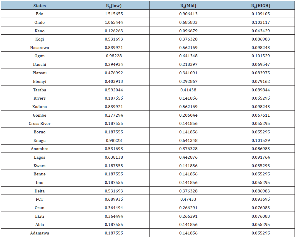
Figure 4 explains the behavior of R0 at varying intervention levels, explained earlier in Table 3. This implies that at low intervention, Edo and Ondo has their R0 greater than 1, which implies instability in the states. This could be accounted for by the proximity between the two states (Edo state shares boundary with Ondo state). Furthermore, R0 is stable at both moderate intervention and high intervention in other states. This means that at moderate intervention, Lassa fever dies off gradually over a while.

Attached separately to keep the formatting intact

Formulating the Compartment into Differential Equation

Existence and positivity of solution

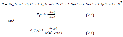
Since the formulated models monitor changes in the human and rodent populations; the parameters are assumed to ber nonnegative for all t ≥ 0, therefore the system of equations in (1) to (7) was analyzed within a feasible region R of biological interest. Theorem 1: The feasible region R defined by {Sh(t,ai),Eh(t,ai),Ih (t,ai),Rh (t,ai),Sr(t,ej),Er(t,ej),Ir(t,ej) ∈ R7 :

with initial conditions Sh(0,ai) ≥ 0,Eh(0,ai) ≥ 0,Ih(0,ai) ≥ 0,Rh(0,ai) ≥ 0,Sr(0,ej) ≥ 0,Er(0,ej) ≥ 0,Ir(0,ej) ≥ 0 is positive invariant for system (1) through (7).

Proof: If the total population size is given by

Nh(t, ai) = Sh(t, ai) + Eh(t, ai) + Ih(t, ai) + Rh(t, ai) and the total size of rodent population is

Nr(t, ej) = Sr(t, ej) + Er(t, ej) + Ir(t, ej)

Then one sees from (1) through (7) that

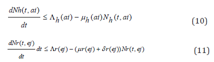
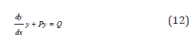
Considering the above equations as a linear differential we have that:

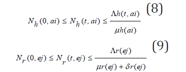
Solving the differential inequalities (8) and (9) one after the other gives

so that

this implies

and

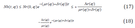
so that

this implies

Taking the limits as t → ∞ gives

and

Thus, the following feasible region

Stability of the disease-free equilibrium

A disease-free equilibrium point is a steady-state solution where there is no LASSA fever infection. Solving the system of equation (3.1)-(3.7) in the absence of disease, we obtain the following equilibrium point,

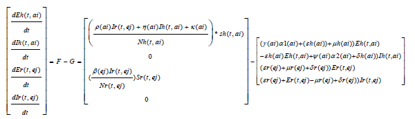
We obtain a basic reproduction number R0 by expressing (1) to (7) as the difference between the rate of new infections in each infected compartment denoted by F and the rate of transfer between each infected compartment denoted by G

The Jacobian matrices JF and JG of F and G are found about π0 Where, T = JFJ-1G From the Jacobian matrices JF and JG of F and G found about π0, we have R0, the maximum eigenvalue of T

Local stability of a disease-free equilibrium

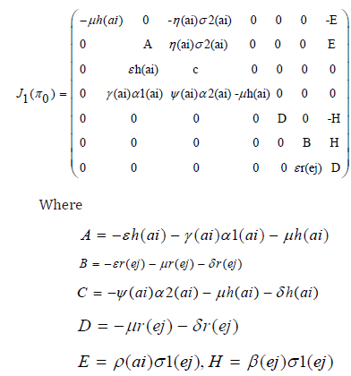
Theorem 2: The disease-free equilibrium for the system (1) - (7) is locally asymptotically stable if R0(ai)<1 and unstable if R0(ai) >1. Proof:. The Jacobian of system (1) through (7) evaluated at the disease-free equilibrium point is

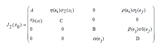
The first, fourth and fifth columns have diagonal entries, therefore the diagonal entries −µh(ai) twice and −µr(ej) − δj (ej) are three of the eigenvalues of the Jacobian matrix i.e λ1 =−µh(ai), λ2 = −µh(ai) and λ3 = −µr(ej) − δr(ej) . Thus, we find the remaining eigenvalues by excluding the corresponding columns and rows. The remaining eigenvalues are obtained from the submatrix

The eigenvalues of the matrix J2(π0) are the roots of the characteristic equation

Further simplification of c 0 in terms of R 0(a) gives

C0 = b1b2 (1 − R0 (a))

The Routh-Hurwitz criterion follows since all the roots of the polynomial (6) have negative real part if and only if the coefficient ci are positive and matrices Hi>0 for i = 0,1,2. From (7) it is shown that c1>0 and c2>0 since all b′ is are positive. Moreover, if R0(a)<1 it is follows from (8) that c0>0. All the eigenvalues of the Jacobian matrices J(π0) have negative real part when R0(a)<1 and the disease-free equilibrium point is locally asymptotically stable. However, when R0(a)>1, we see that c0<0 and by Descartes’ rule of signs ,there is exactly one sign change in the c2,c1,c0 of coefficient of the polynomial (6). So, there is one eigenvalue with the positive real part and the disease-free equilibrium point is unstable. At this juncture, we can now infer from the equation (8) that there exists a c0 such that and since R0(a) majorizes R0(ai) the system (1) - (7) is locally asymptotically stable if R0(ai)<1 and unstable if R0(ai)>1.

Existence of endemic equilibrium

We shall use the theorem below to show that the developed model (1) - (7) has an endemic equilibrium point Ee. The endemic equilibrium point is a positive steady state solution when the disease persists in the population. Theorem 3: LASSA fever model (1) - (7) has no endemic equilibrium when R0(a)<1 and a unique endemic equilibrium exist when R0(a)>1.

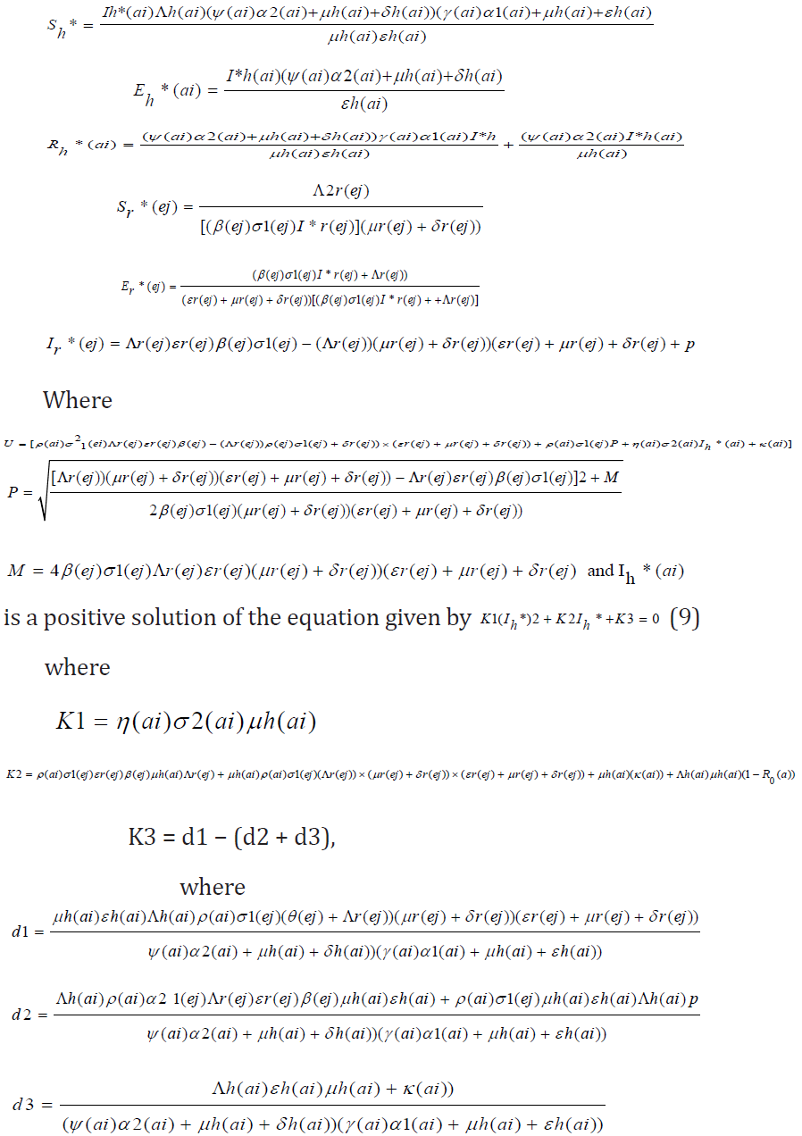
Proof: Let Ee = (sh*, Eh*, Ih*, Rh*, Sr*, Er*, Ir*, ) be a non-trivial equilibrium of system (1) - (7) i.e. all component of Ee are positive. At the steady state, we get

It is clearly seen that K1>0. When R0(a)<1 , one sees that K2>0. However, when R0(a)>1, then K2<0 and endemic equilibrium exists. Finally, K3<0 if d1<(d2 + d3).

Sensitivity analysis

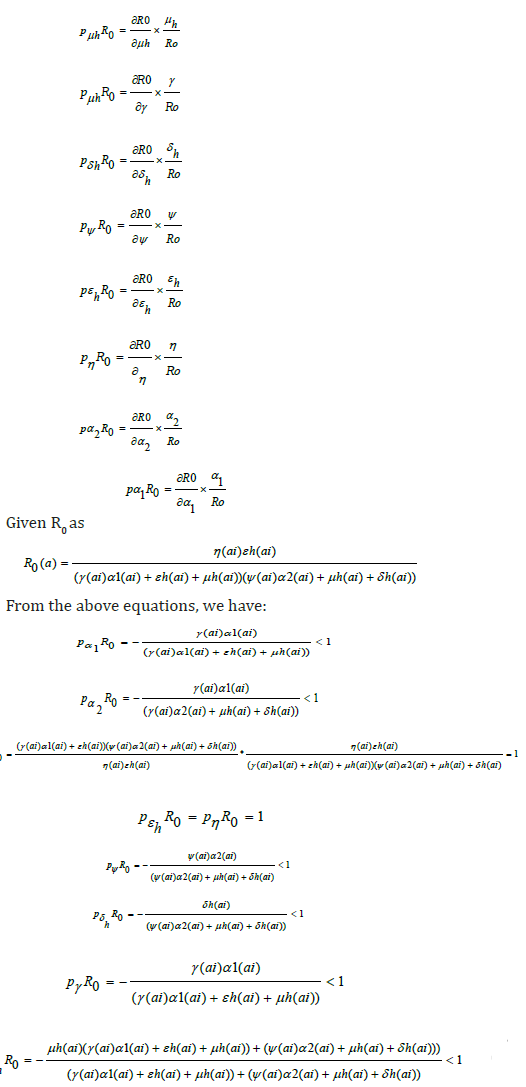
Sensitivity analysis is a statistical technique that can be applied to mathematical or computational models to provide insight on how uncertainty in the input variables affect the model outputs and which input variables tend to drive variation in the outputs. For models such as this infectious disease model, where the output is intended to inform decision-makers, uncertainty in the output can be disconcerting, as a single value is not given. However, the benefit is that a range of output values reveals a suite of possible model outcomes. It is sometimes of value to put in place robust policies that avoid the worst that can happen, although the policies may not achieve optimal results. If mitigating policies and alternatives are included and parameterized within the model, then sensitivity analysis can provide estimates of how responsive key output measures are with respect to these inputs. Sensitivity analysis tells us how important each parameter is to disease transmission. Such information is crucial not only for experimental design, but also to data assimilation and reduction of complex nonlinear models. Sensitivity analysis is commonly used to determine the robustness of model predictions to parameter values, since there are usually errors in data collection and presumed parameter values [19-24]. It is used to discover parameters that have a high impact on and should be targeted by intervention strategies. Sensitivity indices allow us to measure the relative change in a variable when a parameter changes. The normalized forward sensitivity index of a variable with respect to a parameter is the ratio of the relative change in the variable to the relative change in the parameter. When the variable is a differentiable function of the parameter, the sensitivity index may be alternatively defined using partial derivatives. Here, we present the sensitivity analysis of our Model using the formula given as

From the equations above, the most sensitive parameters are η(a1) and εh which are Effective transmission rate in susceptible humans by infected humans and Progression rate of humans from the exposed state to the infectious state respectively. This implies that an increase or decrease in these parameters will cause an instability in R0, as a result of its direct effect on R0.

Data presentation and analysis

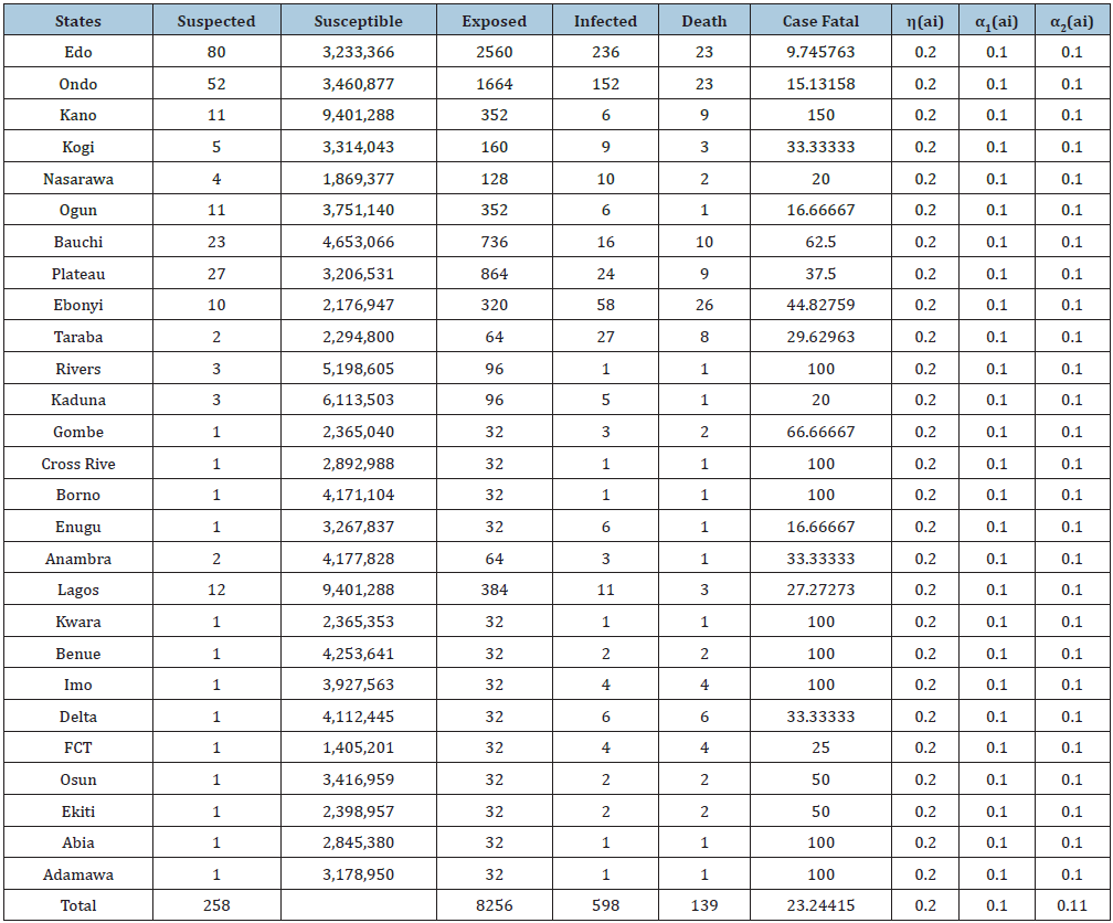
The data below was extracted from the National Center of Disease Control’s weekly Epidemiological Report on LF up till February 2019. Susceptible is the population of the state where LASSA fever has Incidence, Exposed is a percentage of the susceptible which is a function of effective transmission rate, Infected is a population reported confirmed, while death, is the death of confirmed cases (Figures 4-9); (Tables 1-4).

Figure 5: Multiple bar chart illustrating the behavior of R0 for every state at varying intervention levels.


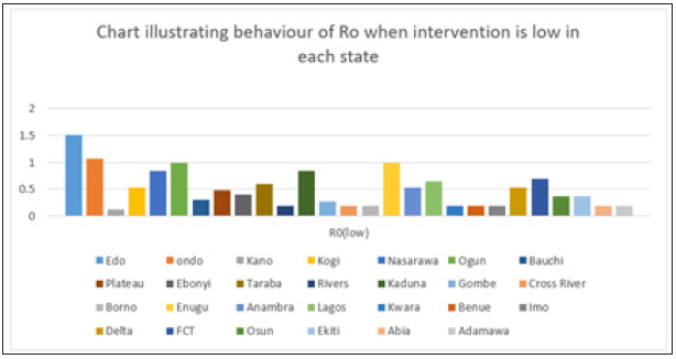
Figure 6: Simple bar chart illustrating R0 at low intervention for every state at varying intervention levels.


Considering Figure 6 above, at low intervention, Lassa fever becomes epidemic at Edo and Ondo state, while the Ro is less than 1for other states which implies stability.

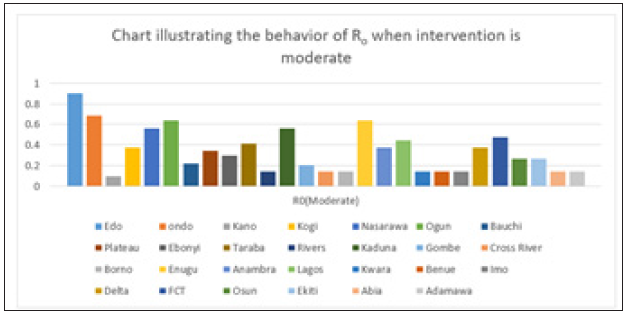
Figure 7: Simple bar chart illustrating R0 at moderate intervention for every state at varying intervention levels.


Considering Figure 7 above, at moderate intervention, Lassa fever becomes stable with the Ro is less than 1 for all states which implies stability.

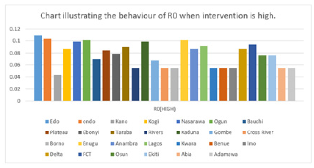
Figure 8: Simple bar chart illustrating R0 at maximum intervention for every state at varying intervention levels.


Considering Figure 8 above, at high intervention, Lassa fever becomes stable with the Ro as low as 0.11 for all states which implies stability.

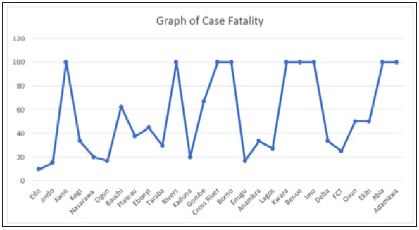
Figure 9: Case fatality of LASSA Across The Nigerian State.


Figure 9 explains case fatality as the percentage of those that are infected that eventually die. It is important to note that Kano, Rivers, Cross Rivers, Borno, Kwara, Benue, Imo, Abia and Adamawa, has 100% fatality with low case of incidence, while compared to states like Edo states,Ogun state, with higher cases of incedence have low case fatality. This could only be pointed to the factor of proximity to diagnostic centers, which exists only in three states in Nigeria. Literarily cases that occur in states closer to diagnostics centers gets fast intervention, as the disease can easily be diagnosed, and detected.

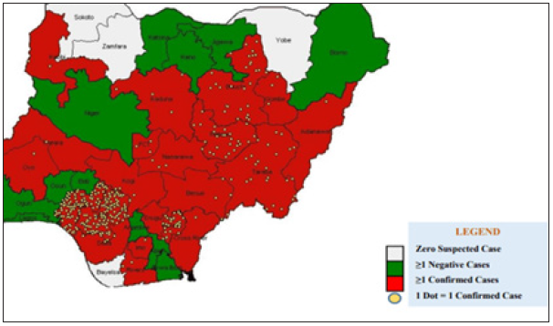
Figure 10: The distribution of confirmed Lassa fever cases in Nigeria.


Figure 10 above validates the first discovery that proximity to diagnostic center affect the case fatality of Lassa fever patient. Population of confirmed cases are more in Edo and Ondo state, which are closer to Lagos Diagnostic center, while places like Plateau, Bauchi, Gombe, are closer to irrua Diagnostic center at Jos.

Table 1: Extracted Data from the National Center of Disease Control (NCDC) and Susceptible is the population of the state where Lassa fever has Incidence, Exposed is a percentage of the susceptible which is a function of effective transmission rate, Infected is a population reported confirmed, while death, is the death of confirmed cases.


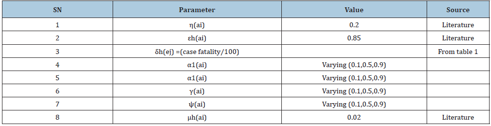
Table 2: Parameter structure used in the models and their sources and explains the values of parameters used in the estimation of R0.


Table 3: Basic Reproduction number obtained across the state at three different levels of parameter and iterates the estimates of R0 gotten from the data collected from NCDC and parameters in Table 2.


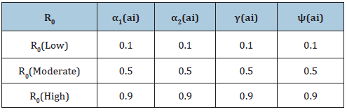
Table 4: Assumed Parameters of R0 at three different levels and above explains the varying degrees of interventions to reduce/stop Lassa fever which are captured by the above four parameters (Treatment rate of exposed humans α1(ai) Treatment rate of infected humans α2(ai), Diagnostic factor of exposed humans γ(ai) proportion of exective treatment of infected humans ψ(ai) )in all the compartments of the model. The values are varied at 0.1, which indicated little or no intervention, 0.5: which indicated medium intervention, while 0.9 indicates maximum intervention at these levels.


Summary

Considering the available data and the result of the sensitivity analysis, varying the values of four parameters which all signify effective and ineffective intervention, towards the eradication of the disease, showed a drastic reduction in R0, as the intervention level increased. It is also important to note that the lines with 100% fatality are states with first incidence of disease, especially places far away from diagnostic centers. This utmost depicts low diagnostics which is a function of proximity to test centers, which are in three places in Nigeria; National reference laboratory in Abuja, Lagos university teaching hospital (LUTH), Irrua specialist teaching hospital (ISTH), Institute of LASSA fever research and control (ILFRC), Irrua, Edo state, Nigeria.

Conclusion

The modification of the transmission dynamics between susceptible compartment to Exposed compartment by the removal of effective contact rate and interaction rate in reservoir has a positive effect on the R_0. At minimum intervention, Edo state and Ondo state has their R0>1 which implies instability. This means in time; LASSA fever will be epidemic in the two state. At mid intervention, R0<1 for all the states, which implies a steady state for the disease with minimal intervention. At maximum intervention R0 reduces to the barest below 0.2 in all states, which implies a steady state for the disease. Therefore, Proximity to diagnostic centers effective healthcare model has major part to play in eradicating LASSA fever in Nigeria.

References

  1. Polyanin D, Manzhirov AV (2007) Mathematics for Engineers and Scientists, USA.
  2. Safronetz D, Lopez JE, Sogoba, Traone SF, Raffel SJ, et al. (2010) Detection of Lassa virus. Mali Emerg Infect Dis 16(7): 1123-1126.
  3. Dietz K, Heesterbeek JA (2000) Bernoulli was ahead of modern epidemiology. Nature 408(6812): 513-514.
  4. Walker DH, Cormick JH, Johnson KM, Webb PB, Kombakano G, et al. (1982) Pathologic and virologic study of fatal Lassa fever in man. Am J Pathol 107(3): 349-356.
  5. Eames KT, Keeling MJ (2003) Contact tracing and disease control. Proc Biol Sci 270(1533): 2565-2571.
  6. Edmunds WJ, Medley GF, Nokes DJ (1999) Evaluating the cost-effectiveness of vaccination programmes: A dynamic perspective. Stat Med 18(23): 3263-3282.
  7. Ferguson NM, Cummings DA, Fraser C, Cajka JC, Cooley PC, et al. (2006) Strategies for mitigating an influenza pandemic. Nature 442(7101): 448-452.
  8. Ferguson NM, Keeling MJ, Edmunds WJ, Gani R, Grenfell BT, et al. (2003) Planning for smallpox outbreaks. Nature 425(6959): 681-685.
  9. Grundmann H, Hellriegel B (2006) Mathematical modelling: a tool for hospital infection control. Lancet Infect Dis 6(1): 39-45.
  10. Hadeler KP, Waldstatter R, Worz Busekros A (1988) Models for pair formation in bisexual populations. J Math Biol 26(6): 635-649.
  11. Hale JK (1969) Ordinary differential equations. USA.
  12. Hamer WH (1906) Epidemic disease in England the evidence of variability and persistency of type. Lancet 1: 733-739.
  13. Hethcote HW, Yorke JA (1984) Gonorrhea transmission dynamics and control. USA.
  14. Murray JD (2001) An introduction mathematical biology. USA.
  15. James TO, Abdulrahman S, Akinyemi S, Akinwale NI (2005) Dynamics transmission of Lassa fever disease. International Journal of Innovation and Research in Education Sciences 2(1): 2349-5219.
  16. Li MY, Mouldoney JS (1995) Global Stability for the SEIR model in epidemiology. Mathematical Biosciences 125(2): 155-164.
  17. Mouldoney JS (1990) Compound matrices and ordinary differential equations, Rocky Mountain. Journal of Mathematics 20: 857-872.
  18. Mccormick JB, Webb PA, Krebs JW, Johnson KM, Smith ES (1987) A prospective study of the epidemiology and ecology of lassa fever. Journal of Infectious Diseases 155: 437-444.
  19. Onuorah MO, Akinwande NI, Nasir MO, Ojo MS (2016) Sensitivity analysis of Lassa fever model. International Journal of Mathematics and Statistic Studies 4: 30-49.
  20. Ogbu OE, Ajuluchukwu CJ, Uneke CJ (2007) Lassa fever in West Africa sub-region: An overview. Journal of Vector Borne Diseases 44: 1-11.
  21. Tomori O, Fabiyi A, Sorungbe A, Smith A, Cormick JB (1998) Viral hemorrhagic fever antibodies in Nigeria populations, Am J Trop Med Hyg 38(2): 407-410.
  22. Okuonghae R, Okuonghae DA (2006) Mathematical model for Lassa fever. Journal of the Nigeria Association of the Mathematical Physics 10: 457-464.
  23. Venden D, Watmough J (2002) Reproduction numbers and subthreshold endemic equilibria for compartment models of disease transmission. Math 180: 29-48.
  24. Winn A, Walker (1975) The pathology of human Lassa fever, Bull. World Health Organ 52(4-6): 535-545.
For more articles in Infectious disease Journals
Please click on below link: https://crimsonpublishers.com/cjmi/

Wednesday, August 10, 2022

The Implementation of Animal Assisted Therapy Programs in Substance use Disorders_Crimson Publishers

The Implementation of Animal Assisted Therapy Programs in Substance use Disorders by Benali A in Psychology and Psychotherapy: Research Studys_Psychology open access jorunals


Abstract

Animal Assisted Therapy (AAT) is presented as a complementary program that, together with rehabilitation treatment, favors the clinical evolution of the patients who participate in it. The integration of AAT in the healthcare and health field has been increased in recent years; highlighting the growing interest in AAT programs, along with numerous studies that examine the benefits and efficacy of different therapeutic approaches with animals [1,2], after different and novel pilot experiences in substance use disorders [3], it seems that TAA is an intervention approach with utility in the rehabilitation processes of people with disorder substance use.

Keywords: Animal assisted therapy; Substance use disorders; Motivation; Therapeutic intervention

Introduction

AAT is a coordinated, programmed and structured therapeutic intervention, where the collaboration of an animal with specific qualities is included, as a characteristic element of the rehabilitation process. All the treatment must be designed, documented and evaluated by the personnel who carry out the program, together with the professionals of the resource in which it is applied [4-6]. The AAT differs from other animal activities (animal assisted activities, service animal programs and animal assisted education), primarily because the process of intervention of the animal and responsible specialist is supervised and subject to the action of the reference professional that includes such therapy within the rehabilitation treatment [4]. In recent years, the scientific community has been making an important effort to show how the use of animals in clinical intervention processes helps improve the physical, psychological and social health of patients. AAT is presented as adjunctive therapy to standard therapeutic programs, contributing, in a supplementary way, to achieving the objectives proposed in the treatment [6,7].

AAT Benefits

The performance of AAT programs are included within joint therapies that enhance the benefits achieved by the patient by participating in standard rehabilitation programs [8,9]. This is because interactions with the animal stimulate the release of oxytocin, improving patients' perceived mental health, behavior, therapeutic intervention and, finally, the relationship of well-being with the professionals of the resource [3,6]. In AAT the animal is integrated into psychotherapeutic treatment with the aim of promoting the increase of the physical, psychosocial and cognitive functions of the patients. The benefits provided are summarized in the following areas: identification and affective regulation (emotions, mood and motivation); achieving healthy self-esteem; social skills learning; improvement of the socialization process; and lastly, the stimulation of cognitive functions [3,5]. The benefits provided can be used in many healthcare and healthcare settings, favoring patients with various pathologies. Table 1 shows the main animals used as co-therapists in rehabilitation programs, along with the diversity of psychotherapeutic intervention settings where TAA programs are implemented [1].

Table 1: Animal species and areas of AAT intervention.

Source. Own elaboration


AAT

Main animals used Horses, dolphins, farm animals (cows, donkeys, goats, sheep, chickens, pigs, rabbits, ducks ...), cats, dogs and pets. Different areas of intervention Neurorehabilitation, education, mental health, geriatrics, gerontology, prisons, hospitals, addictions and oncology.

Implementation of AAT in substance use disorders

Once the benefits of incorporating TAA are known, the possible utility in the framework of rehabilitation in addictions is raised; where TAA programs complementary to the treatment program are temporarily integrated, mainly with the collaboration of therapy dogs, which participated as therapeutic tool [2]. The sessions usually are structured following a standard scheme: it began with an introduction in which the animal therapy technician begins the topic and objectives pursued in the session, and the patients participate in observation activities to learn the handling of the animal [8]. After this section, patients actively experiment and interact with the animal, working on the objectives set. Finally, it concludes with the evaluation of the participants accompanied by the therapist and the specialist in charge to assess the achievements obtained [5].

In the course of the successive evaluations, an improvement is sought in the therapeutic alliance with the professionals who are part of the therapeutic team of the resource; greater adherence to treatment, with the aim of increasing the motivation of patients who interacted with the animal [3,9]. Other perceived benefits, and no less important, should be presented, such as: the feeling of psychological well-being linked to a better management of anxiety, anxiety or craving, since interaction with the animal facilitates the identification and regulation of the present emotional state [2,7]. Finally, a second group of benefits is detected, which includes the improvement of perceived social support, the learning of coping strategies and the best adaptation to the residential resource [3,8].

Conclusion

It is valued that the implementation of AAT, as a complement to the therapy given in addiction rehabilitation centers, can be beneficial, since it favors the motivation, participation and involvement of patients [10]. AAT facilitates a proactive attitude in the patient, which as it acquires new benefits during the course of the sessions using interaction with the animal, manages to improve adherence to treatment, facilitates the therapeutic relationship, and ultimately, is a multisensory element of intervention that favors the evolution and achievement of psychotherapeutic treatment goals [2,11]. Thus, the need to continue intervening in this line is considered, in order to scientifically study the usefulness and validity of AAT as a therapeutic complement to the treatment of substance use disorders and behavioral addictions.

References

  1. Mauriño GPA, Luz AL, Babot M, Lacasa F, Álvarez Segura M (2017) La aplicación de la terapia asistida con animales en la salud mental infarto-juvenil. Revista de Psicología Universidad de Antioquia 9(2): 177-188.
  2. Casey J, Csiernik R, Knezevic D, Ebear J (2018) The impact of animal-assisted intervention on staff in a seniors residential care facility. International Journal of Mental Health and Addiction 16(5): 1238-1248.
  3. Montolio MM, Sancho Pelluz J (2019) Animal assisted therapy in the residential treatment of dual pathology. International Journal of Environmental Research and Public Health 17(1): 120.
  4. Huertas MC, Nieves ICR, Álvarez M (2014) Desarrollo de la terapia asistida por animales en la psicologí Informes Psicológicos 14(2): 125-144.
  5. Rubio RD, Loscertales AA, Obís MMB, Obís PB, López APS (2017) Terapia asistida por animales. Animal-supported therapy. Revista Española de Comunicación en Salud 8(2): 254-271.
  6. Uhlmann C, Nauss C, Worbs A, Pfund U, Schmid P (2019) Effects of an animal-assisted intervention on psychiatric in-patient addiction treatment-a pilot study. Fortschr Neurol Psychiatr 87(5): 305-311.
  7. Abreu T, Figueiredo AR (2015) Paws for help: Animal-assisted therapy. European Psychiatry 30(1): 1651.
  8. Álvarez AA, Tobio TG, Roca VO, Facal VCA, Moldes VI, et al. (2013) Efectos de la terapia asistida con animales en personas con daño cerebral o lesión medular. Trauma (Spain) 24(2): 109-116.
  9. Contalbrigo L, De Santis M, Toson M, Montanaro M, Farina L, et al. (2017) The efficacy of dog assisted therapy in detained drug users: A pilot study in an Italian attenuated custody institute. Int J Environ Res Public Health 14(7): 683-699.
  10. Klemetsen MG, Lindstrom TC (2017) Animal-assisted therapy in the treatment of substance use disorders: A systematic mixed methods review. Human-Animal Interaction Bulletin 5(2): 90-117.
  11. Duque CM, Lozano CA, Castro RH, Marín CA (2019) Educación asistida con perros. Inclusión & Desarrollo 6(2): 15-23.
For more articles in Psychology open access jorunals
Please click on below link: https://crimsonpublishers.com/pprs/

Tuesday, August 9, 2022

Developing Therapies that Prevent Progenitor Cell Migration for Preventing the Development and Further Growth of Endometriotic Lesions- Future Prospects-A Short Communication_Crimson Publishers

Developing Therapies that Prevent Progenitor Cell Migration for Preventing the Development and Further Growth of Endometriotic Lesions- Future Prospects-A Short Communication by Kulvinder Kochar Kaur in Perceptions in Reproductive Medicine_international peer reviewed open access journals


Short communication

Endometriosis represents a frequent as well as benign gynecological disease that is defined as the presence of endometrial tissue (glands as well as stroma) in sites external to the normal endometrium as well as rest of the parts of uterus [1]. About 10% of women of childbearing age have endometriosis with the rate escalating in recent years. There is an immediate need for getting efficacious therapies. Earlier we had reviewed on the etiopathogenesis ,management of Endometriosis as well as Adenomyosis in infertile patients ,effect on oocytes along with Endometriosis in adolescents and effectively managing both pain as well as infertility along with preventing risk of malignancy in endometriosis but still no permanent solution is coming hence the endeavor to keep looking for newer avenues [2-8].

It is known that Endometriosis is an estrogen associated disease ,along with neovascularization having a crucial part in its establishment ,propagation as well as recurrence .It has been shown that via mobilization as well as recruitment of endothelial progenitor cells (EPC’s) estrogen has significant role in neovascularization [9,10]. Further estrogen can induce the endometriotic lesions escalating [11,12]; (Figure 1 & 2), with some recent studies illustrating that therapy with estrogen facilitates vasculogenesis in endometriotic lesions via enhancing amounts of EPC’s [13]. Chemokine receptor 4(CXCR4) is a key manipulator regarding homing of EPC’s [14], while Li et al. [13], demonstrated that estrogen escalates the recruitment of EPC’s to the areas of damage through the stromal cell derived factor(SDF1)-CXCR4 pathways in myocardial infarction(MI) [11]. Certain studies have illustrated that estrogen increases the SDF1/CXCR12 synthesis in human Endometrial stromal cells (HESCs)in a time as well as dose-based fashion [11]. Nevertheless, the part as well as mode of SDF1/CXCR12 in endometriosis is not understood.

CK2 represents a serine/threonine kinase made up of 2 catalytic (α or α’)subunits as well as 2 regulatory (β) subunits, that assemble in the form of a tetramer.CK2 can phosphorylate substrates to control a lot of biological events [15]. Earlier Feng et al. [16], showed that CK2 controls angiogenesis of endometriotic lesions along with a protein kinase CK2 inhibitor inhibits the development of new blood vessel’s as well as growth of endometriotic lesions. Still it was not clear what is the insight in CK2 controls of EPC’s at the time of vasculogenesis in endometriotic lesions [17]. Endometriosis has been known to be crucially based on actively going angiogenesis and thus new blood vessels represent a mark of the lesions. Trying to get methods of blocking this angiogenesis as well as vasculogenesis might help in innovative ways of tackling endometriosis. Bone marrow (BM)-derived endothelial cells aid in vasculogenesis, that includes the ones for endometriosis as well as uterus at the time of pregnancy [18]. In view of their part in endometriosis lesions as well as recruitment during active disease, these endothelial progenitor cells are good targets for therapeutic intervention.

Figure 1: Courtesy ref no-12-Localization of human endometrial mesenchymal stem cells. (A-C) Immunofluorescence images of human endometrium showing perivascular identity of human eMSCs. a. Co-localization (white arrows) of CD146 and platelet-derived growth factor receptor beta (PDGF-Rβ) in pericytes of venules and possibly capillaries in the functionalism stroma. The x/z and y/z planes are shown on the far right and underneath the merged images demonstrating co-localization of the two surface markers. b. Perivascular SUSD2 expression (white arrows). c. ATP-binding cassette, subfamily G member 2 (ABCG2) and αSMA co-staining showing perivascular and endothelial identity of SP cells. The white dotted lines indicate the junction between the endometrium (en) and myometrium (my) and yellow dotted line indicates the luminal surface (lu) of the uterine epithelium. (D) Schematic showing location of stem/progenitor cells identified in the human endometrium. Epithelial progenitor cells are postulated to be a subpopulation of cells located in the base of the glands in the basalis, identified by SSEA-1. Sushi domain containing-2+ (SUSD2+) eMSCs are perivascular cells. eMSC co-expressing CD146 and PDGFRβ/CD140b are most likely pericytes, as they are located adjacent to endothelial cells in vessels (v) in both the basalis and the functionalism. SP cells are a heterogeneous population comprising CD31+ endothelial cells and CD140b+CD146+ pericytes. Scale bar in (A)=50μm. (A) Reprinted with permissions from Schwab and Gargett [12]. (C) Reprinted with permissions from Masuda et al. (2010). (D) Adapted from Gurung et al. (2015).


Figure 2: Courtesy ref no- 12-Schematic describing the hypothesis that endometrial stem/progenitor cells shed in neonatal uterine bleeding may play a role in early onset endometriosis. Neonatal uterine bleeding occurs in 5% of neonates. It is hypothesized that retrograde neonatal bleeding occurs because thick mucus obstructs the long neonatal cervix. Fragments of shed endometrial tissue are postulated to contain an endometrial epithelial progenitor cell (pink) and a perivascular MSC (pink) together with niche cells. These rapidly adhere to the neonatal mesothelium, invade and/or become contiguous with the mesothelial lining where they remain quiescent for ∼10 years. Rising estrogen (E2) levels associated with thelarche and menarche reactivate the stem/progenitor cells to initiate growth of endometriosis lesions on the surface of or below the peritoneal mesothelium, resulting in early onset endometriosis. Reprinted with permissions from Gargett et al. (2014).


Both in endometrial as well as decidual physiology as well as pathology like endometriosis bone marrow (BM)-derived cells have a proven part. Stem cells via BM engraft normal endomerium and help in this endometrium repair following injury. BM-derived stem cell become decidual cells along with support pregnancy playing a necessary role [19]. Thus these BM-derived stem cells get transported within the circulation along with getting recruited to along with engrafted in endomerium is a part of normal physiology, CXCL12 represent a strong chemoattractant which results in migration as well as engraftment of BM cells in normal endomerium as well as Endometriosis [19]. Taylor’s group earlier demonstrated that endometriosis synthesis large amounts of CXCL12 (also called stromal derived factor 1[SDF1] [20]. Estrogen activates the synthesis as well as liberation of CXCL12. BM cells which get moved to e endometriosis by the attraction of CXCL12 are a subpopulation of endothelial progenitor cells. These endothelial progenitor cells are circulating cells which stick to the endomerium at the areas of hypoxia or ischemia and aid in new vessel development. These stem cells getting incorporated is key for blood vessels growth as well as progression of endometriosis.


The control of CXCL12 expression by the protein kinase CK2 has been found by Zhao et al. [21]. The same group earlier demonstrated that CK2 controls angiogenesis within these Endometriosis lesions. Blockade of this enzyme avoids development of new blood vessels as well as growth of Endometriosis. This ubiquitous as well as constitutively active serine threonine kinase has been shown to be controlled via estrogen, whose activity thus escalates CXCL12. This CXCL12 attracts endothelial progenitor cells, by a mode by which these cells get homed to endometriosis lesions. By blocking this path at any junction blocks endothelial cell migration, depletion of estradiol(E2). On blocking CK2 action with its potent as well as selective inhibitor CX4945, or with usage of CXCL12 receptor antagonist AMD3100 all inhibit this signaling path. In the same way Taylor showed in animal models of endometriosis recently regarding AMD3100 inhibits stem cell migration to endometriosis and thus results in regression of these lesions [22]

Conclusion

Will therapy with agents that block CK2 have the capacity as treatment of endometriosis? CK2 represents a pleiotropic protein kinase, that controls lot of growth as well as survival paths in normal cells as well as disease. Though CK2 inhibitors CX4945 has been formed for treatment of cancers ,with its nonspecific effect on practically all tissues, risk of off target actions is high, and thus makes it unlikely to be used for endometriosis therapy .But this gives a lot of lessons regarding blockade of signaling path that ends in endothelial cell engraftment of endometriosis. Inhibition of CXCL12. Action by using AMD3100 holds promise and has realistic new way of Endometriosis therapy as shown in murine models of Endometriosis [23]. More significant, we get insight that estrogen depriving controls CK2, CXCL12 as well as endothelial cell migration. Progestin therapy does not add this benefit. The therapies used currently for decreasing Estrogen are Gn RH agonists, aromatase inhibitors. Though this suddenly is not possibly help in future forming agents which block CK2 to be included as agents for Endometriosis therapy regimens we have got insight that standard therapies will inhibit endothelial cell engraftment as well as lesion expansion. Considering non hormonal therapy CXC R4 antagonist have>specificity as well as less toxic as compared to CK2 inhibitors, but action on pregnancy is not well known. Decrease of estrogen will also block cell engraftment. Thus, E2 deprivation remains gold standard for Endometriosis therapy till now.

References

  1. Giudice LC (2010) Endometriosis. N Engl J Med 362: 2389-2398.
  2. Kulvinder Kochar K, Allahbadia GN, Singh M (2017) Current controversies and future challenges in endometriosis therapy. EC Gynaecology 1: 9-13.
  3. Kulvinder Kochar K, Allahbadia GN, Singh M (2016) An update on pathophysiology and medical management of endometriosis. Advances in Repr Sci 4(2): 53-73.
  4. Kulvinder Kochar K, Allahbadia GN, Singh M (2017) Meeting the challenges of endometriosis associated pain-newer options for future and research directions. BAOJ Bioinfo 1: 9.
  5. Kulvinder Kochar K, Allahbadia GN, Singh M (2019) An update on diagnosis and management of adolescent endometriosis -A short communication. Acta Scientific Paediatrics 2(5): 48-50.
  6. Kulvinder Kochar K, Allahbadia GN, Singh M (2019) Dissecting the reduced ovarian reserve seen with ovarian enometriomas with that caused by surgical excision of ovarian enometriomas-what comes first. Surg Med Open Acc J 2(2).
  7. Kulvinder Kochar Kaur, Allahbadia GN, Singh M (2020) How can we optimize the granulose cell function of oo¬cytes from an endometriosis patient to im¬prove the reproductive outcomes-A short communication?. Perception in Reproductive Medicine 3(4).
  8. Kulvinder Kochar Kaur, Allahbadia GN, Singh M (2020) An update on the various theories of the origin of uterine adenomyosis-A systematic review. Archives of Urology 3(1): 31-45.
  9. Streblow K, Werner N, Berweiler J, Link A, Dirnagl U, et al. (2003) Estrogen increases bone marrow derived endothelial progenitor cells production and diminishes neointima formation. Circulation 107(24): 3059-3065.
  10. Auth RJ, Nenicu A, Nickels RM, Menger MD, Laschke MW (2016) Estrogen stimulates homing of endothelial progenitor cells to endometriotic lesions. Am J Pathol 186: 2129-2142.
  11. Tsutsumi A, Okada H, Nakamoto N, Oka Moto R, Yasuda K, et al. (2011) Estrogen induces stromal cell derived factor-1(SDF1/CXCR12) production in human endometrial stromal cell: A possible role of endometrial epithelial cells. Fertil Steril 95: 444-447.
  12. Gargett CE, Schwab KE, Deane JA (2016) Endometrial stem/progenitor cells: The first ten years. Human Reprod Update 22(2): 137-163.
  13. Li H, Liu J, YeX, Zhang X, Wang Z, et al. (2013) 17β-Estradiol enhances the recruitment of bone marrow derived endothelial progenitor cells into infar cted myocardium by inducing CXCR4 expression. Int J Cardiol 162(2): 100-106.
  14. Sainz J, Sata M (2007) CXCR4 a key modulator of vascular progenitor cells. Arterioscler Thromb Vasc Biol 27: 263-265.
  15. Shehata M, Schnabi S, Demirtas D, Hilgarth M, Hilgarth M, et al. (2010) Reconstitution of PTEN activity by CK inhibitors and interference with the PIK3/Akt cascade counteract the antiapoptotic effect of human stromal cell in chronic lymphocytic leukaemia. Blood 116: 2513-2521.
  16. Feng D, Welker S, Korbel C, Auth RJ, Menger MD, et al. (2012) Protein kinase CK2 is a regulator of angiogenesis of Endometriotic lesions. Angiogenesis 15(2): 243-252.
  17. Montenarh M (2014) Protein kinase CK2 and angiogenesis. Adv Clin Exp Med 23(2): 153-158.
  18. Tal R, Dong D, Shaikh S, Manallapalli R, Taylor HS (2019) Bone marrow-derived endothelial progenitor cells contribute to vasculogenesis of pregnant mouse uterus. Biol Reprod 100(5): 1228-1237.
  19. Tal R, Shaikh S, Pallavi D, Tal A, Giraldez LF, et al. (2019) Adult bone marrow progenitors become decidual cells and contribute to embryo implantation and pregnancy. PLoS Biol 17(9): e3000421.
  20. Li F, Alderman MH, Tal A, Manallapalli R, Coolidge A, et al. (2018) Haematogenous dissemination of mesenchymal stem cells from Endometriosis. Stem Cells 36(6): 881-890.
  21. Zhao RM, Feng D, Zhuanf G, Liu Y, Chi S, et al. (2020) Protein kinase CK2 participates in estrogen mediated endothelial cell homing to Endometriosis lesions through stromal cells in an SDF1- CXC R4 dependent manner. Fertil Steril 113(5): 1067-1079.
  22. Plochino N, Manallapalli R, Shaikh S, Habata S, Tal A, et al. (2020) CXC R4 or CXC R7 antagonists treat endometriosis by reducing bone marrow cell trafficking. J Cell Mol Med 24: 2464-2474.
  23. Taylor HS (2020) Bone marrow in the pathophysiology of Endometriosis. Fertil Steril 113: 942-942.
For more articles in international peer reviewed open access journals
Please click on below link: https://crimsonpublishers.com/prm/

Visual-cognitive Skills and Physical Qualities in Elite Soccer: Practical Considerations for Training and Return-to-Play Protocols: Crimson Publishers

Visual-cognitive Skills and Physical Qualities in Elite Soccer: Practical Considerations for Training and Return-to-Play Protocols by Lukasz...