Crimson Publishers High Impact Journals

Thursday, March 24, 2022

Application of Physical Factors in Complex Etiopathogenetic Therapy Patients with Coronavirus-19_Crimson Publishers

Application of Physical Factors in Complex Etiopathogenetic Therapy Patients with Coronavirus-19 by Bitsoev Vladimir Dodtievich in Techniques in Neurosurgery & Neurology_Journal of Neurosurgery


Abstract

Fundamental scientific research of domestic and foreign scientists strongly suggests that the progress of medicine is impossible without the widespread use of modern physical factors in the diagnosis, prevention, treatment and rehabilitation of almost all nosologically forms of diseases from newborns to old age of patients. Each micro-and macro-organism has individual bioenergetic characteristics corresponding to its type, which is the main condition for normal life activity of the organism. In the case of “alien” bioenergetic characteristics, specific biological processes inherent in this organism are disrupted, which leads to its death [1]. The biopotential of each person is strictly individual in both normal and pathological conditions. The degree of deviation of the biopotential corresponds to the stage of development of the disease, i.e. the formation of intermediate States of the body with a violation of its supramolecular structures.

Keywords: Etiopathogenetic therapy; Supramolecular structures; Coronavirus; COVID-19

Introduction

Natural and preformed physical factors occupy an important place in medicine of the XXI century, as they are important for the diagnosis, treatment, prevention, and medical rehabilitation of patients.

Medicine should be developed according to this concept

"The level of medical excellence determines the progress of conservative treatment of patients with any pathology other than traumatology." Surgery should not be a factor in determining the progress of medicine. It is forced and will yield to the conservative method of treatment constantly with different speed and scale. Based on the progress of physical and technical Sciences, it is necessary to replace the concept of "mechanism of action of the physical factor" with "mechanism of interaction of the physical factor and the whole organism" [1-9]. Already from the moment of contact of the physical factor and the organism, intermediate States of both sides are formed with the release of energy (for the continuation of the cyclic process). In the Russian and world literature, we have not found scientific studies of their significance for the diagnosis, effectiveness and correction of treatment, prevention, and medical rehabilitation. This is especially important in Oncology for monitoring and managing the treatment process [3,5]. For the body, any external influence is a violation of its integrity and therefore the system of rapid disposal of it is instantly activated. Even your own blood outside the vascular bed is a foreign body for the body. That is, there is an instantaneous transfer of the work of all organs and systems of the body to an abnormal mode [4,5]. It should be noted that at the moment of development of medical science there are no methods for determining the time level of the cycle of each system at the time of exposure to the body of a medicinal substance, in its transition States at the supramolecular level and when all systems return to normal mode of life [10-15]. In this regard, it is difficult to imagine the advantage of a particular drug, and in particular for a particular patient. The lack of data in the chronological sequence of the drug in its path to achieving the goal calls into question the advantages of drug therapy over physical therapy.

It is known that " the interaction of a drug with the body is studied in two aspects: how it affects the body (pharmacodynamics) and what happens to it in the body (pharmacokinetics). Pharmacodynamics studies the localization, mechanism of action and pharmacological effects of medicinal substances. Pharmacokinetics studies the patterns of absorption, distribution, and elimination of medicinal substances in the human and animal bodies [2].  At the same time, it should be noted that the speed, scale, content, and time of formation of intermediate formations of a pharmacological drug in the body for each patient are strictly individual. All drugs in interaction with the body before their introduction into medical practice, in accordance with pharmacodynamics, pharmacokinetics should be studied by fast, harmless, and highly informative methods [5]. Compliance with this concept contributes to ensuring a high therapeutic effect, primary and secondary prevention of diseases, prevention of complications and side effects on the body.

This is not observed in medical and pharmaceutical practice due to the lack of research methods for pharmacological preparations at the supramolecular level [6]. The biopotential for each person is strictly individual both in norm and in pathology. In this regard, any nosology in everyone causes a deviation of its biopotential in accordance with the stage of development of the disease, i.e. the formation of intermediate States of the body with certain violations of its supramolecular structures. This, in turn, determines the clinical picture at the time of examination of the patient and is the leading condition for choosing the right treatment tactics for any specialist doctor, so that the regression of the disease is accompanied by the restoration of destroyed supramolecular structures, excluding new gross violations at any level of the entire body [6,7]. This concept is not the main principle for drug therapy, due to the lack of highly informative research methods: frequency, dose, mechanism of action of pharmacological drugs at the supramolecular level. It is known that the final characteristic of any drug at the supramolecular level is "energy", which is difficult to dose and regulate for therapeutic purposes. The impact of energy from any physical factor is dosed and regulated (physio dynamics) using nanotechnology and its path can be freely traced to each molecule of the whole organism (physio kinetics) without disrupting supramolecular structures, without negative consequences by means of a Nano sensor.(patents for inventions: Russia # 2675006, Germany # 20 2017 006 896.)

Modern pharmaceutical science does not have such a high level of control over the path of a medicinal substance in the body. "The conversion of the energy of photons, light particles, into electrical energy takes place in several stages," explains Professor Christoph Well, head of the IFG Institute. First, light is absorbed on the surface of the light-sensitive material [11]. Under the influence of the energy of photons of light, the electrons leave their places, leaving in their place electronic holes, with which they immediately form quasiparticles called polaritons. These polaritons exist only for a very short time, moving to the boundaries of the material, where they break up into electrons and holes, which continue to move further on their own. And the future fate of these charge carriers already depends on the nature of the light-sensitive material used" [11]. In this regard, any therapeutic effect on the body should be considered a trigger for restoring homeostasis, connecting its own internal systems. After studying the officially proposed and published Russian media medicines designed to combat coronavirus-19, we must admit not only their ineffectiveness, but sometimes even harm. Their original assumptions are clearly wrong. In fact, physiotherapy is seen as a more appropriate approach. It is based not only on the stated considerations, but also on half a century of experience in use.

Highly effective methods of physical therapy include

Light Therapy of the device "BIOPTRON"

Figure 1: Light Therapy (PVIP) performed by the “BIOPTRON-COMPACT” device-5cm.


Figure 2: Light Therapy (PVIP). The device “BIOPTRON PRO”-11cm.


Figure 3: Light therapy (PVIP) conducted by the Bioptron PRO apparatus-11cm.


Its spectral range-480-3400 nm-reproduces the dominant types of Solar radiation on Earth-visible and IR radiation, under the influence of which the body absorbs and uses radiant energy. Polychromatic visible and infrared polarized (PVIP) light activates the enzymes nicotinamidadenin-dinucleotide phosphate-oxidase (NADP-oxidase) and nucleotide containing biopteroflavoprotein-NO-synthesis, localized in the cell membrane and using the surrounding oxygen produce its active forms-superoxide anion, hydrogen peroxide, hydroxyl radical and nitric oxide (NO) [15]. They conduct a light signal from the surface of the irradiated cell to its nucleus, affecting specialized intracellular mechanisms for conducting the activation signal (protein phosphorylation, the state of calcium channels, the content of calcium in the cell, etc.). The enzymes responsible for the formation of ROS and NO, as themselves and intermediaries, are found in cells and tissues, in all types of white blood cells, platelets, endothelial and smooth muscle cells of blood vessels. It was found that nitric oxide-NO, is an important part of the mechanism of blood vessel dilation and platelet aggregation, without which phototherapy could hardly be highly effective [10,14]. After daily 5-10 irradiations, the number of mononuclear leukocytes-monocytes and lymphocytes-circulating in the blood increases by 14-17%. 30 minutes after the first exposure to PVIP light, Pro-inflammatory cytokines-tumor necrosis factor (TNF-α), interleukins-IL-6, IL-2, and IL-12-disappear from the circulating blood. So, at the initial increased content of TNF-α, it falls by 30 times, IL-8-by 4-6 times, IL-2-by 4-10 times and IL-12-by 12 times, by the end of the course [14,16]. Simultaneously, the plasma content of anti-inflammatory cytokines-IL-10 and transforming growth factor-TFR-β1 increases [14]. A feature of PVIP-light phototherapy is a rapid 6-fold increase in the blood of the most important immunomodulator-interferon-γ (IFN-γ). The most important function of this cytokine is to activate cellular immunity (the functional state of monocytes, macrophages, natural killers, and cytotoxic T-lymphocytes), which primarily increases the body's antiviral and antitumor resistance [13] (Figures 1-3).

Application of dry carbon dioxide baths "Reabox"

Dry carbon dioxide baths (SUV) - a method of percutaneous therapeutic action of carbon dioxide on a patient whose body is located up to the neck level in a specially equipped box. Application (SUV) "Reabox" provides non-invasive, i.e. does not violate the integrity of the skin, the introduction of carbon dioxide, which distinguishes this method from CO2 injections. Direct action of carbon dioxide on the respiratory center. The excitation of the respiratory center is not caused by carbonic acid itself, but by an increase in the concentration of hydrogen ions due to an increase in its content in the cells of the respiratory center. The specificity of carbonic acid as a respiratory center pathogen was revealed by the experiments of Frederick and Holden, who found that h+ and HCO3 ions pass poorly through the cell membrane, and undifferentiated carbonic acid passes well: undifferentiated H2CO3 diffuses into the cells of the nerve center, which dissociates already in the nerve cells, releasing the irritating H+ion. Faster diffusion into cells than other acids is a specific feature of carbonic acid, and this is associated with a stronger irritating effect on the respiratory center [12,15]. Hyperventilation for a short time (several tens of minutes) leads to death due to the loss of carbon dioxide by the body. Humoral regulation of respiration, the role of carbon dioxide, oxygen, and blood pH in this process. The main respiratory stimulant is CO2. Blood pH also plays an important role in the regulation of respiration. When the pH of arterial blood decreases in comparison with the normal level (7.4), lung ventilation increases, and when the pH increases above the norm, ventilation decreases. Increasing the content of CO2 in the blood stimulates respiration both by reducing the pH and directly by the action of CO2 itself [12,15]. The effect of CO2 and H+ ions on respiration is mediated mainly by their action on special structures of the brain stem that have chemosensitivity (Central chemoreceptors are part of the blood-brain barrier; low sensitivity threshold). It was found that a decrease in the pH of the cerebrospinal fluid by only 0.01 is accompanied by an increase in pulmonary ventilation by 4l/min. [15]

Lack of O2 can be a respiratory stimulant in the case of barbiturates as narcotic drugs, because in this case, the sensitivity of the respiratory center to CO2 is suppressed. Breathing pure oxygen (O2) in patients with reduced sensitivity to CO2 is very dangerous, because when the O2 voltage increases in the arterial blood the last respiratory stimulant (lack of O2) is eliminated in the blood and respiratory arrest may occur. In such cases, it is necessary to use an artificial respiration device (Table 1).

Table 1: The normal ranges of total CO2 in the blood should be as follows.


Extremely high frequency therapy (EHF) is the therapeutic use of millimeter-wave electromagnetic waves

The experience of using it for more than 30 years shows high efficiency in the treatment of a wide range of diseases, including cancer patients. Extremely high frequencies occupy the range of 30-300 GHz (the wavelength range is 10-1 mm). The peculiarity of this frequency range is that millimeter radiation of cosmic origin is almost absorbed by the earth's atmosphere, so the biological evolution of all living organisms took place with a very small natural EHF electromagnetic background. This, apparently, explains the active influence of low-intensity millimeter radiation on a person. The following wavelengths are most used in EHF therapy: 4.9mm (60.12 GHz), 5.6 mm (53.33 GHz), and 7.1 mm (42.19 GHz) [8]. Low-intensity millimeter radiation refers to non-ionizing radiation, i.e. it cannot have a destructive harmful effect on the biological tissues of the body, and therefore it is safe. A specific feature of EHF exposure is its normalizing nature, i.e. EHF radiation normalizes only the physiological parameters of a number of States of the body that deviate from it: it increases the values of reduced indicators and reduces the values of inflated values. Parameters that are normal do not respond to radiation of the body with a millimeter field. That is, the features of EHF therapy as non-invasiveness, lack of Allergy to EHF radiation, drug-free therapy contribute to the normalization of intracellular energy of any cell in the whole body (Figures 4-6).

Figure 4: Dry carbon dioxide baths using the “Reabox” device.


Figure 5: IR spectroscopy of the inner surface of the left elbow joint spectrum of the left elbow joint area before treatmen spectrum of the left elbow joint area after RayForce treatment


Multifunctional device for spot infrared and magnetic therapy for effective pain relief (Rayforce)

IR wavelength: 850nm. Magnet power: 1000 Gauss. Charging: from sunlight and artificial light. IR-therapy. It is proved that waves of different ranges affect the body in different layers and levels. IR radiation has the greatest penetration depth. In physiotherapy, waves are used in the range from 780 to 1400nm, i.e. short, penetrating the tissues to a depth of 5cm. The effect of IR radiation is aimed at accelerating the physical and chemical processes reactions: the processes of tissue repair and regeneration are stimulated, the vascular network expands, blood flow accelerates, cell growth increases, biologically active substances are produced, and white blood cells are sent to the lesion site. The reserve functions of the body are awakened. Permanent magnetic field (PMP) improves microcirculation, stimulates healing processes, activates immunological reactions, has anti-inflammatory and sedative effects [16,17]. Experimental studies were conducted at the scientific center for fiber optics (ncvo) of the Russian Academy of Sciences, Moscow. New non-toxic, non-hygroscopic silver halide light guides with low optical losses in a wide spectral range of 3-15 microns have been developed by the ntsvo staff, allowing to obtain skin spectra in vivo with a good signal-to-noise ratio even on the uncooled standard DNGS Fourier spectrometer Bruker. Results of RayForce treatment effectiveness based on IR spectroscopy data. This experiment confirmed the high therapeutic effectiveness of the RayForce device: For rice.5 IR spectroscopy shows the absence of pain in the left elbow joint after the treatment of RayForce device in the form of a complete restoration of the spectrum in the form and amplitude of light transmission, as well as correction of morphological changes in this area of exposure in the wavelength range of 970-1400nm. Based on the data in Fig.6. according to IR spectroscopy, there is reason to assert that the right elbow joint in the experiment was healthy and should be considered IR spectroscopy of the right elbow joint control.

Figure 6: IR spectroscopy of the inner surface of the right elbow joint. spectrum of the right elbow joint area before treatment spectrum of the right elbow joint area after RayForce


Symptoms of coronavirus, the first signs of COVID-19.

  1. Elevated temperature
  2. Dry cough
  3. Dyspnea
  4. The tightness in his chest
  5. Runny nose
  6. Weakness
  7. Undue fatigability

The lung involvement by coronavirus the structure of human lungs is cellular. They consist of tiny bubbles saturated with air - the alveoli. Each such alveola is surrounded by capillaries, through which, in fact, carbon dioxide is removed from the blood and oxygen is supplied. Red blood cells (red blood cells) are responsible for their transport through tissues and organs. Alveolar cells that participate in gas exchange are of two types: type I. Thin. Oxygen passes through them; Type II. A surfactant is isolated - a substance that envelops the alveola and protects it from damage. The coronavirus attacks mainly type II cells. Spiked proteins on its surface are bound by angiotensin converting enzyme 2 (APF2) on their surface. So, the virus "breaks" the protection and gets inside the cell, starting to replicate its RNA. The host cell soon dies, and the coronavirus spreads to neighboring cells and thus gradually affects the lungs. Naturally, our immune system does not sit still and actively produces macrophage cells. The result of this struggle is the death of the alveoli and a decrease in the turnover of gas exchange. This continues until the so-called alveolar collapse occurs and the acute respiratory distress syndrome begins. In severe inflammation, fluid rich in inflammatory proteins enters the bloodstream and spreads to other organs and tissues. This is how the systemic inflammatory response syndrome (SARS) develops, followed by septic shock and multiple organ failure. The incubation period of COVID-19 is from 2 to 14 days, during which time there are no symptoms. Preventive physical therapy should be started as soon as a coronavirus infection is suspected.

  1. light Therapy with the "BIOPTRON" device
  2. Impact on the face from 10 cm to 5 minutes
  3. On the neck area with 10 cm 5 minutes
  4. On the inter-scapular area with 5 cm 10 minutes
  5. On the plantar surfaces of the feet from 5 cm to 5 minutes
  6. On Palmar surfaces from 5 cm to 5 minutes
  7. Twice a day, daily, the course of treatment is 14 days
  8. Dry carbon dioxide baths "Reabox"
  9. CO2 concentration of 18-20%, 15 minutes, 1 time per day, treatment course of 14 days
  10. EHF therapy, the device " Yav”
  11. Paravertebral inter-scapular area with two points on both sides, the distance between the points is 10cm
  12. Epigastric region
  13. Paravertebral at the level of the VII cervical vertebra
  14. Emitters: 4.9 mm (60.12 GHz), 5.6 mm (53.33 GHz) and 7.1 mm (42.19 GHz) for 3 minutes per field 1 time per day, daily, 14 days of treatment

Therapeutic breathing exercises 5 minutes, 1 time a day, daily, 14 days of treatment. In case of complications, the physiotherapist plans of daily physiotherapy with hourly correction individually for each patient according to the state of the clinic. Physical therapy is performed in combination with medication. The proposed physiotherapy plan provides indications and contraindications at the supramolecular level, as well as for children from two years of age with a 50% reduction in the exposure time of each method, i.e. if an adult is 5 minutes, then children are 2.5-3 minutes. When establishing the diagnosis of "pneumonitis", the oxygen-helium mixture should be inhaled according to the developed method of academician A. G. Chuchalin from the AKGS-31 apparatus of the Minsk research Institute of radio materials.

Conclusion

Given the characteristics of coronavirus (COVID-19) infection, its differences from other known viruses are

  1. suddenness of occurrence
  2. high speed, scale and unhindered distribution
  3. program selectivity of penetration into the intracellular space
  4. Consistency of the striking nature at the supramolecular level of chronically weakened organs and systems, considering their biological age.

The wave origin of coronavirus-19, that is, based on quantum mechanics (entanglement), should be assumed. In this regard, it should be argued that a global solution to the problem of neutralizing the damaging insidious actions of the virus (COVID-19) is possible at the level of quantum physics and can only be done by a group of scientific physicists led by professor Lukin Mikhail Dmitrievich of the United States Harvard University. The physiotherapeutic methods proposed above for the prevention, treatment, and rehabilitation of patients with coronavirus (COVID-19) infection are also consistent with quantum physics, since their mechanism of action on the whole organism is identical to quantum touch, so they should be included in the program for combating coronavirus infection (COVID19).

References

  1. Aldersons AA (1985) Mechanism of electrodermal reactions. USA.
  2. Bertram GK (1998) Translation from english edited by Doctor of Medical Sciences, prof. Basic and clinical pharmacology 1(2).
  3. Bitsoev VD (2012) System of restorative treatment of degenerative dystrophic lesions of the spine: abstract of Doctor of Medical Sciences. Russia.
  4. Bitsoev VD (2015) New direction in the study of the role of interaction of the body and physical factors in complex therapy of patients. Russia.
  5. Bitsoev VD (2015) The Application of corrective underwater spinal traction with underwater phototherapy to the spine pathology. Clinical Medicine Research 6(4): 204-213.
  6. Bitsoev VD (2016) New approach in investigating the role of interaction between an organism and physical factors in complex patient therapy. Russia.
  7. Garyaev P (1995) Wave genetic code. USA.
  8. Devyatkov ND, Golant MB, Betsky OV (1987) Instructions for the use of a therapeutic installation for the treatment of millimeter-wave radiation of non-thermal intensity. USA.
  9. Jean ML (1998) Supramolecular chemistry. Nauka.
  10. Malkoch AV, Maydannik VG, Kurbanova EG (2000) The Physiological role of nitric oxide in the body (Part 1).
  11. Oldenburg Michael (2001) Photon up-conversion at crystalline organic-organic heterojunctions. Advanced Materials pp. 65.
  12. Ryabov GA, Azizov YM (2002) The Role of nitric oxide as a regulator of cellular processes in the formation of multiple organ failure. Educational and scientific center of the MC UD of the President of the Russian Federation, Moscow.
  13. Savinov VA, Samokhvalov EG (1997) Human energy System. Moscow.
  14. Samoylova KA (203) The Mechanism of anti-inflammatory, immunomodulating, wound healing and normalizing metabolism of the light of the device "BIOPTRON”, Moscow.
  15. Sosunov AA (2000) Nitric Oxide as an intercellular mediator". Saransk.
  16. Ulashchik VS (1994) Essays on general physiotherapy. pp. 198.
  17. https://www.orbitlonglife.ru/zhizn-bez-boli/orbit-rayforce/

For more articles in Journal of Neurosurgery
Please click on below link: https://crimsonpublishers.com/tnn/

Wednesday, March 23, 2022

Temperature Influence on the Process of the Gelation of the Engine Oil_Crimson Publishers

 Temperature Influence on the Process of the Gelation of the Engine Oil by Krzysztof Biernat in Progress in Petrochemical Science_Petrochemical Science journals


Abstract

The article discusses the process of gelation of engine oils and its influence on exploitative properties. The results of research on the propensity of engine oils for gelation at low and high temperatures are presented. The influence of various factors such as: base oil, vegetable oil, FAME and soot on the gelling process was determined. It was found that the cause of gelation of oils, depending on the operating conditions, oil components, may be several factors independent of each other. These factors are discussed in the article.

Keywords: Gel; Gelling process; Engine oil; Oil components; Vegetable oil; Fame; Soot

Introduction

In the 1990s, the problem of forming a gelatinous (gel) oil structure in the operation of vehicles powered by diesel oil appeared in Poland. However, the problem of gelation of motor oils was already noticed in Europe in the 1980s. At that time, it was assumed that this could be related to the introduction of a new category of engine oils, including the CD class according to API. The quality class CD introduced then a new generation of additives improving detergents and dispersants. In addition, the process of limiting sulfur content in diesel oil started, which was initially thought to have adversely affected the performance of engine oil. In Poland, the phenomenon of gelation was observed mainly in the late autumn and early spring periods, i.e. during periods in which large temperature fluctuations could occur. In winter or summer, when the temperature jumps were not large, this problem did not occur. In addition, the problem of gelation included engine oils of various quality and viscosity grades of known domestic and foreign producers. A characteristic feature of gelation is the rapid increase in kinematic viscosity at both 40°C and 100°C, the other parameters do not change much in relation to fresh oil.

The problem of gelation is a very unfavorable phenomenon for the engine. Gelled oil completely loses its operational functions, i.e. it does not provide lubrication, does not dissipate heat, does not neutralize acidic products originating, among others from incomplete burning of fuel, etc. which leads to engine seizure. Based on the collected literature on the operational problems of motor oils, including the phenomenon of gelation, several factors can be distinguished that can cause the formation of a gelatinous oil structure. The most important of them are:

  1. Soot content
  2. Base oil composition
  3. Content of vegetable oil or fatty acid methyl esters

But in addition to the factor that triggers the gelling process, temperature also plays a key role. Under the same atmospheric conditions, during the operation of vehicles, some oils show a greater tendency to form a gel while others are less and undoubtedly related to the temperature, not only of the engine itself, but also of the ambient temperature.

Gel Structure

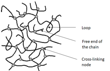
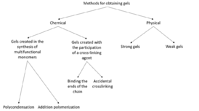
A gel is a colloidal system that consists of at least two components. Each of the components forms a separate continuous phase extending over the entire volume of the mixture. The gelling agent forms a rigid, branched, porous network spreading in the liquid constituting the second component of the gel, causing it to immobilize [1]. The gel structure consists of network nodes, loops, and free ends of chains. The diagram of the gel structure is shown in Figure 1 [2,3]. The division of gels is related to the type of interactions responsible for creating a rigid network, and thus with the type of gelling agent. There are chemical gels in which in the process of gel formation gelling agent molecules create covalent bonds between them, and physical gels in which the molecules of the gelling agent are bound by much weaker intermolecular interactions, mainly hydrogen bonds and electrostatic, dipoledipole interactions, van der Waals, as well as hydrophobic. The physical gels are thermally reversible, because the change of external parameters such as temperature and pressure lead to the disintegration of the gel network. In contrast, chemical gels are formed in the polycondensation, polymerization and copolymerization of multifunctional monomers or because of introducing a cross-linking agent into the polymer. The chemical (covalent) bonds formed in this way create nodes of the network [4]. The best-known methods for obtaining gels are shown in Figure 2 [4,5].

Figure 1: Gel structure scheme.


Figure 2: Chemical and physical methods of obtaining gels.


Gelation Temperature and Gelation Index

Multigrade engine oils have been introduced to reduce the viscosity of the oil at low temperatures to facilitate engine starting. With the easier start-up of the engine at low temperatures, there was a problem with the oil pumpability. This problem turned out to be much more serious because the lack of pumpability of the oil could lead to engine damage. In bench tests, it was found that at low temperatures we have two types of problems related to pumpability:

  1. High viscosity that limits flow
  2. Gelation of the oil subjected to long-term low temperature, which has been termed "air bonding", because the oil pump is aerated because of sucking oil from the oil sump along with the air [6].

Research on oil-air binding led to the development of a method for testing the tendency of oil to gel using a Brookfield scanning viscometer. The STB method as an international standard ASTM D 5133 allowed the study of the tendency of oil to form a gel. The method consists in heating the tested sample to 90 °C, and then slowly cooling it, while continuously measuring the viscosity [7]. Based on this method, you can designate:

  1. Gelation Temperature
  2. Gelation Index

The literature data show that the highest gelation and consequent air retention were found at temperatures much higher than those associated with limited flow, for the same class of SAE motor oils. Modern multi-grade motor oils require a maximum gelation index value of 12. This is the value at which the resulting gels in the engine oil do not endanger the engine's operation. The value of the gelation index above 12 indicates that the oil is highly prone to gel formation in the entire volume and can lead to engine failure [7].

Factors Influencing Oil Gelation Process at Low Temperatures

In determining how the lubricant will behave at low temperatures and the likelihood of gelation, several factors must be considered. These include the composition of the base oil, the paraffin content, the flow temperature, and the depth of the refined base oil. All these factors will have a significant impact on gelation and lubricant properties at low temperatures.

The effect of the composition and depth of the process of refining the base oil and the content of paraffins on the gelation process

Crude oil naturally contains some amounts of paraffinic compounds, which may affect the oil's tendency to form the gel at low temperatures. The more paraffins or paraffin compounds in the base oil, the greater the tendency of the oil to gel. Most paraffins can be removed by refining but cannot be removed completely. During the dewaxing process, most paraffins are reduced or the paraffin structure is converted into a different structure with better low temperature properties. Usually, the more refined base oil, the higher the viscosity index and better low temperature properties [8]. Most of the oils produced on mineral bases tend to thicken significantly, as paraffin particles crystallize at low temperatures. This, in turn, extends the time needed to pump oil through the entire engine and requires more work of the starter and the battery. Therefore, considering the tendency to gel, the best mineral bases are API Group II and III oils. The API II base oils are oils obtained through the use of hydrogen catalytic processes, such as hydrotreating and mild hydrocracking during processing of vacuum distillates of crude oil, whereas API Group III base oils are oils obtained through the use of deep hydrocracking processes and hydro isomerization of distillates vacuum and hydro isomerization of paraffins [9]. Therefore, API base oils of Group II and III have a lower volatility and lower flow temperatures.

Summarizing the above considerations, it can be concluded that the less paraffinic compounds and/or the deeper refining of the base oil, the lower the tendency to gel the finished oil at low temperatures.

Effect of flow temperature and oil solidification temperature on the gelation process

The flow temperature and freezing point are important physicochemical parameters that affect the gelling process, especially for oil that will run at low temperatures. The solidification temperature is the temperature at which the oil will not flow under the influence of gravity. When the oil is cooled, the paraffins remaining in the oil begin to crystallize and solidify with each other, so that the liquid becomes more and more dense until it stops flowing. Therefore, for vehicles that will operate at extremely low temperatures, one should choose lubricating oil that has the lowest freezing point to avoid problems due to reduced flow at low temperatures [8]. Lubricants operating at low temperatures should, in addition to a highly refined oil base and the appropriate package of refining additives, also contain a depressant, an additive that lowers the freezing point.

The tendency of engine oils to gelation at low temperatures

A study of the propensity of engine oils for gelation at low temperatures was carried out by scanning using a Brookfield viscometer (STB) in accordance with ASTM D 5133-15. The test was carried out in the temperature range from -5 °C to -40 °C at a shear rate of 0.2s-1 [10]. The measurements were carried out for currently produced fresh engine oils and with the addition of fatty acid methyl esters (FAME):

  1. LC - mineral engine oil SAE 15W/40 API SF/CD
  2. LM - mineral engine oil SAE 15W/40 API SJ/CF
  3. LSP - synthetic engine oil SAE 5W/40 API SN/CF

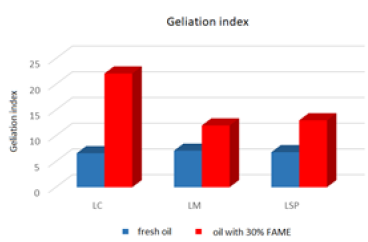
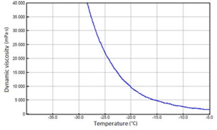
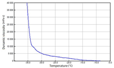
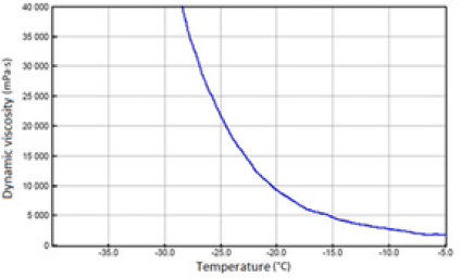
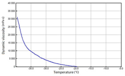
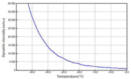
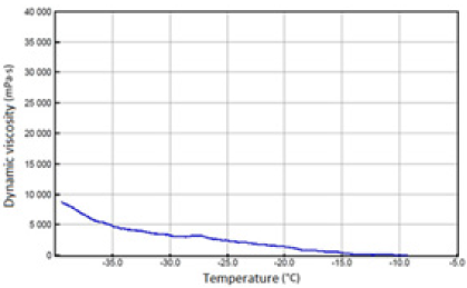
The above-mentioned engine oils and mixtures prepared with their use containing 30% FAME were tested for the determination of gelatinization by the STB method. Based on the dynamic viscosity dependence on the temperature, the gelation index was determined, which is shown in Figure 3. In Figures 4-9, the dependence of dynamic viscosity on the temperature of engine oils is shown. Dependence of dynamic viscosity on temperature. Based on the obtained test results, it can be concluded that commercial oils have a low tendency to gelation. The gelation index ranged from 6.6 to 7.1. This indicates that the oils tested belong to weakly gelling oils. However, the addition of 30% FAME caused an increase in the gelation index in relation to oils not containing FAME. This indicates that oils containing an addition of fatty acid methyl esters show a very high tendency to gelation. Based on these studies, it can be concluded that fatty acid methyl esters are a gelling agent. At elevated temperatures, ester bonds in FAME break, thanks to which free radicals are formed, which build into the structure of the oil creating permanent bonds in the nodes of the network. Under the influence of cooling, the oil begins to thicken to form a gel structure [11].

Figure 3: Gelation index of engine oils.


Figure 4: Fresh SAE 15W/40 API SF/CD oil.


Figure 5: SAE 15W/40 API SF/CD oil with 30% FAME.


Figure 6: Fresh SAE 15W/40 API SJ/CF oil.


Figure 7: SAE 15W/40 API SJ/CF oil with 30% FAME.


Figure 8: Fresh SAE 5W/40 API SN/CF oil.


Figure 9: SAE 5W/40 API SN/CF oil with 30% FAME.


Factors Influencing Oil Geliation Process at High Temperatures

The engine oil used on the one hand is exposed to high temperatures and products of incomplete fuel combustion that arise during operation of the engine. On the other hand, it can be diluted with fuel, which speeds up oil oxidation processes due to the content of the biocomponent in it. Additional hazards that may cause sudden, premature degradation of engine oils cause adverse interactions that may occur between oil and fuel, and usually resulting from the antagonistic effects of not always compatible components contained in both products [12]. The progressive oxidation leads to thickening of the oil and raising its acid number. Over time, the oil begins to become more and more saturated with soluble and insoluble oxidation products, which causes that the effectiveness of dispersing and detergent additives is insufficient to keep them suspended in oil, tar and sludge are deposited on engine components [13]. There are various toxic compounds and solid particles in the engine exhaust, the main ingredient being soot. Part of the soot does not burn and deposits on the walls of the cylinder. This soot along with the oil film is scraping through the rings and gets into the engine oil. Soot particles can also come from the destruction of oil [13].

The effect of soot on the process of gelling engine oil

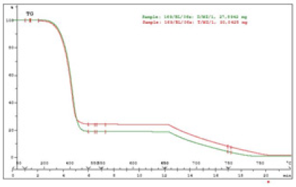
The main reason for oil thickening is usually its oxidation, thermal degradation, the presence of glycol or the accumulation of soot. The tests carried out at the Institute showed that gelled oils, apart from the increase in kinematic viscosity, had the basic parameters of physicochemical properties at the level of fresh oils. The only difference was the huge amount of soot in these oils, the content of which was 17 to 25% (w/w). The soot content tests were carried out using the thermogravimetric method in the atmosphere of nitrogen and air. Thermogravimetric curves for 2 gelled oils that contain soot are presented at Figure 10. Based on thermogravimetric curves, the soot content was calculated as the difference in weight loss of the sample in mass% after its decomposition under nitrogen and air [14].

Figure 10: Thermogravimetric curves TG oil samples D/MZ/1 and T/MZ/1.


In samples marked:

  1. D/MZ/1 (SAE 15W40 API CG-4), 17.6% (w/w) was found
  2. T/MZ /1 (SAE 10W40 API CF)22.2% (w/w) was found

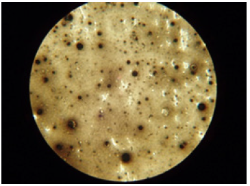
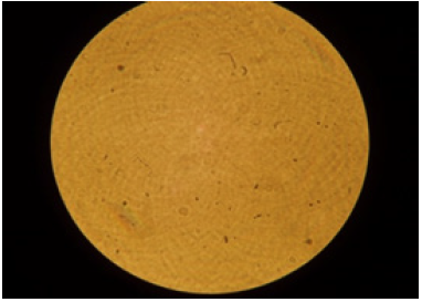
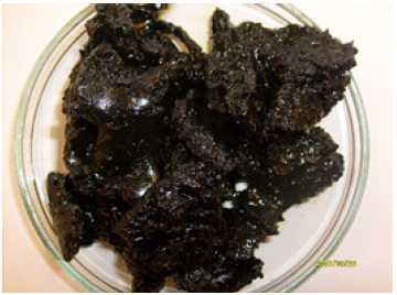
For the oil sample D/MZ/1, a photo was taken under an electron microscope to determine whether the soot formed agglomerates in the entire oil volume. The black spherical spots shown in Figure 11 show that the soot in the oil accumulated in the form of agglomerates. However, Figure 12 shows fresh oil that does not contain soot. It was observed that the gelling problem occurs when the engines are fed with low soot and PAH fuels. Soot gets partially into the engine oil from the exhaust gases in which it is dispersed. Dispersed small particles of soot do not affect the increase in the kinematic viscosity of the engine oil. However, soot particles tend to agglomerate, resulting in soot aggregates that lead to a rapid increase in viscosity and even gel. A characteristic feature of the gel resulting from agglomeration of soot in engine oil is a significant increase in viscosity of the oil at temperatures of 40 °C and 100 °C. Figure 13 shows gelled engine oil containing high amounts of soot [14].

Figure 11: Soot agglomerates in engine oil.


Figure 12: Fresh oil without soot.


Figure 13: Gelled oil containing soot.


The effect of vegetable oil additive on the process of gelling engine oil

The process of gelling engine oils can also be caused by the addition of vegetable oil. Under the influence of the high temperature prevailing in the engine polymerization of unsaturated triglyceride bonds and oxidation of vegetable oil occurs, which in consequence may lead to device failure, e.g. by gelling motor oil.

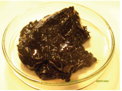
Each vegetable oil, regardless of its origin, easily undergoes aging. How fast this process takes place depends on the amount of unsaturated fatty acids. The fattier acids containing double and triple bonds, the aging process proceeds faster. The factors that accelerate the oxidation process are temperature and oxygen contained in the air. Motor oil containing vegetable oil has a characteristic floral aroma, and its gel is a yellow-brown color. The tests carried out for the content of fatty acids also confirmed the presence of triglycerides of fatty acids - the main components of vegetable oils. Figure 14 shows gelled engine oil containing added vegetable oil [14].

Figure 14: Gelled oil containing vegetable oil.


Conclusion

The literature data show that the temperature has a very large effect on the gelling process of engine oils, which was confirmed in laboratory tests. Based on the conducted tests it can be concluded that the reason for the process of gelling of engine oil:

  1. At low temperatures is:
    1. Paraffin, which cannot be completely removed from the base oil, because paraffin hydrocarbons are a component of crude oil. The gelation process is often associated with the precipitation of paraffins, especially at low temperatures
    2. The tendency to retain air and thus the increase in viscosity, which hindered the flow of oil,
    3. Too high flow and solidification temperature
    4. Addition of a gelling agent, e.g. fatty acid methyl esters or other compounds containing hydroxyl groups of the glycol type
    5. At high temperatures it is:
  2. Agglomeration of soot in engine oil
  3. Addition of vegetable oil

Most problems related to the thickening of engine oil, regardless of temperature, can be avoided by monitoring the degree of engine oil contamination during operation (especially gelling substances, sediments, solid particles, and soot). Routine oil analysis can help detect problems with oil gelling before significant engine damage occurs.

References

  1. Jaworska M, Vogt O (2013) Pochodne sorbitolu i celulozy jako czynniki żelujące. Chemik 67(3): 242-349.
  2. Przygocki W, Włochowicz A (2001) Fizyka polimeró PWN Wydawnictwo Naukowe Poland.
  3. Rubinstein M, Colby RH (2003) Polimer Physics, Oxford UK.
  4. Kavanagh Gaynor M, Simon B Ross Murphy (1998) Rheological characterization of polymer gels. Prog Polym Sci 23(3): 533-562.
  5. Caprotti R (1998) Harm free use of diesel additives. SAE Technical Paper Series 982569.
  6. http://zmienolej.pl/pl/blog/kompendium-wiedzy/jak-ocenic-jakosc-oleju-silnikowego
  7. Selby TW, Müller GC (2008) Thermal history of the engine oil and its effects on low temperature pumpability and gelation formation, paper, SAE International.
  8. http://www.machinerylubrication.com/Read/30447/combat-oil-gelation
  9. Beran E (2008) Wpływ budowy chemicznej bazowych olejów smarowychna ich biodegradowalność I wybrane właściwości eksploatacyjne. Prace Naukowe Wydziału Chemicznego Politechniki Wrocławskiej Monografie 5(5): 178.
  10. ASTM D 5133-15-Standard test method for low temperature, low shear rate, viscosity/temperature dependence of lubricating oils using a temperature- scanning technique.
  11. Ziółkowska M, Biernat K (2018) Skłonność olejów silnikowych do tworzenia żelu w niskich temperaturach. Przemysł Chemiczny 97(6): 938-941.
  12. Urzędowska W, Stępień Z (2012) Wybrane zagadnienia dotyczące zmian właściwości silnikowego oleju smarowego w eksploatacji. Nafta-Gaz 68(12): 1102-1110.
  13. Jakóbiec J, Budzik G (2007) Czynniki mające wpływ na stopień degradacji oleju silnikowego wokresie eksploatacji. Archiwum Motoryzacji 3: 209-216.
  14. Ziółkowska M (2009) Proces żelowania olejów silnikowych w czasie eksploatacji pojazdów zasilanych olejem napędowym. Przemysł Chemiczny 88(3): 268-271.

For more articles in Petrochemical Science journals
Please click on below link: https://crimsonpublishers.com/pps/

Tuesday, March 22, 2022

The Maze that is Not Maze: It is Not Filling the Gap!_Crimson Publishers

 The Maze that is Not Maze: It is Not Filling the Gap! by Ovidio A Garcia Villarreal in Surgical Medicine Open Access Journal_Journal of Surgery and Medicine


Abstract

When properly performed, the true Maze procedure is highly effective to eliminate any type of atrial fibrillation or flutter. Since non-paroxysmal atrial fibrillation is sustained by macro-reentrant circuits located anywhere in both atria, a full bi-atrial lesion pattern is the best way to approach all possible causes in one single step. As we move into less invasive surgical techniques, we must more clearly maintain the scope of the Maze procedure. Difficult approaches are oftentimes the source for incomplete, unsuccessful, and frustrated forms of the Maze. The two only alternative energy sources enabling full transmural lesions in the atrial myocardium are bipolar radiofrequency as well as cryothermia. Standardization of the Maze procedure as a full bi-atrial lesion pattern as well as the proper selection in the alternative energy source is the keystone to get the best outcome.

Keywords: Atria; Atrial fibrillation; Bipolar radiofrequency; Cryolesion; Maze procedure

Introduction

On October 13, 1986, the formal initiation of surgery for Atrial fibrillation in a human took place [1]. Although it was not really a Maze procedure, as we know it today, this was the predecessor of the true Maze. However, the first Maze I procedure, recognized as such, it took place on September 25, 1987. Since then, the Maze procedure has evolved considerably. However, the secret for the Maze’s success has consisted in knowing how to preserve its essence as a “full bi-atrial lesion pattern” [2]. We discuss in this paper the essence and basic aspects of the Atrial Fibrillation (AF), as well as the principles about how the Maze procedure works. This will enable us to get a general overview in order to understand the current approaches of the Maze procedure and how effective they are.

Basic Considerations

AF is a reentry arrhythmia. Nevertheless, two basic and general conditions are necessary to produce AF. The first one is the triggers located mainly up to 90% of times into and around the Pulmonary Veins (PV), which are generating AF [3]. The second required condition is at least one macro re-entrant circuit sustaining the AF [3]. The first one leads the second one. Under normal conditions, when the atria are not electrically remodeled, the macro re-entrant circuit cannot be sustained for a long time. These circuits fade away by themselves. In this way, PV triggers can initiate a new macro re-entrant circuit over and over again. This intermittent behavior alternating with normal periods of sinus rhythm is the so-called “paroxysmal AF”. On the contrary, once these macro re-entrant circuits persist in the atria for more than seven days, the atrial myocardium becomes electrically remodeled, and the circuits can stay for longer in a continuous way to the point that PV triggers are no needed anymore to produce AF. Now, the AF becomes self-sustained by means of these macro re-entrant circuits located anywhere in the both atria. This continuous arrhythmia is known as “non-paroxysmal AF” (persistent, long-standing persistent, or permanent AF) [3].

Getting the hang of the maze procedure

One of the most important concepts discovered by Dr. Cox and his working group has been the minimum diameter size of the macro re-entrant circuit to produce AF of 6 cm or larger than this in the human atria. In such a way that if two separate incisions or burn lines are placed in parallel to less than 6 cm far away one from the other, so there will not be enough space for the development of the macro re-entrant circuits [4]. Moving from the theory into the practice, in the setting of the paroxysmal AF, PV isolation is highly effective to eliminate AF. Triggers located into and around the PV are isolated in 90% of times [5]. Nonetheless, when approaching any form of non-paroxysmal AF (persistent, long-standing persistent) PV isolation is not enough to eliminate AF [6,7]. A much more complete procedure addressing not only the PV isolation but also all possible involved macro re-entrant circuits is required for this purpose. The Maze procedure provides an excellent option. This surgical procedure offers an outstanding solution thanks to its full bi-atrial lesion pattern approaching all possible macro re-entrant circuits in one-go, in one-single step [8].

The problem with the maze that is not a true maze

In the light of the foregoing, we have to be very precise and extra cautious when selecting the lesion set pattern for surgical ablation of AF. All possible macro re-entrant circuits are located anywhere in both atria. They are not exclusive for the left atrium. Said that, in cases of non-paroxysmal AF, it is easy to understand that the only option to surgically treat this type of AF is by using a pattern as comprehensive as possible. If any incomplete procedure is utilized, effectiveness drops off dramatically. So, what does it happen when analyzing the meta-analyses and systematic reviews regarding the effectiveness of the maze procedure? Why does it seem not to be so effective? Well, to answer this question, we must tackle two extremely important issues. The first one is linked to the lack of standardization for the maze procedure [9,10]. There is actually a lack of standardization in the way of selecting the pattern for the maze procedure. From my view, according to the concept explained above, the best way to use the Maze procedure is as a full bi-atrial lesion pattern. This is the only possible way to break up all possible macro re-entrant circuits in both atria. The author has already made quite clear that the pulmonary vein and antrum isolation is not sufficient for this purpose.
In order to prove this hypothesis, the author isolated by surgical means the PV in one hundred patients in a way that no room for doubt about the transmurally of the isolation. In this series, only 30 % of patients remained in sinus rhythm after a 5-year follow-up [6]. The second one arises when gathering some articles meeting special criteria to be analyzed as part of a meta-analysis. It is quite frequent to fall into the error of accounting as Maze some cases that are not a true Maze. The less complete the maze, the worse the final outcome. It has given rise to the wrong perception that the Maze is not so effective when analyzing as a whole regardless of whether or not the procedure it is complete. By way of explanation, after analyzing all studies included in a meta-analysis about surgical ablation for AF, when analyzing the bi-atrial lesion set group, only one author out of eight (24 of 238 total patients) had correctly performed the bi-atrial Maze procedure. All other authors had used monopolar RF, electrocautery energy, incomplete PV box lesion set, or not so clear if monopolar or bipolar the procedure [11]. Another example is the meta-analysis by Li et al. [12]. They conclude that there is no difference in sinus rhythm conversion between left-sided or biatrial surgical ablation groups. However, it is worth stating out that when analyzing the normal sinus rhythm recovery beyond 1 year, for example, out of 7 studies matching criteria, in only 3 of them were correctly performed the Maze procedure.
A wide of variety of lesion sets as well as in the use of alternative energy sources other than bipolar RF or cryothermia are the most common errors. So, the final understanding must be the need of a real standardization to perform the Maze procedure. In one hand, the need of always using a full bi-atrial lesion pattern, regardless the underlying pathology originating the AF [9,10]. On the other hand, it is widely accepted that there are only two alternative energies to get a full and consistent transmurality lesions on the atria, namely, cryolesion and bipolar radiofrequency ablation [13].

Less invasive techniques are not glittering at all

In an attempt to make simpler the original maze procedure, some changes and adjustments have been made in order to achieve desired results. One of them, the main one, has been to replace the surgical incisions by burn lines using the aforementioned energies. This is the so-called Cox-maze IV procedure. This took place in 2002, by using monopolar RF as alternative energy source to surgically treat AF. Unfortunately, transmural lesions on the atrial myocardium are not consistent when using this type of energy. After FDA cleared the bipolar clamp for surgical ablation of AF in 2003, industry launched the widespread use of this device in 2004 [14]. It was quickly concluded that only cryolesion and bipolar radiofrequency caused effective transmurality lesions on the atria [13]. Since then, the scope of the maze procedure has been worldwide extended. Despite the original Cox-maze III procedure remains as the gold standard [15], the Cox-maze IV procedure offers a reasonable option [16]. Still, great invasiveness has been indicated as the chief reason to be reluctant to use it.

The Maze procedure has moved toward less invasive techniques as mini-invasive approaches. In this setting, we can find three main groups, viz, the Cox-maze IV endocardial approach through a right mini-thoracotomy (on pump), the epicardial total thoracoscopic approach (off-pump), and the combined epicardial-endocardial approach, so-called hybrid Maze procedure (off-pump) [17]. The endocardial maze procedure has an effectiveness similar than the Cox-maze IV procedure because both of them are following the full bi-atrial lesion pattern by using just bipolar RF ablation as well as cryolesion over such a very critical areas (coronary sinus, mitral and tricuspid annuli) [16]. The epicardial approach is carried out by a bilateral thoracoscopy. It is an off-pump procedure. Nevertheless, bipolar RF has just a limited application, as the bipolar clamp is unable to reach all the necessary critical areas. A so-called transpolar pen is preferred to burn the mitral line, as well as the superior and inferior connecting lines at the box lesion in the left atrium [18]. Given the fact that the transpolar pen is not a true bipolar device, the penetration range of burn is wall-thickness dependent. In more than 6.7 mm of wall thickness, as often found, transmurally becomes incomplete [19]. But on the other hand, catheter-based techniques by electrophysiologists are highly effective to find some electric gaps on the burn lines. When combined epicardial and endocardial approaches (off-pump), the new hybrid maze procedure comes up [20]. Nonetheless, it is fairly worth highlighting this hybrid procedure can become as highly effective as the Cox maze IV procedure if adhered to the original full bi-atrial lesion pattern of the original maze procedure [20]. The major challenge has been the education in understanding the importance of using as much as possible the full bi-atrial lesion pattern in this kind of techniques. By way of explanation, the main source for failures after hybrid maze procedure has been the lack of completeness in the lesion pattern set [17,20]. However, when properly performed, the true hybrid maze IV procedure can become the gold standard for the stand-alone persistent AF once the catheter-based techniques have failed (class of recommendation IIA, level of evidence B) [21].

Conclusion

In conclusion, most of the time, failures after the maze procedure are the result of technical errors in the selection of an incomplete lesion pattern, as well as in the selection and way of using the alternative energy sources. As a general rule, the more complete the lesion pattern, the higher the success after Maze procedure, irrespective the percutaneous or surgical approach. Special interest must be focused on performing the full bi-atrial lesion pattern as much as possible, regardless the underlying disease causing the AF.

References

  1. Cox JL (2011) The first maze procedure. J Thorac Cardiovasc Surg 141(5): 1093-1097.
  2. García VOA (2020) Why maze procedure is so effective. Let’s get straight down to business. Cir Card Mex 5(1): 6-8.
  3. Cox JL (2003) Atrial fibrillation I: A new classification system. J Thorac Cardiovasc Surg 126(6): 1686-1692.
  4. Cox JL, Boineau JP, Schuessler RB, Kater KM, Ferguson TB, et al. (1995) Electrophysiologic basis, surgical development, and clinical results of the maze procedure for atrial flutter and atrial fibrillation. Adv Card Surg 6: 1-67.
  5. Haïssaguerre M, Jaïs P, Shah DC, Takahashi A, Hocini M, et al. (1998) Spontaneous initiation of atrial fibrillation by ectopic beats originating in the pulmonary veins. N Engl J Med 339(10): 659- 666.
  6. Garcia VOA (2015) Pulmonary vein isolation for persistent atrial fibrillation. Long-term results. Asian Cardiovasc Thorac Ann 23(6): 665-669.
  7. Garcia VOA (2016) eComment. Pulmonary vein isolation is not enough to treat long-standing persistent atrial fibrillation. Interact Cardiovasc Thorac Surg 23(2): 299.
  8. Cox JL (1991) The surgical treatment of atrial fibrillation. IV. surgical technique. J Thorac Cardiovasc Surg 101(4): 584-592.
  9. García VOA (2018) Standardization in maze procedure: A step towards a better future. J Thorac Dis 10(Suppl 33): S3887-S3889.
  10. Ad N (2015) The importance of standardization in surgical ablation for atrial fibrillation. J Thorac Cardiovasc Surg 151(2): 399-401.
  11. Clure MGR, Belley CEP, Jaffer IH, Dvirnik N, An KR, et al. (2018) Surgical ablation of atrial fibrillation: A systematic review and meta-analysis of randomized controlled trials. Europace 20(9): 1442-1450.
  12. Li H, Lin X, Ma X, Tao J, Zou R, et al. (2018) Biatrial versus isolated left atrial ablation in atrial fibrillation: A systematic review and meta-analysis. Biomed Res Int 2018: 3651212.
  13. Ad N, Damiano RJ, Badhwar V, Calkins H, Meir M, et al. (2017) Expert consensus guidelines: Examining surgical ablation for atrial fibrillation. J Thorac Cardiovasc Surg 153(6): 1330-1354.
  14. Garcia VOA (2012) eComment. Alternative energy sources in surgery for atrial fibrillation. Interact Cardiovasc Thorac Surg 15(1): 128.
  15. Stulak JM, Suri RM, Burkhart HM, Daly RC, Dearani JA, et al. (2014) Surgical ablation for atrial fibrillation for two decades: Are the results of new techniques equivalent to the Cox maze III procedure? J Thorac Cardiovasc Surg 147(5): 1478-1486.
  16. Ruaengsri C, Schill MR, Khiabani AJ, Schuessler RB, Melby SJ, et al. (2018) The cox-maze IV procedure in its second decade: Still the gold standard? Eur J Cardiothorac Surg 53(Suppl 1): i19-i25.
  17. Je HG, Shuman DJ, Ad N (2015) A systematic review of minimally invasive surgical treatment for atrial fibrillation: A comparison of the cox-maze procedure, beating-heart epicardial ablation, and the hybrid procedure on safety and efficacy. Eur J Cardiothorac Surg 48(4): 531-540.
  18. Laar C, Geuzebroek GS, Hofman FN, Van PBP (2016) The totally thoracoscopic left atrial maze procedure for the treatment of atrial fibrillation. Multimed Man Cardiothorac Surg. p. mmv043.
  19. Sakamoto S, Voeller RK, Melby SJ, Lall SC, Chang N, et al. (2008) Surgical ablation for atrial fibrillation: The efficacy of a novel bipolar pen device in the cardio plegically arrested and beating heart. J Thorac Cardiovasc Surg 136(5): 1295-1301.
  20. Cox JL, Churyla A, Malaisrie SC, Pham DT, Kruse J, et al. (2019) A hybrid maze procedure for long-standing persistent atrial fibrillation. Ann Thorac Surg 107(2): 610-618.
  21. Badhwar V, Rankin JS, Damiano RJ, Gillinov AM, Bakaeen FG, et al. (2017) The society of thoracic surgeons 2017 clinical practice guidelines for the surgical treatment of atrial fibrillation. Ann Thorac Surg 103(1): 329-341.

For more articles in Journal of Surgery and Medicine
Please click on below link: https://crimsonpublishers.com/smoaj/

Monday, March 21, 2022

Adult Ovarian Granulosa Cell Tumor: Role of Systemic Chemotherapy and Surgical Treatment_Crimson Publishers

 Adult Ovarian Granulosa Cell Tumor: Role of Systemic Chemotherapy and Surgical Treatment by Nour Tashtush in Investigations in Gynecology Research & Womens Health_Scholarly articles for women's health journal


Abstract

Ovarian granulosa cell tumors (GCT) are rare ovarian neoplasms. The main treatment for GCT is surgical. The volume of operation depends on the stage of the disease and the age of the patient. The possibility of maintaining fertility in patients of reproductive age in the early stages of the disease and the feasibility of lymphadenectomy are discussed. The positive effect of repeated cytoreductive operations for relapse was demonstrated. Role of chemotherapy in the treatment of GCT, due to the rarity of GCT and the late recurrence of the disease, is still not defined. Adjuvant chemotherapy is not shown to protect against relapse in patients with adult type (AGCT). Hormone therapy is considered to be a promising direction for the palliative treatment of AGCT relapses.

Keywords: Granulosa cell tumors; Relapse; Surgical treatment; Fertility-saving treatment; Chemotherapy; Hormone therapy; Aromatase inhibitors

Introduction

Ovarian tumors are a heterogeneous group of tumors, including epithelial and non-epithelial ovarian tumors. Non-epithelial ovarian tumors account for approximately 10% of all ovarian cancers, including malignant germ cell tumors and and sex cord-stromal tumors. Sex cord-stromal tumors consist of a heterogeneous group of neoplasms with diverse clinicopathological features and biological behavior. GCT are rare sex cord-stromal tumors. Their frequency is approximately 3%-5% of all malignant ovarian tumors, and the incidence is from 0.6 to 2.1 cases per 100000 women per year [1]. According to the histological features, GCT are divided into two types: adult and juvenile [2]. Both adult and juvenile types GCT, due to the uncertainty of their malignant potential, until recently, according to the 2003 WHO histological classification, were referred to borderline tumors due to the relatively high 5-and 10-year survival [3]. In the 2014 WHO histological classification, GCOS were divided into borderline, which included the GCT of the juvenile type, and the malignant GCT of the adult type [4].

Despite the definition of AGCT not as borderline, but as low-grade malignant tumors, there are no specific recommendations for the surgical and drug treatment in such patients. This is primarily due to the late recurrence of the disease, requiring long-term follow-up of patients, as well as the rarity of these tumors and conflicting data on prognosis factors.

Surgical treatment

The main treatment is surgery (hysterectomy with bilateral salpingoophorectomy). It was shown significant positive effect of full staging at an early stage and primary treatment in a specialized clinic on the indicator of five-year relapse-free survival [5]. Multivariate analysis of multicenter retrospective study with data on 102 patients with AGCT of an adult type did not find the effect of surgical treatment options on the recurrence rate of the disease [6]. Fertility-saving unilateral salpingooophorectomy is one of the options for surgical treatment in young patients with stage IA [7,8]. Stage of the disease is an independent prognostic factor for the occurrence of a relapse of the disease [9,10]. For advanced disease it is recommended to perform optimal interval cytoreduction after chemotherapy, but lymphodissection is not recommended because according to the authors it did not affect the recurrence rate of the disease, and metastases were not found in histological examination of the removed lymph nodes [5].

Drug treatment

The role of drug treatment in AGCT is under discussion. Increasing of relapse-free survival and time to progression has been reported in high-risk patients receiving adjuvant chemotherapy [11,12]. Other studies have not found a positive effect of chemotherapy on relapse rates, even in the early stages of AGCT [13,14]. In AGCT patients with stage IC, there were no differences in relapse-free survival between patients with or without adjuvant chemotherapy [14]. Adjuvant chemotherapy is not shown to protect against relapse in patients with adult type AGCT [8]. The most widely used first-line adjuvant treatment regimen in patients with AGCT is BEP regimen. Platinum-based chemotherapy is currently used for patients with advanced stages or a recurring disease, with a total response rate of 63% to 80%. Combination chemotherapy with taxes and platinum appears to be a suitable regimen for further research [15]. The gynecological oncology group (GOG) is currently conducting a randomized phase II trial, the results of which are expected in 2024, by comparing the effect of the TC regimen with the BEP regimen on progressive survival in adult GCT patients (ClinicalTrials.gov Identifier NCT01042522). It is expected that the TC regimen may be associated with relatively lower toxicity and similar non-progressive survival compared to the BEP regimen.

Hormone therapy based on progestogens (megestrol) and gonadotropin-releasing hormone agonists [16-18] is considered to be a promising direction for the palliative treatment of GCT relapses. It was also shown promising results of the use of aromatase inhibitors in relapses of AGCT [19,20]. The efficacy of targeted therapy, particularly, mTOR inhibitors and tyrosine kinase inhibitors in the treatment of GCT, is currently being investigated [21,22].

Discussion

GCT are rare low-grade malignant sex cord-stromal tumors with indolent behavior. The etiology of GKO, like most ovarian epithelial tumors, is unknown. GKO of the ovaries are distinguished by their ability to secrete sex steroid hormones (estrogens), and glycoprotein hormone inhibin, which are used in the clinic as tumor markers. So, the majority of patients are diagnosed at early stage [6,23]. The main risk factors of AGCT include nulliparity, fatness, oral contraceptives and family cancer history. The recent studies provided powerful evidences that fork head box protein L2 (FOXL2), PI3K/AKT signaling pathway, TGF-β signaling pathway, Notch signaling pathway and etc. were involved in granulosa cell tumor through influencing cell proliferation and apoptosis [24]. More and more clinical data show that FOXL2 mutation is the main factor in AGCT. So, understanding the FOXL2 regulation mechanism is instrumental to develop new prevention and therapy methods [25]. The main treatment is surgery [6-8]. Most clinicians agree that in patients of a young and young age with stage IA disease can be limited to unilateral adnexectomy with adequate surgical staging, which allows these women to maintain reproductive function [7,8]. At the pre- and postmenopausal age, preference is given to radical surgery in the amount of extirpation of the uterus with appendages and removal of the momentum with mandatory optimal surgical staging [5,6,8,9,15]. Stage of the disease is an independent prognostic factor for the occurrence of a relapse of the disease [9,10]. Adjuvant chemotherapy is not shown to protect against relapse in patients with adult type AGCT [8]. High recurrent rate is the most critical factor for GCT death. At present, the most important problem lies in the early diagnosis and prevention of recurrence. Studies showed that hormones play a critical role in the pathogenesis and treatment of GCT, especially in some ineffective cases for radiotherapy and chemotherapy [24,25]. Targeting drugs for signal pathway in the subsequent chemotherapy can significantly improve the survival rate of patients [21,22,24,25].

Conclusion

There were not found significant difference in the overall and relapse-free survival of patients with ovarian AGCT, depending on the options of surgical and drug treatment they underwent. Additional multicenter randomized trials are needed to clarify the effectiveness of the various options for surgical and drug treatment of adult GCT patients.

References

  1. Bryk S, Pukkala E, Martinsen JI, Kallio LU, Tryggvadottir L, et al. (2017) Incidence and occupational variation of ovarian granulosa cell tumours in Finland, Iceland, Norway and Sweden during 1953-2012: A longitudinal cohort study. BJOG 124(1): 143-149.
  2. Young R (2005) Sex cord-stromal tumors of the ovary and testis: their similarities and differences with consideration of selected problems. Modern Pathology 18: 81-91.
  3. Tavassoli FA, Devilee P (2003) Pathology and genetics of tumours of the breast and female genital organs. In: Tavassoli FA, Devilee P (Eds.), World Health Organization Classification of Tumours. IARC press, France, p. 432.
  4. Kurman RJ, Carcangiu ML, Herrington CS, Young RH (2014) WHO classification of tumours of female reproductive organs. IARC, Lyon, France.
  5. Mangili G, Ottolina J, Gadducci A. Giorda G, Breda E, et al. (2013) Long-term follow-up is crucial after treatment for granulosa cell tumours of the ovary. Br J Cancer 109(1): 29-34.
  6. Lee IH, Choi CH, Hong DG, Song JY, Kim YJ, et al. (2011) Clinicopathologic characteristics of granulosa cell tumors of the ovary: A multicenter retrospective study. J Gynecol Oncol 22(3): 188-195.
  7. Iavazzo C, Gkegkes ID, Vrachnis N (2015) Fertility sparing management and pregnancy in patients with granulosa cell tumour of the ovaries. Journal of obstetrics and gynaecology: The journal of the Institute of Obstetrics and Gynaecology 35(4): 331-335.
  8. Wang D, Xiang Y, Wu M, Keng S, Jiaxin Y, et al. (2018) Is adjuvant chemotherapy beneficial for patients with FIGO stage IC adult granulosa cell tumor of the ovary? Journal of Ovarian Research 11: 25.
  9. Park JY, Jin KL, Kim DY, Kim JH, Kim YM, et al. (2012) Surgical staging and adjuvant chemotherapy in the management of patients with adult granulosa cell tumors of the ovary. Gynecol Oncol 125(1): 80-86.
  10. Thomakos N, Biliatis I, Koutroumpa I, Sotiropoulou M, Bamias A, et al. (2016) Prognostic factors for recurrence in early stage adult granulosa cell tumor of the ovary. Arch Gynecol Obstet 294(5): 1031-1036.
  11. Munem A, Bahrani B, Mehdi I, Kamona A, Nadas AM, et al. (2012) Aromatase inhibitors--a viable option for recurrent granulosa cell tumour of ovary: overview and case report. J Pak Med Assoc 62(5): 505-507.
  12. Meisel JL, Hyman DM, Jotwani A, Zhou Q, Rustumd NR, et al. (2015) The role of systemic chemotherapy in the management of granulosa cell tumors. Gynecol Oncol 136(3): 505-511.
  13. Nosov V, Silva I, Tavassoli F, Adamyan L, Eisner R, et al. (2009) Predictors of recurrence of ovarian granulosa cell tumors. Int J Gynecol Cancer 19(4): 628-633.
  14. Mangili G, Ottolina J, Cormio G, Loizzi V, Iaco P, et al. (2016) Adjuvant chemotherapy does not improve disease-free survival in FIGO stage IC ovarian granulosa cell tumors: The MITO-9 study. Gynecol Oncol 143(2): 276-280.
  15. Colombo N, Parma G, Zanagnolo V, Insinga A (2007) Management of ovarian stromal cell tumors. Journal of Clinical Oncology 25(20): 2944-2951.
  16. Briasoulis E, Karavasilis V, Pavlidis N (1997) Megestrol activity in recurrent adult type granulosa cell tumour of the ovary. Ann Oncol 8(8): 811-812.
  17. Kauppila A, Bangah M, Burger H, Martikainen H (1992) GnRH agonist analog therapy in advanced/recurrent granulosa cell tumors: Further evidence of a role of inhibin in monitoring response to treatment. Gynecol Endocrinol 6(4): 271-274.
  18. Hardy RD, Bell JG, Nicely CJ, Reid GC (2005) Hormonal treatment of a recurrent granulosa cell tumor of the ovary: Case report and review of the literature. Gynecol Oncol 96(3): 865-869.
  19. Korach J, Perri T, Beiner M, Davidzon T, Fridman E, et al. (2009) Promising effect of aromatase inhibitors on recurrent granulosa cell tumours. Int J Gynecol Cancer 19(5): 830-833.
  20. AlHilli MM, Long HJ, Podratz KC, Gamez JN (2012) Aromatase inhibitors in the treatment of recurrent ovarian granulosa cell tumors: Brief report and review of the literature. J Obstet Gynaecol Res 38(1): 340-344.
  21. Rico C, Laguë MN, Lefèvre P, Tsoi M, Devillers A, et al. (2012) Pharmacological targeting of mammalian target of rapamycin inhibits ovarian granulosa cell tumor growth. Carcinogenesis 33(11): 2283-2292.
  22. Jamieson S, Fuller PJ (2015) Tyrosine kinase inhibitors as potential therapeutic agents in the treatment of granulosa cell tumors of the ovary. Int J Gynecol Cancer 25(7): 1224-1231.
  23. Khosla D, Dimri K, Pandey AK, Mahajan R, Trehan R (2014) Ovarian granulosa cell tumor: Clinical features, treatment, outcome, and prognostic factors. N Am J Med Sci 6(3): 133-138.
  24. Yang AD (2018) Ovarian adult-type granulosa cell tumor: Focusing on endocrine-based therapies. International Journal of Endocrine Oncology 5(2).
  25. Li J, Bao R, Peng S, Zhang C (2018) The molecular mechanism of ovarian granulosa cell tumors. J Ovarian Res 11(1): 13.

For more articles in Scholarly articles for women's health journal
Please click on below link: https://crimsonpublishers.com/igrwh/

Friday, March 18, 2022

The mystery of Styracura schmardae stingrays from the Brazilian Amazon coast_Crimson Publishers

 The mystery of Styracura schmardae stingrays from the Brazilian Amazon coast by Ana Rita Onodera Palmeira Nunes in Examines in Marine Biology & Oceanography_Journal of Oceanography and Marine science


Abstract

The Atlantic Chupare stingray Styracura schmardae is a rare species with a range distribution from the Gulf of Campeche, in Mexico, to the state of Ceará, in Brazil. Herein we discuss the information gap of the S. schmardae population in the Brazilian Amazon coast (BAC), a place surrounded by mysteries.

Introduction

The Brazilian Amazon coast (BAC) is a large area comprising the states of Amapá, Pará and Maranhão, which suffers the influence of countless estuaries, voluminous rivers and dense mangrove forests. The coastline presents certain sui generis characteristics, such as a macrotidal regime with amplitudes reaching up to 7.5 m, a tropical weather with average temperatures above 27 °C throughout the year and a rainy season with a mean precipitation rate of about 2000 mm/year [1]. Due to the particularities of this environment, the local ichthyofauna displays significant physiological plasticity, and many species present adaptations to significant salinity variations [2], a striking condition of BAC estuarine systems due, mainly to the local rain volume [3]. However, ecology assessments usually focus on the importance of abundant species, rather than the role of rare species in various types of environments. Nevertheless, rare species presents restricted and specific relationships to certain types of environments or correspond to equally neglected endemic species [4].

In this context, the Atlantic Chupare stingray Styracura schmardae is considered a rare species throughout its distribution area, which ranges from the Gulf of Campeche, in Mexico, to the state of Ceará, in Brazil, including the Lesser and Greater [5,6]. Currently, the Styracura genus, plays an important role as a sister group of the Potamotrygoninae subfamily, composed by the freshwater rays of South America, based on morphological and molecular analyses [7,8,9]. Even though the Styracura genus was recently relocated, on a provisional basis, from the Dasyatidae family to the Potamotrygonidae family, it is possible that this genus may belong to a family of its own, since it inhabits in marine environments [9].

In Brazil, occurrence records are available only for S. schmardae [6,10-14], with no publications on its biology and ecology, except for information available in gray literature. This lack of information appears to occur globally, as this species is currently classified as Data Deficient (DD) by the International Union for Conservation of Nature’s Red List (IUCN) [5]. Thus, a great mystery surrounds S. schmardae, since a BAC population is known, but specifically, where does it reside? Important ecological information on this species is being collected in the central Bahamas, located in the Northwest Atlantic Ocean [15], probably comprising a distinct population from the BAC, considering that the migration process of these demersal rays is difficult to occur, since individuals would have to find shallow migration routes or cross significantly deep locations.

The reduced number of records in Brazil is due not only to the southern limit of S. schmardae’s distribution, but also due to its environmental specificities, considering that records are available only for estuarine environments undergoing significant influence from the mouths of the Amazon rivers, supporting low salinity values [12]. In addition, few fishers in the region know how to fish this animal, which requires specific knowledge comprising type of bait and hook, as well as location, moon phase and tidal dynamics. However, it is almost common knowledge that S. schmardae offers a higher risk for fisher accidents, as it is the only stingray that swims towards humans when caught in artisanal fishing (Palmeira-Nunes, unpublished data). This behavior led the species to be popularly known as Tatá in the state of Pará, an onomatopoeia in Portuguese for such a swimming movement.

In addition to its rare occurrences and restricted habitats, another mystery comprises adult specimen records, since, with the exception of a 2000 mm Disk Width (DW) observed in a specimen captured in Venezuela [16], all other records are for immature animals. Where are the adults of this species? Obviously, it is much more difficult to transport an adult individual belonging to a large species to an ichthyological collection than a young one, considering that S. schmardae appears to reach maturity at ~ 1000 mm DW (Palmeira-Nunes, unpublished data). However, no records are available, even considering current tools that allow for the cataloguing of live or freshly caught animals. In addition, little is known about the ecology of registered juvenile specimens, although they are indeed captured and consumed [5].

The series of unpublished studies from the central Bahamas will certainly shed new light on the population structure of S. schmardae in the Caribbean, since that is the largest population registered for this species until today [15]. However, a significant knowledge gap in this regard still remains for the BAC, exactly where the closest populations to the sister group restricted to the freshwater environment would be. We encourage researchers in the region to publish ecological data on this species, even if scarce, or to seek a network of contacts so that this puzzle may begin to be solved.

References

  1. Souza Filho PWM (2005) Costa de manguezais de macromaré da Amazônia: Cenários morfológicos, mapeamento e quantificação de áreas usando dados de sensores remotos. Revista Brasileira de Geofísica 23: 427-435.
  2. Barthem R (1985) Ocorrência, distribuição e biologia dos peixes da baía de Marajó, estuário amazô Boletim do Museu Paraense Emilio Goeldi. Série Zoologia 2: 49-69.
  3. Cervigón F (1985) La Ictiofauna de las Aguas Costeiras Estuarinas del Delta del Rio Orinoco en la Costa Atlántica Occidental, Caribe. In: Yáñez AA (Ed.), Fish community ecology in estuaries and coastal lagoons: Towards an ecosystem integration. Mexico, 5: 57-78.
  4. Wosnick N, Palmeira ARO, Nunes JLS (2019) E-letter-pinocchioland: The role of the Brazilian Amazonian coast in elasmobranch conservation. Science 334: 1.
  5. Charvet AP, Almeida MP (2006) Himantura schmardae. The IUCN Red List of Threatened Species.
  6. Jucá Queiroz B, Santander N, Medeiros J, Nascimento RS, Furtado NFCP, et al. (2008) Cartilaginous fishes (class Chondrichthyes) off Ceará State, Brazil. Western equatorial atlantic-an update. Arquivos de Ciências do Mar 41(2): 73-81.
  7. Carvalho M, Loboda TS, Silva JPCB (2016) A new subfamily, Styracurinae, and new genus, Styracura, for Himantura schmardae (Werner, 1904) and Himantura pacifica (Beebe & Tee-Van, 1941) (Chondrichthyes: Myliobatiformes). Zootaxa 4175(3): 201-221.
  8. Lovejoy NR, Birminghan E, Martin AP (1998) South American rays came in with the sea. Nature 396: 421-422.
  9. Last PR, White WT, Carvalho MR, Séret B, Stehmann MFW, et al. (2016) Rays of the world. CSIRO Publishing, Melbourne.
  10. Camargo M, Isaac V (2001) Os peixes estuarinos da região norte do Brasil: Lista de espécies e considerações sobre sua distribuição geográ Boletim do Museu Paraense Emílio Goeldi, Nova Serie, Zoologia 17(2): 133-157.
  11. Gomes UL, Gadig OBF (2003) Família Somniosidae. In: Menezes NA, Buckup PA, Figueiredo JL, Moura RL (Eds.), Catálogo das Espécies de Peixes Marinhos do Brasil. São Paulo, Brazil p. 160.
  12. Almeida MP, Charvet A, Rincon G, Barthem R (2008) Occurrence report of Himantura schmardae (Chondrichthyes: Dasyatidae) off northern Brazil. Arquivos de Ciências do Mar 41(2): 90-94.
  13. Rosa RS, Gadig OBF (2014) Diversidade dos Chondrichthyes marinhos no Brasil. Arquivos de Zoologia 45: 89-104.
  14. Jimenez E, Lima D, Amaral M, França T, Costa M, et al. (2017) Peixes da zona costeira do Parque Nacional do Cabo Orange, estuário amazônico, Amapá, Brasil. Macapá 90.
  15. O Shea R, Ward CRE, Brooks EJ (2017) Range extension in styracura (= Himantura) schmardae (Caribbean Whiptail Stingray) from the Bahamas. Caribbean Naturalist 3(8): 1-388.
  16. Cervigón F, Cipriani R, Fischer W, Garibaldi L, Hendrickx M, et al. (1994) Características generales de la ictiofauna. In: Claro R (Ed.), Ecología de los peces marinos de Cuba, pp. 55-70.

For more articles in Journal of Oceanography and Marine science
Please click on below link: https://crimsonpublishers.com/eimbo/

The Efficacy of Sap Obtained from Commiphora gileadensis Against Cadmium-Induced Liver Toxicity in Rats: Crimson Publishers

The Efficacy of Sap Obtained from Commiphora gileadensis Against Cadmium-Induced Liver Toxicity in Rats by Amal S Soliman in Research in Med...