Crimson Publishers High Impact Journals

Monday, January 10, 2022

Theory of the Evolution of Multimorbidity: It is Created, In A Small Part but Significant, by the Medical Intervention Itself_Crimson Publishers

 Theory of the Evolution of Multimorbidity: It is Created, In A Small Part but Significant, by the Medical Intervention Itself by Jose Luis Turabian in COJ Reviews & Research_Journal of Research and Review


Abstract

The morbidity evolution is uncertain. Not only in relation to the most obvious relationships, such as the evolution of bacterial resistance to antibiotics or genetic susceptibility to disease. Rather, this evolution of diseases over time and between countries is not explained in biologic changes but in contextual changes. And technological changes work to our advantage, but also to our detriment. There is a creation of new diseases, which result in giving pharmacological treatments for minor problems, increasing concern about future diseases in healthy populations, and converting personal and social problems into diagnosable health disorders and in need of pharmacological treatment. There is a medicalization of symptoms and risk factors. There is a recruitment of a greater number in diagnoses of chronic diseases through new detection and diagnosis technologies and the new disease definitions, as well as the equalization of risk factors with disease. It leads to overdiagnosis, over-treatment, multimorbidity and polypharmacy. This brings a dramatic increase in adverse drug reactions and drug-drug interactions. It is admitted that the mechanism due to causation, associations and links between diseases accounts for half of the reasons for accumulation of diseases, but the mechanism for iatrogenesis and complication accounts for another 10% of the causes of accumulation of diseases. Although it has long been known that the most important relationship on health is in psychosocial factors, current bio-technological evolution leads a new theory that complements the previous: multimorbidity and disease in general, in a small part but significant, can be created by the medical intervention itself. But it must bear in mind, that this mechanism could be prevented, so the presence of multimorbidity is not a natural and forced consequence of nature.

Keywords: Health care; Health expenditures; Drug-related side effects and adverse reactions; Overdiagnosis; Multimorbidity; Polypharmacy

Introduction

Biomedical development, especially focused on technology and drugs, has played a very important role in most of the fundamental advances in medicine, in the last 50 years. Scientific advances applied to health have increased people's survival. In addition, new medical technologies, interventions, innovative medical devices, advances in biochemistry, biomedical engineering and medical informatics, and medications for acute and chronic diseases are being developed and introduced with unprecedented speed, and it is virtually guaranteed, that science and engineering biomedical will be even more important in the future [1,2].

However, there are dark parts in those bright perspectives. There is an epidemic of diagnoses and treatments. The prevalence of the disease is growing rapidly in societies with high-tech medicine. On the other hand, the feeling of being sick grows while global health seems to improve dramatically. The feeling of being sick is greater the better the objective data, such as life expectancy [3,4].

The morbidity evolution is uncertain. Not only in relation to the most obvious relationships such as the evolution of bacterial resistance to antibiotics or genetic susceptibility to disease. Rather, this evolution of diseases over time and between countries is not explained in genetic changes but in contextual changes, and technological changes work to our advantage, but also to our detriment.

In this way, there is a creation of new diseases, which result in giving pharmacological treatments for minor problems, increasing concern about future diseases in healthy populations, and converting personal and social problems into diagnosable health disorders and in need of pharmacological treatment [5].

Many processes that were previously foreign to medicine have been explained and treated by doctors. The recruitment of a greater number in diagnoses of chronic diseases through new detection and diagnosis technologies and the disease definitions, as well as the equalization of risk factors with disease, leads to overdiagnosis, over-treatment, multimorbidity and polypharmacy. There is a medicalization of symptoms and risk factors, such as in the pre-diabetes, where a war against "prediabetes" has created millions of new patients and a tempting opportunity for the pharmaceutical industry [6], or as the increasing criteria of primary prevention treatments for cardiovascular diseases with statins, according to which the number of people susceptible to treatment increases between 30-60% [7].

On the other hand, high-risk prevention strategies have the disadvantage of their inability to prevent diseases in a large part of the population with a relatively small average risk and where most cases of disease originate. Expanding the criteria that justify individual preventive interventions lead to the treatment of larger and healthier strata of the population [8].

All these phenomena, which are rooted in current biomedical practice, bring with it the increase in multimorbidity. Multimorbility is the presence of two or more long-term conditions, with the overlapping of mental, cardiovascular, diabetes, cancer and respiratory diseases. Multimorbidity is now a generalized phenomenon that affects the health of populations throughout the world, with the highest burden among individuals or disadvantaged subpopulations, having become a serious public health problem due to its negative consequences on the quality of life, the greater tendency to disability and mortality, polypharmacy, and cost of utilization of health services, and that give place to a considerable burden of care. Multimorbidity is not simply a problem of chronological ageing, neither it is randomly distributed [9,10].

The increase in multimorbidity and polypharmacy brings with it a dramatic increase in adverse drug reactions and drug-drug interactions. Cascade prescription occurs when a new drug is prescribed to "treat" an adverse reaction caused by another drug, based on the mistaken belief that a new medical condition has developed.

Adverse events associated with cascade prescription occur when the second drug increases the severity of the adverse reaction produced by the first drug, or when the second drug exposes the patient to the occurrence of new adverse reactions. Of every 100 courses of drug treatment, there are 20 adverse drug reactions, between 5 and 25 of clinically observable drug-drug interactions and between 15 and 50 potential drug-drug interactions, which arrive to 100 in geriatric patients [11-13].

It has been found that the application of individual disease guidelines in a patient with five chronic conditions would result in the prescription of 19 doses of 12 different medications, taken at five time points during the day, and that carries the risk of 10 concomitant interactions or adverse events. Care that is "significantly better" can be significantly worse and a nightmare for the patient [14].

The study of the natural history of the accumulation of diseases in patients, proposes a new theory: multimorbidity, in a small but significant part, is created by the medical intervention itself. It is admitted that the mechanism due to causation, associations and links between diseases accounts for more than half of the reasons for accumulation of diseases, and the mechanism for iatrogenesis and complication accounts for another 10% of the causes of accumulation of diseases.

And we must bear in mind that both mechanisms could be prevented, so the presence of multimorbidity is not a natural and forced consequence of nature. The accumulation of health problems in people can be prevented, on the one hand, by acting on causation mechanisms, associations and links between diseases, which occur in the initial stages of pathological processes, but mainly by avoiding iatrogenesis and complication, which occurs in the final phase of pathobiography, which could eliminate more than 10% of the accumulated health problems in patients with multimorbidity [15].

It should be clear that the increase in health demand, the frequency of patient visits to the doctor's office, the increase in morbidity and multimorbidity, and the feeling of "discomfort" in people despite reducing mortality, cannot be resolved by increase in the supply of resources, nor with the increase in biomedical interventions and pharmacological treatments, but on the contrary, the organizational characteristics of the biomedical practice and the biomedical technical actions are causes, not negligible, of the inefficiency, increase of the health demand, increased costs and iatrogenesis.

It is the existing trends of medicalization that increase the prevalence of the disease and its treatment giving rise to polytherapy with its consequences of disease that theoretically requires more pharmacological interventions and consequently more disease, etc. This catastrophic loop occurs, for example, when drug treatment, from the beginning, is carried out with associations of medications, with the use of higher doses of medications, and/or with detection and preventive advice that are not based on scientific evidence, etc.

Although it has long been known that the most important relationship on health is in psychosocial factors [16], a new theory on the accumulation of diseases and the creation of multimorbidity and net disease is proposed: multimorbidity and disease in general, in a small part but significant, can be created by the medical intervention itself.

References

  1. Turabian JL (2017) Metaphors to think about technological tools and patients care in family medicine. Res Med Eng Sc 2(4).
  2. Turabian JL (2018) Medical advancements associated with biomedical science must be so slender, as powerful, and as serene as a gothic cathedral. Br Biomed Bull 6(1): e309.
  3. Sen A (2002) Health: Perception versus observation. BMJ 324(7342): 860-861.
  4. Illich I (2017) The obsession with perfect health. Journal for Cultural Research 21(3): 286-291.
  5. Moynihan R, Brodersen J, Heath I, Johansson M, Kuehlein T, et al. (2019) Reforming disease definitions: A new primary care led, people-centred approach. BMJ Evid Based Med 24(5):170-173.
  6. Piller C (2019) Dubious diagnosis. Science 363(6431): 1026-1031.
  7. Fort A (2018) How do I use the new cholesterol guidelines? Clinician Reviews 28(10): 18-19.
  8. Chiolero A, Paradis G, Paccaud F (2015) The pseudo-high-risk prevention strategy. Int J Epidemiol 44(5): 1469-1473.
  9. Turabian JL, Perez FB (2016) A way of helping “Mr. Minotaur” and “Ms. Ariadne” to exit from the multiple morbidity labyrinth: The “Master Problems”. Semergen 42: 38-48.
  10. Turabian JL (2018) Notes for a theory of multimorbidity in general medicine: The problem of multimorbidity care is not in practice, but in the lack of theoretical conceptualization. Journal of Public Health and General Medicine 1(1): 1-7.
  11. Hennessy S, Flockhart DA (2012) The need for translational research on drug-drug interactions. Clin Pharmacol Ther 91(5): 71-73.
  12. Kolchinsky A, Lourenço A, Li L, Rocha LM (2013) Evaluation of linear classifiers on articles containing pharmacokinetic evidence of drug-drug interactions. Pac Symp Biocomput pp.409-420.
  13. Turabian JL (2019) Approach to the epidemiology of drug interactions in primary health care. The visible part of a dangerous great iceberg growing rapidly. Epidemol Int J 3(2): 000126.
  14. Mangin D (2012) Beyond diagnosis: Rising to the multimorbidity challenge. BMJ 344: e3526.
  15. Turabian JL (2018) Longitudinal study of a series of cases on trajectory of the chain of accumulating health problems in certain people. Am J Family Med 1(1): 1001.
  16. Wilkinson R (2000) Mind the gap-hierarchies, health and human evolution. London, UK.

For more articles in Journal of Research and Review
Please click on below link: https://crimsonpublishers.com/cojrr/

Friday, January 7, 2022

Clinical Considerations of Using Extra Acupoints and Traditional Acupoints_Crimson publishers

 Clinical Considerations of Using Extra Acupoints and Traditional Acupoints by Tong Zheng Hong in Advancements in Bioequivalence & Bioavailability_Bioequivalence open access journals


Abstract

The clinical uses of extra and traditional acupoints represent the application of acupuncture theories. However, the issues like whether or not to combine these two acupoints, timing, and efficacy deserve attention in practice. Expected outcomes cannot occur if Patter identification is not followed.

Keywords: A-shi acupoints; Extra acupoints; Traditional acupoints; Pattern

Introduction

The efficacy of acupuncture for pain relief has been verified with scientific evidence, which enables acupuncture to become popular and accepted in the past years around the world. In addition, acupuncture has also evolved into one of the frequently used healthcare medicines in the West, even though it has only been viewed up to now as the complementary or the integrative medicine in the West [1]. Looking at the historical developments of acupoints, we can understand that acupoints are discovered accidently in the daily life with the specific indications and actions, which are first accepted and recognized as A-shi acupoints. Accepted gradually with time, these Ashi acupoints are systematically analyzed for the indications and actions, and finally incorporated into the meridians.

Acupoints used clinically at present are generally categorized as:

A. Acupoints on the traditional fourteen meridians and

B. Acupoints not located at the traditional fourteen meridians, such as A-shi acupoints and extra acupoints [2].

In addition to the traditional acupoints located on the fourteen meridians, extra acupoints have also gained more attentions. Whether or not the combination of traditional acupoints and extra acupoints can be more effective actually remains uncertain. This article aims to present issues in the clinical uses for acupuncturists to consider.

Issues in clinic

Pattern identification, the process to identify the disharmony between the systems, is truly the unique concept that plays a key in the diagnosis and treatment. Patterns are derived from the Yin- Yang and the Five Elements theories, which sees the human body as a whole miniature of the universe and emphasizes the similarity of the Zang-Fu organs, based on the inductive logic thinking. In other words, the treatment outcomes cannot be expected if an acupuncturist does not strictly follow the specific requirements of the Yin-Yang and the Five Elements theories.

Application of Ying-Yang

Yin-Yang theory highlights the balance of the Zang-Fu organs. In this theory, the traditional twelve meridians are divided into Yin and Yang meridians, which are subcategorized as three hand and foot Yin and Yang meridians. A disease is the presentation of imbalanced Yin-Yang, in which Yin is defined as materials to fuel the organs and Yang refers to functions of the Zang-Fu organs [3]. The five-shu acupoints located on traditional twelve meridians are also divided into Yin-Yang in accordance with the Five Elements theory. The combination of LI 4 and Liv 3 in (Table 1) is the typical application of Yin-Yang, which is called the Four Gates. Unfortunately, this theory cannot apply to the extra acupoints and A-shi acupoints only for they are not incorporated into the traditional twelve meridians.

Table 1:The Four Gates protocol.


Combination or not

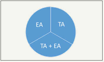
In general, an acupuncturist in practice may face the challenge, considering using the traditional acupoints only, the extra acupoints only, or the combining both traditional and extra acupoints, including A-shi acupoints, as presented in the three-acupointselection protocol in (Figure 1). The indications of extra and A-shi acupoints remains uncertain without scientific evidence; therefore, it is strongly suggested to consider the traditional acupoints first based on the Yin-Yang and the Five Elements theories in the server conditions. The golden rule “Less needling for best outcomes” should be born in mind in order not to make patients uncomfortable. Palpation that facilitates acupuncturists to make diagnosis seems to be the feasible technique for acupuncturists to consider when to combine extra acupoints and A-shi acupoints with traditional acupoints and only select traditional acupoints [4].

Figure 1:Three-acupoint-selection protocol. Note: TA for traditional acupoints on the meridians; EA for extra and A-shi acupoints


Timing

The priority of selecting traditional acupoints and extra and A-shi acupoints for needing is challenging. It is suggested that palpations should be conducted first before making the decisions. In the Decision-making model, the priority can be determined with the palpations.

Efficacy

Whether or not manipulations of sedation and tonification on extra and A-shi acupoints can be used for excess or deficiency patterns deserve attention. The manipulation of lifting-thrusting stimulated on Zusanli (ST36) increased the temperature of the skin [5], which suggests that the scientific evidence of the manipulations is needed to verify if the same result can be applied to extra and A-shi acupoints.

Conclusion

In terms of cost, convenience, and time saving, acupuncture is the top priority for patients. Contemporary scientific evidence shows acupuncture is effective for pain relief, etc.; however, it is surely that expected outcomes and results cannot be obtained if pattern based on TCM is not accurately identified and strictly followed in scientific research and practicing. Without practical guidelines discussed in the literature for acupuncturists to follow in clinical practice, it is expected the issues presented in this paper may be helpful and attract more attentions.

References

  1. Hong TZ (2018) Notes for clinical use of extra acupoints. J Complement Med Alt Healthcare 8(1): 555728.
  2. Hong TZ (2018) Reminders for clinical application of extra acupoints. Curr Trends Biomedical Eng & Biosci 16(3): 555939.
  3. Hong, TZ (2017) Principles of protocol to treat heart blood deficiency. J Complement Med Alt Healthcare 4(4): 555644.
  4. Hong TZ (2018) Decision-making model proposed in use of extra/a-shi acupoints with traditional acupoints. Advancements Bioequiv Availab 2(2).
  5. Huang, Tao Huang, Xin Zhang, Weibo Jia, Shu Yong Cheng, et al. (2013) The influence of different acupuncture manipulations on the skin temperature of an acupoint. Evidence-Based Complementary and Alternative Medicine p. 5.

For more articles in Bioequivalence open access journals
Please click on below link: https://crimsonpublishers.com/abb/

Thursday, January 6, 2022

Relationship Between Arrhythmias and Level Activity of Athlete’s-Role of HRMs_Crimson Publishers

 Relationship Between Arrhythmias and Level Activity of Athlete’s-Role of HRMs by Robert Gajda in Examines in Physical Medicine and Rehabilitation: Open Access_Rehabilitation medicine journal impact factor

Keywords

Arrhythmias; Exercise; Heart Rate Monitors (HRMs); Endurance sport; Sudden cardiac death

Introduction

Among all papers devoted to the relationship between arrhythmias and endurance training, the study Andersen et al. is particular value [1]. The study includes all Swedish citizens completing the famous Swedish skiing event “Vasaloppet,” enduring 90 kilometers of strenuous cross-country skiing. The participants are ranging from elite to recreational athletes and their training status (measured as maximal oxygen consumption) is closely related to their finishing time. The authors completed data about more than 4,400 participants of this strenuous ski marathon during the period 1989-1998.

Accounting for age, socioeconomic status and education, they observed a higher incidence of arrhythmias in cross-country skiers with a long history of endurance training [1]. Compared to those who had completed only one single race, those who had completed 7 or more races had a 29% higher risk of a subsequent arrhythmia. Further, elite athletes finishing at 100- 160% of the winning time had a 37% higher risk of arrhythmias than recreational athletes finishing at more than 241% of the winning time. The associations were mainly driven by the most common types of arrhythmia: atrial fibrillation and brady arrhythmias. The authors did not find any significantly increased incidence of potential lethal ventricular arrhythmias with any of the exposures.

In another paper published in 2015 in BMJ, Andersen and co-workers presented a very unique analysis about the associations of exercise capacity and muscle strength in late adolescence with the risk of vascular disease and arrhythmia based on long-term observation of 1.1 million young Swedish men who participated in mandatory military conscription between 1972 and 1995. During a median follow-up of 26 years, more than 26,000 vascular disease events and more than 17,300 arrhythmia events were recorded. The study revealed that exercise capacity and muscle strength in late adolescents are independently associated with a lower risk of vascular disease and arrhythmia. Exercise capacity had a U-shaped association with risk of arrhythmia driven association with risk of atrial fibrillation and a U-shaped with bradyarrhythmia. Thus, the benefit of a lower risk of vascular events with higher exercise capacity (hazard ratio for vascular events of 0.67) was not outweighed by a higher risk of arrhythmia [2, 3].

Other risk factors of arrhythmias in athletes comprise age, exercise intensity and above all-exercise duration. A threshold for the increased risk of atrial fibrillation is 1500 hours of exercise per life. Atrial volume is a strong predictor of AF (athletes with AF have a larger leftatrial volume than those without) in older athletes (39±9 years old) and in veterans [4-7].The etiology and pathophysiology of AF in athletes are not clear. Increased activity of the parasympathetic vagal system is the most important modulator and trigger of AF in athletes, whereas a substrate has still been a subject of debate. Structural remodeling of atrial myocardium in response to permanent pressure and volume overloading (“overtraining syndrome”), inflammation and fibrosis are the most probable causative factors [8].

Ventricular tachyarrhythmias, identified in elite athletes without cardiovascular abnormalities, are frequent expressions of the “Athlete’s Heart Syndrome” (up to 70%), though they are not related to the presence or magnitude of training-induced LV hypertrophy [9]. Brief deconditioning (5-6 months) usually results in the resolution of arrhythmias in athletes without organic heart disease [9]. Many authors of older and recently published original papers and reviews agreed that Supra Ventricular Tachyarrhythmias (SVT) in sportsmen are rare and usually benign. This kind of benign arrhythmia may appear with palpitations, weakness, lightheadedness, and even syncope. Frequent SVT may impair athletic performance. Sinus node reentry tachycardia is an uncommon finding in athletes. The average heart rate is between 130 and 140bpm. Atrial tachycardia is extremely rare in athletes. The atrial rate is generally between 150 and 200bpm [10-12].

The widespread use of sport Heart Rate Monitors (HRMs) contributes to the “catching” of an increasing number of tachyarrhythmias among both symptomatic competitive athletes and amateurs [13]. Especially in the case of “professionals” it can be a life-saving factor [14]. Although heart rate monitors do not recognize the type of arrhythmia, their worrying indications in conjunction with clinical symptoms allow for a fairly preliminary diagnosis [15]. In the future, the technological development of sport heart rate monitors will definitely provide further solutions for athletes allowing to determine the type of arrhythmias if they appear in sports training [16].

References

  1. Andersen K, Farahmand B, Ahlbom A, Held C, Ljunghall S, et al. (2013) Risk of arrhythmias in 52 755 long-distance cross-country skiers: A cohort study. Eur Heart J 34(47): 3624-3631.
  2. Andersen K, Rasmussen F, Held C, Martin N, Per T, et al. (2015) Exercise capacity and muscle strength and risk of vascular disease and arrhythmia in 1.1 million young Swedish men: Cohort study. BMJ 351: h4543.
  3. Hoogsteen J, Bennekers JH, Wall EE, Hemel NM, Wilde AA, et al. (2004) Recommendations and cardiological evaluation of athletes with arrhythmias: Part 1. Neth Heart J 12(4): 157-164.
  4. Nielsen JR, Wachtell K, Abdulla J (2013) The relationship between physical activity and risk of atrial fibrillation-A systematic review and meta-analysis. J Atr Fibrillation 5(5): 789.
  5. Everett BM, Conen D, Buring JE, Moorthy MV, Lee IM, et al. (2011) Physical activity and the risk of incident atrial fibrillation in women. Circ Cardiovasc Qual Outcomes 4(3): 321-327.
  6. Eijsvogels TM, Fernandez AB, Thompson PD (2016) Are there deleterious cardiac effects of acute and chronic endurance exercise?. Physiol Rev 96(1): 99-125.
  7. Hong KL, Glover BM (2018) The impact of lifestyle intervention on atrial fibrillation. Curr Opin Cardiol 33(1): 14-19.
  8. Olshansky B, Sullivan R (2014) Increased prevalence of atrial fibrillation in the endurance athlete: Potential mechanisms and sport specificity. Phys Sportsmed 42(1): 45-51.
  9. Biffi A, Maron BJ, Giacinto B, Porcacchia P, Verdile L, et al. (2008) Relation between training-induced left ventricular hypertrophy and risk for ventricular tachyarrhythmias in elite athletes. Am J Cardiol 101(12): 1792-1795.
  10. Biffi A, Maron BJ, Verdile L, Fernando F, Spataro A, et al. (2004) Impact of physical deconditioning on ventricular tachyarrhythmias in trained athletes. J Am Coll Cardiol 44(5): 1053-1058.
  11. Viitasalo MT, Kala R, Eisalo A (1984) Ambulatory electrocardiographic findings in young athletes between 14 and 16 years of age. Eur Heart J 5(1): 2-6.
  12. Verdile L, Maron BJ, Pelliccia A, Spataro A, Santini M, et al. (2015) Clinical significance of exercise-induced ventricular tachyarrhythmias in trained athletes without cardiovascular abnormalities. Heart Rhythm 12(1): 78-85.
  13. Gajda R (2019) Extreme bradycardia and brady arrhythmias at athletes. What will technology development bring as a help to diagnosis them? Res Inves Sports Med 5(4).
  14. Gajda R, Biernacka EK, Drygas W (2018a) Are heart rate monitors valuable tools for diagnosing arrhythmias in endurance athletes?. Scand J Med Sci Sports 28(2): 496-516.
  15. Gajda R, Biernacka EK, Drygas W ( 2019) Atrial Fibrillation in athletes-easier to recognize today? Res Inves Sports Med 5(4).
  16. Gajda R, Biernacka EK, Drygas W (2018b) “The problem of arrhythmias in endurance athletes: are heart rate monitors valuable tools for diagnosing arrhythmias?”, Horizons in World Cardiovascular Research. Nova Science Publishers, New York, USA 15: 1-64.

For more articles in Rehabilitation medicine journal impact factor
Please click on below link: https://crimsonpublishers.com/epmr/


Wednesday, January 5, 2022

Fibrosing Mediatinitis with Intracardiac Extention: A Rare Crippling Ailment-Case Report_Crimson Publishers

 Fibrosing Mediatinitis with Intracardiac Extention: A Rare Crippling Ailment-Case Report by Mariah Obino in Open Journal of Cardiology & Heart Diseases_American Journal of Cardiology


Abstract

The exact cause of fibrosing mediastinitis is not fully understood, but it is not inherited. Many cases are linked to a specific type of fungal infection called histoplasmosis. Treatment depends on which structures of the mediastinum are affected, the severity of the scarring and, in some cases, the cause of the condition.

Other less common causes of fibrosing mediastinitis include radiation therapy, autoimmune disease, tuberculosis, certain medications, Behcet’s disease, and inflammatory diseases such as sarcoidosis in roughly 10-20% of cases, the cause of fibrosing mediastinitis cannot be determined.

Keywords: Mediastinitis, Fibrosing mediastinitis, Sclerosing non-Hodgkin’s lymphoma, Intra-Cardiac tumors

Introduction

It is commonly seen in young patients with clinical features of obstruction of the superior vena cava, pulmonary vessels, airways or esophagus [1]. Fibrosing mediastinitis is an infiltrative disorder known to encase mediastinal structures [2] seen commonly in young patients. It can often be mistaken for a tumor such as nodular sclerosing Hodgkin’slymphoma; understanding the imaging appearance and clinical presentation is therefore vital in avoiding misdiagnosis [1]. Fibrosing mediastinitis is also known as mediastinal fibrosis or sclerosing mediastinitis. This is a rare benign disorder caused by x of acellular collagen and fibrous tissue within the mediastinum that may mimic a malignant process on imaging. Two types of fibrosing mediastinitis have been described:

1. Idiopathic/non-granulomatous fibrosing mediastinitis which is an idiopathic reaction to autoimmune syndromes, use of methysergide or radiation therapy to the mediastinum.

2. Granulomatous fibrosing mediastinitis is almost always an idiosyncratic/abnormal immunological response to infectious and inflammatory conditions most commonly tuberculosis and histoplasmosis in different endemic regions [2].

Fibrosing mediastinitis has also been classified as focal and diffuse. The focal type is seen on imaging (CT or MRI) as a localized, calcified mass in the mediastinum or in the pulmonary hila. The diffuse type appears as a diffusely infiltrating, non-calcified mass traversing multiple mediastinal compartments. It is commonly seen in young patients with clinical features of obstruction of the superior vena cava, pulmonary vessels, airways or esophagus [1].

Materials and Methods

17-year-old HIV negative male, living in care home presented to a national tertiary hospital with cough, difficulty in breathing, dyspnea and fever over the past 1 year. He also had bilateral deep venous thrombosis and was on tuberculosis medication and warfarin for suspected TB and pulmonary embolism respectively.

He was referred to our institution after there was no improvement despite treatment. Full haemogram done at our institution was unremarkable except for an Hb of 10.1g/dl. Other investigations done for inflammatory process like CRP, ESR, and for TB like sputum, genetic analysis was inconclusive. The patient also underwent an echocardiogram which showed a markedly dilated right atrium with a mass adherent to the base, and dilated inferior vena cava, severe right ventricular dilatation and severe tricuspid regurgitation.

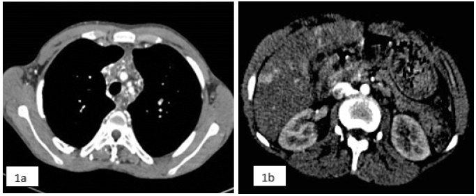
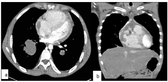
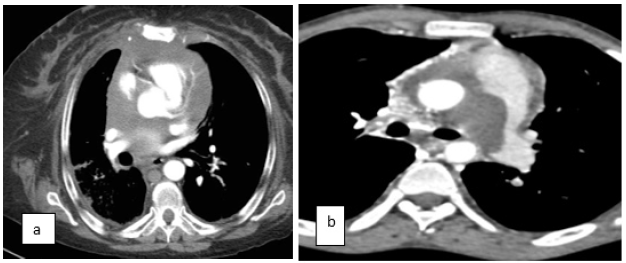
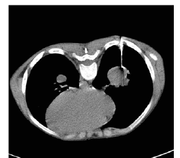
The patient was referred to the radiology department for a CT chest to further evaluate the cardiac findings as well as to evaluate the chest for evidence of tuberculosis. CT Chest showed multiple small nodes in the superior mediastinum, an ill-defined soft tissue mass extending caudally from the superior mediastinum encasing the blood vessels resulting in occlusion of the superior vena cava. The soft tissue mass extended to the right atrium, pulmonary trunk and right pulmonary artery terminating in the apical segment of the right lower lobe. A similar smaller mass was seen in the course of the left lower lobe pulmonary artery. Multiple small collateral blood vessels were seen in the mediastinum secondary to the SVC occlusion. Abnormal enhancement in the liver due to collateral flow through the vein of Sappy was also seen.

CT scan chest with contrast [Figures 1-4]

This was thought to represent an aggressive sarcoma arising from the blood vessels and the patient underwent a CT guided biopsy of the mass in the right lower lobe which showed benign fibrous tissue with no evidence of malignancy. Zn staining for TB was also negative. Due to the perceived non-concordance of the histology findings with the imaging findings, a repeat biopsy was carried out which revealed similar findings on histology. The patient then underwent an open mediastinal biopsy under cardiopulmonary bypass as the tumor mass was extending intra cardiac [Figure 5].

Figure 1:a) CT scan of the chest with contrast shows a) mediastinal mass (with no calcifications) encasing the mediastinal vessels with formation of collaterals due to resultant compression of the SVC; b) Abnormal hepatic enhancement (veins of sappy).


Figure 2:a) Mediastinal mass infiltrating the right atrium; b) Coronal image of the same.


Figure 3:a) SVC not visualized as a result of the mediastinal mass encasing it completely at this level; b) CT lung window showing left bronchial stenosis as a result of the mediastinal mass.


Figure 4:a) CT scan shows encasing of the left pulmonary artery by the mediastinal mass; b) Nodular sclerosing lymphoma mimicking idiopathic fibrosing mediastinitis (images from our institution) note absence of calcifications.


Figure 5:CT guided biopsy of the right lower lobe of right lung.


Surgical findings [Figure 6]

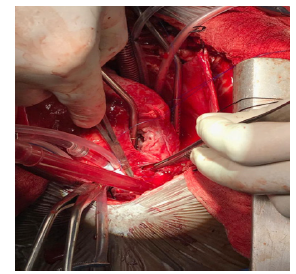
Chest opened by midline sternotomy. Cardiopulmonary bypass achieved with difficulty after cannulating Aorta, and Right Atrium as the whole of RA was filled with the mass, which was extending to the SVC, IVC, and PA. It was thick and fibrous and was densely infiltrating the myocardium. The initial impression was that, it could be an infiltrating malignancy. Hence tissue biopsy was taken from RA and MPA, and with routine closure followed by weaning off the pump. In the post-operative period, he was on high inotropes, and had features of cardiac failure. He succumbed on the 3rd post op day.

Histology [Figure 7]

A diagnosis of idiopathic fibrosing mediastinitis was made.

Figure 6:Per operative picture shows fibrosing mass inside RA extending to SVC, IVC and MPA.


Figure 7:Histologic findings included dense fibrosis, chronic lympho-plasmacytic infiltrate, and widespread calcification.


Discussion

There is no cure for fibrosing mediastinitis, and the benefits of current treatments are unclear. In general, treatment options depend on which structures of the mediastinum are affected (i.e. the heart, large blood vessels, windpipe, esophagus, and lymph nodes), the severity of the scarring and, in some cases, the cause of the condition. For example, fibrosing mediastinitis caused by histoplasmosis may be treated with antifungal medications while cases caused by autoimmune disorders may respond best to corticosteroids. Other treatment options include surgery to removed scarred tissue and local therapies to treat specific problems (stents, laser therapy etc.). The CT findings are in keeping with diffuse type fibrosing mediastinitis which was confirmed on biopsy. The following findings were seen in our patient and have been described in literature and case reports [1-3]:

1. Superior vena cava obstruction with multiple collaterals including enhancing hepatic pseudo lesions representing collateral venous flow through the vein of Sappey.

2. Encasement of mediastinal structures including the ascending aorta without narrowing.

3. Features of right heart failure, [3] enlarged right ventricle, bowing of the interventricular septum to the left, backflow of contrast into the inferior vena cava and hepatic veins.

4. Narrowing of the left main bronchus.

The left main pulmonary artery was compressed, while its branches were encased without any stenosis. The right main pulmonary artery was markedly narrowed proximally and completely infiltrated distally with non-visualization of its branches. These appearances must be differentiated from thromboembolic disease as well as pulmonary artery sarcoma [4]. The clue in this case was extensive involvement of the thoracic inlet and anterior mediastinum not usually seen in thromboembolic disease and the young age of the patient without other risk factors like a known underlying malignancy. However, the history of recurrent DVT is concerning for superimposed thrombosis. Intracardiac masses are a rare entity and the extension of the disease process into the right atrium is unusual and made diagnosis difficult in this patient. Only one previous case of intracardiac mass due to fibrosing mediastinitis has been reported [5]. Fibrosing mediastinitis usually results from an excessive host response to a prior infection that involved the mediastinal lymph nodes. Confirmatory diagnosis of mediastinal fibrosis involves surgical biopsy with a good sample showing only fibrosis without evidence of malignancy. This is important in excluding differential diagnoses that include; sclerosing non- Hodgkin lymphoma, nodular sclerosing Hodgkin disease and pulmonary artery sarcoma [2,4]. Additional tests are usually done to rule out any granulomatous causes like histoplasmosis and tuberculosis. In our patient ZN staining for acid-fast bacilli was negative; although whether tuberculosis was the cause of the fibrosis could not be ascertained as the patient had been on anti-TB medication. It should also be noted that definitive histopathologic proof of infection by demonstrating organisms within affected tissues is absent in many cases [1].

Conclusion

Idiopathic fibrosing mediastinitis is a rare entity and there are only few case reports in the literature. It can rarely extend into the cardiac chambers presenting as an intracardiac mass [5] is even rare. It has a propensity to infiltrate the pulmonary vasculature which has to be differentiated from pulmonary embolism. Extensive mediastinal involvement without calcification in the diffuse type of mediastinal fibrosis can mimic nodular sclerosing Hodgkin’s lymphoma. Familiarity with the various imaging features of fibrosing mediastinitis is important for prompt and accurate diagnosis and may preclude an invasive life-threatening biopsy procedure if images are interpreted in the correct clinical context.

References

  1. Rossi SE, McAdams HP, Rosado-de-Christenson ML, Franks TJ, Galvin JR, et al. (2001) Fibrosing mediastinitis. Radiographics 21(3): 737-757.
  2. McNeeley MF, Chung JH, Bhalla S, Godwin JD (2012) Imaging of granulomatous fibrosing mediastinitis. Am J Roentgenol 199(2): 319-327.
  3. Seferian A, Steriade A, Jaïs X, Planché O, Savale L, et al. (2015) Pulmonary hypertension complicating fibrosing mediastinitis. Medicine (Baltimore) 94(44): e1800.
  4. Jeong N, Seol SH, Kim IH, Kim JY (2016) Pulmonary artery sarcoma-Multimodality imaging. J Clin Imaging Sci 6: 45.
  5. Pereira-da-Silva T, Galrinho A, Ribeiro A, Jalles N, Martelo F, et al. (2013) Intracardiac mass due to fibrosing mediastinitis: The first reported case. Can J Cardiol 29(8): 1015.

For more articles in American Journal of Cardiology
Please click on below link: https://crimsonpublishers.com/ojchd/

Tuesday, January 4, 2022

Triple Noncontiguous Thoracic Spine Fractures Treated with Hybrid 3D Intraoperative CTNavigated Minimal Invasive Fixation Technique: A Case Report_Crimson Publishers

 Triple Noncontiguous Thoracic Spine Fractures Treated with Hybrid 3D Intraoperative CTNavigated Minimal Invasive Fixation Technique: A Case Report by Patrick Misson in Techniques in Neurosurgery & Neurology_Journal of Neurology


Abstract

Introduction: Multilevel noncontiguous thoracic spine fractures are usually due to high-energy trauma and often associated with life threatening organ injuries or neurological deficits. Early posterior percutaneous fixation has shown its efficacy in non-neurologic fractures and for comorbidity management. However, it remains a tricky procedure due to spine instability and anatomic landmark modification. Additional intraoperative CT-assisted navigation might improve precision in such long and complex spine fixation.

Case Presentation: A 26-year old men was admitted to ICU after a very high velocity motorcycle accident at 250km/h. Surprisingly, he had no neurological deficits, but the initial traumatic assessment revealed triple unstable noncontiguous thoracic spine fractures of Th4, Th6 and Th8. The patient was treated with a hybrid minimal invasive approach using an intraoperative 3D CT-scan navigation system and percutaneous pedicle-screwing method. The postoperative CT-Scan shown instrumentation from Th2 to Th10 and demonstrated correction of regional kyphosis. The patient regained his mobility and started to walk 3 days following the surgery. He was discharged with temporary additional thoraco-lumbar corset 7 days after surgery without any postoperative complications.

Discussion: Navigated trans-pedicular screw fixation has proven its superiority in precision. The use of live intraoperative CT-scan with spinous process reference marker provides critical information about spine alignment and pedicular trajectories which facilitate screw placement in fractured or misplaced vertebrae. The addition of standard lateral C-arm can be easily implemented.

Conclusion: Noncontiguous triple fractures of the thoracic spine are an unusual presentation of polytrauma injury. Long and complex thoracic posterior spine fixation is feasible in a safe way using an intraoperative CT-navigated minimal invasive technique in support.

Keywords: Noncontiguous spine fractures; Thoracic spine fixation; Minimal invasive technique; Intraoperative CT-scan navigation

Abbreviations: CT: Computed Tomography; ISS: Injury Severity Score; ICU: Intensive Care Unit; ASIA: American Spinal Injury Association; MRI: Magnetic Resonance Imaging; AO-Spine: Arbeit gemeinschaft für Osteo-synthesefragen Spine.

Introduction

Multilevel noncontiguous thoracic spine burst fractures are rarely reported in the literature [1-5]. They are usually described as floating spine injuries. The reported incidence of noncontiguous spine fractures varies from 1.6 to 19%, with a usual range of 3 to 8%. However, this incidence has been historically underestimated due to overlooked injuries or delayed X-rays diagnosis.  Recent evidences based on routine full-spine CT-scan suggest the actual frequency may be as high as 20% [6]. These injuries are strongly associated with high-energy trauma, mainly fall from high height or traffic accident [7]. The average age at the time of accident is 30,9 years. They are frequently associated with life threatening organ injuries and neurological deficit, resulting in high Injury Severity Score (ISS) [3,4,6,7]. In such clinical situations, early surgical fixation has shown its efficacy compared to conservative treatment. It allows better comorbidity management and leads to better clinical outcomes in terms of mobilization. Recently, percutaneous posterior spinal fixation has become the new gold standard compared to a conventional open approach. It reduces the invasiveness of the procedure and offers better precision [8]. However, percutaneous fixation on multilevel traumatic injuries remains a tricky procedure due to spine instability and anatomic landmark modification. This is particularly the case at thoracic spine level where anatomical variability and the restricted size of the pedicles are limiting the margin of error [8,9]. Additional intraoperative CT-assisted navigation might improve precision in such long and complex spine fixation procedure [8-14]. In the light of the above, we report here the case of a patient with traumatic noncontiguous triple burst fractures of the thoracic spine. To our knowledge, this is the first report of such a long thoracic spine fixation using 3D intraoperative CT-navigated minimal invasive technique for multilevel traumatic fractures. The purposes of this report are twofold. First, it exposes our technical report in this specific case. Secondly, we want to discuss the benefits of intraoperative navigation assistance.

Case Presentation

A 26-year old man was admitted to ICU after a very high velocity motorcycle accident on a speed track. He has been ejected from his motorbike at a speed of 250km/h and hit the tire wall directly with the back. On the hospital admission, he did not show any major abdominal injury but a respiratory insufficiency due to a bilateral hemopneumothorax requiring bilateral pleural drain. Initial neurological evaluation revealed severe back pain but no neurologic deficit. According to the American Spinal Injury Association (ASIA) impairment scale, his neurological status has been estimated as ASIA E. A crush syndrome was also present at the admission but resolved within 24 hours with optimal hydration and urine alkalization.

Radiologic Findings

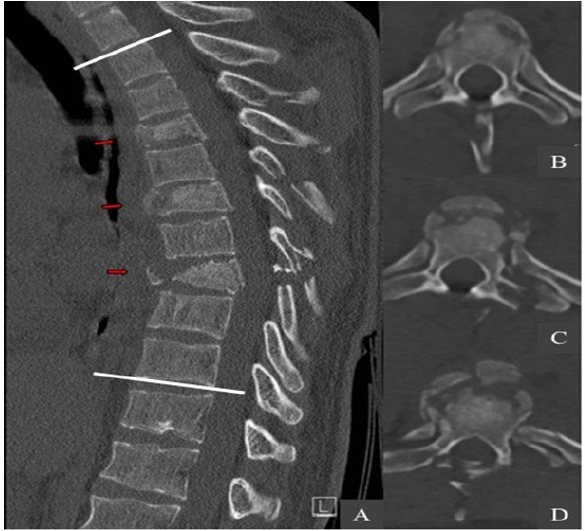
The initial whole-body computed tomography revealed three unstable noncontiguous vertebral body fractures distributed on Th4, Th6 and Th8. These were complicated by the association of multiple fractures of pedicles, spinous and transverse processes at adjacent thoracic spine levels (Figure1). These fractures were graded according to AO Spine classification as it follows: type A4 with 32% vertebral compression on Th4; type A3 with 18% vertebral compression on Th6; type B2 with 75% vertebral compression on Th8 (Table 1). The regional kyphosis between Th2 and Th10 was measured with an angle of 38.5°. The computed tomography also revealed a fracture of the sternal manubrium, multiple bilateral rib fractures from Th5 to Th9 and a left great trochanter fracture. Interestingly the whole spine MRI assessment did not show any spinal cord injury neither epidural lesions. The ISS was measured at 24.

Figure 1: Preoperative CT-scan. (A) The sagittal and axial views show three unstable noncontiguous vertebral body fractures (red arrows) distributed on Th4 (B), Th6 (C) and Th8 (D). The regional kyphosis was measured with an angle of 38.5° between Th2 and Th10 (white lines).


Table 1: Fractures summary according to the AO Spine classification.


Surgical Technique

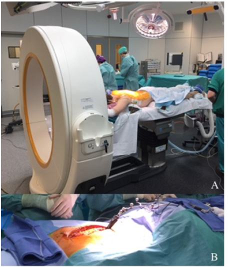
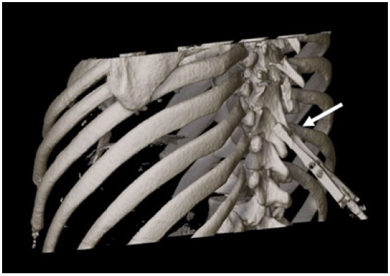
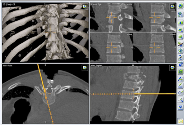
The patient undergone a surgery based on a hybrid minimal invasive approach using an intraoperative 3D CT-scan navigation system and percutaneous pedicle-screwing method. We performed a posterior thoracic spine fixation from Th2 to Th10. The patient was placed in a prone position on a mobile, radiolucent, carbon fiber operating table connected to the Airo ring (Figure 2A). A fluoroscopy was used to check the spinal alignment and to determine the vertebral levels to be instrumented. A medial posterior skin incision was performed from Th1 to Th11 down to the muscular fascia. At this stage, a navigated reference marker clamp was firmly placed with minimal muscle dissection on the spinous process of Th9, which was the closest intact bony structure adjacent to the fractured levels [10] (Figure 2B). A first intraoperative CT-scan acquisition was performed, assessing a good kyphosis correction (Figure 3). The acquired datas were processed thanks to the intraoperative image-guidance system. A series of trans-pedicular channel was performed from Th10 to Th7, based on the 3D navigated CT-scan with a navigated standard Jamshidi needle (Figure 4) K-wires were inserted into the pedicles before insertion of cannulated screws. Additional lateral fluoroscopy was performed at the end of each screw placement in order to verify the screwing accuracy and the absence of bone displacement [12]. A second series of pedicular screwing was then realized using the same procedure under the control of a second CT-scan with a Th4 reference marker clamp. Two 24cm slightly bent titanium rods were placed in a strictly minimal invasive way to connect the screws. A vertebral distraction was applied to the vertebral body of Th8 to restore correct vertebral height and complete the procedure. Further decompressive laminectomy was unnecessary, as the MRI did not highlight any spinal cord compression.

Figure 2: Operating room setting with the Airo ring. (A) Midline incision with navigated reference marker fixed on Th9.


Figure 3: First intraoperative CT-scan acquisition with reference marker fixed on the Th9 spinous process (white arrow).


Figure 4: Intraoperative 3D-navigated screw placement trajectory.


Radiologic and Clinical Outcomes

The postoperative CT-scan demonstrated correction of regional kyphosis with an angle of 32° (Figure 5). Eleven screws were placed completely within the pedicle unit. Two screws (Th4 and Th7 right) were considered as “in-out-in” with a moderate penetration of the external pedicle wall (3-6mm outside the pedicle boundaries)9, hence there was no indication to reposition these. The patient regained his mobility and started to walk three days following the surgery. An additional thoraco-lumbar corset was placed for a period of 6 weeks. He was discharged 7 days after surgery without any postoperative complications and resumed his professional activity a month later.

Figure 5: Postoperative sagittal CT-scan reconstruction of the instrumentation from Th2 to Th10. (A) The regional kyphosis was measured with an angle of 32° (white lines) after the surgery. (B) Posterior (C) and left posterolateral (D) 3D images.


Discussion

The case reported here is a unique presentation of triple unstable noncontiguous thoracic spine burst fractures without any neurologic lesions. Furthermore, the way he was treated by means of intraoperative minimal invasive navigation despite the multitude and the non-contiguous of these fractures has never been reported in the literature. In case of non-neurologic thoracolumbar fractures, it is more and more commonly admitted that a rapid surgical fixation allows a better management compared to conservative treatment. Recent developments of percutaneous techniques have also optimized the treatment of other comorbidities. It allows early mobilization, decrease the complication rate and reduce the duration of hospitalization, as well as it could be appliable to treat various types of fractures [1,2,4,5,7-9]. Although there are still debates, our experience shows that this is also the case for noncontiguous fractures [1-5].  Some factors such as the important length of fixation, anchoring difficulties on a multi-fractured spine and repercussion on spine mobility should be considered. It is not always possible to restore a perfect spine alignment. Reduction as much as possible of the number of fixed levels is a major concern in order to optimize the clinical outcome and spine range of motion. Yet, when the number of intact segments in between two fractured segments that need surgery is lower than 4, it is recommended to connect both fixations in one single instrumentation.1 From the very beginning of the medical care, the objective was to multiply anchor points, especially in between fractured level and even in fractured vertebrae in order to reduce the fixation length and maintain spine mobility. This could also increase the rigidity of the instrumentation and reduce the use of transverse connector. Percutaneous pedicular thoracic spine screwing is not an easy procedure for many reasons: the narrowness of the pedicles, radiological opacification generated by the ribs, anatomical modifications due to the trauma and instability of fractured fragments. The use of intraoperative navigation like the Airo system has recently proved his superiority in terms of precision with a correct placement rate of 95-99% compared to 89-95% with conventional free hand percutaneous technique [8,9,11-13]. Regarding these data, there is no doubt that the 3D-intraoperative navigation technique has the best accuracy. In this specific case, intraoperative navigation has been helpful to securely realize pedicular screwing at many segments and in particular in two fractured vertebrae (Th4 and Th6) 8,9,13. It also allowed the intraoperative control of screws placement and kyphosis correction [8,10-13]. When needed it gives you the possibility of directly replacing misplaced screws and avoid reoperation. However, intraoperative spine navigation remains a difficult and time-consuming procedure that requires a learning curve to be comfortable with [8,9,11-13]. There have been concerns about extended surgical time. Recent series show that the instrumentation setup for iCT navigation requires more time, but each screw placement is faster than with free-hand technique and takes about 2 to 3 minutes. One in the other, the total time for the whole procedure seems comparable [8,9,11,12].

Another main hazard during minimal invasive surgery is the radiation exposure that is greater to the surgeon compared with open spine procedures [9,11]. The use of intraoperative navigation can decrease the global radiation exposure [9,11]. In case of such a long fixation, the medical staff irradiation was significantly reduced. However, patient irradiation is higher with intraoperative CT-scan acquisition than in usual procedure. On the other hand, regarding radiation hazard, each intraoperative acquisition remains lower than a standard diagnostic CT [9,11,13]. Two screws were considered as “in-out-in” (Th4 and Th7 right) without any consequences for spine stability or neurological damage. This inaccuracy could be explained by biomechanical instability, movements of reference marker during trajectory navigation, instrumentation at a large distance from the tracking device or inaccuracy of Jamshidi needle setting [8,9,12,13]. From a technical point of view, some recommendations can be given for the use of intraoperative navigation in long fixation: The use of an additional lateral C-arm fluoroscopy can be easily implemented, even at a later time during surgery, and provide direct radiological information about screws placement or bone fragment displacement [11,12]. This information will be correlated by the surgical team with the image-guided navigation and gives a double check when in doubt or in critical regions. In order to optimize the precision, a new CT-scan acquisition should be performed every 4 to 5 segments allowing the repositioning of the reference marker [10,13]. The setting of the navigated Jamshidi needle must also be checked frequently, ideally at the start of each pedicular sight [11]. Due to the number of percutaneous tubes needed in very long fixation, it is strongly recommended to start the navigation with the level farthest from the camera in order not to be disturbed by the interposition of tubes between Jamshidi needle and the camera.

Conclusion

Noncontiguous triple fractures of the thoracic spine are an unusual presentation of polytrauma injury. Long and complex thoracic posterior spine fixation is feasible in a safe way using an intraoperative CT-navigated minimal invasive technique in support [8-10,12,13]. The use of intraoperative imaging provides critical information about spine alignment and pedicular trajectories which facilitate screw placement in fractured or misplaced vertebrae [13]. It requires optimal surgical preparation and intervention steps must be followed scrupulously in order to avoid classic traps. Finally, this technique can be labelled as “hybrid” as it combines the use of a midline incision with minimal invasive fixation, and also the use of fluoroscopy in addition to intraoperative navigation.

Acknowledgement

Patrick Misson provided the conception and design of the article, drafting the article, revised it critically for important intellectual content, and final approval of the version to be submitted. Didier Martin and Abdelhammid Mendili were responsible for the article critically for important intellectual content. Corentin Delhaye provided radiological interpretation and image management. Jean Pirson provided the anesthesia. Cristo Chaskis provided the revised the article critically for important intellectual content and gave final approval of the version to be submitted. No funding to declare.

Conflict of Interest

The authors declare that there is no conflict of interest.

References

  1. Cho Y & Kim YG (2019) Clinical features and treatment outcomes of acute multiple thoracic and lumbar spinal fractures: a comparison of continuous and noncontinuous fractures. J Korean Neurosurg Soc 62(6): 700-711.
  2. Wittenberg RH, Hargus S, Steffen R, Muhr G, Bötel U (2015) Non-contiguous unstable spine fractures. Spine 27(3): 254-257.
  3. Takami M, Okada M, Enyo Y, Iwasaki H, Yamada H, et al. (2017) Non-contiguous double-level unstable spinal injuries. Eur J Orthop Surg Traumatol 27(1): 79-86.
  4. Seçer M, Alagöz F, Uçkun O (2015) Multilevel non-contiguous spinal fractures: Surgical approach towards clinical characteristics. Asian Spine J 9(6): 889-894.
  5. Kim JK, Moon BJ, Kim SD, Lee JK (2018) Minimal invasive non-fusion technique for the treatment of non-contiguous lumbar burst fractures in young age patient: A case report. Med 97(10): 10-13.
  6. Nelson DW, Martin MJ, Martin ND, Beekley A (2013) Evaluation of the risk of non-contiguous fractures of the spine in blunt trauma. J Trauma Acute Care Surg 75(1): 135-139.
  7. Giorgi H, Blondel B, Adetchessi T, Dufour H, Tropiano P, et al. (2014) Early percutaneous fixation of spinal thoracolumbar fractures in polytrauma patients. Orthop Traumatol Surg Res 100(5): 449-454.
  8. Hubbe U, Kogias E, Vougioukas (2009) Image guided percutaneous trans-pedicular screw fixation of the thoracic spine. A clinical evaluation. Acta Neurochir 151(5): 545-549.
  9. Allam Y, Silbermann J, Riese F, Greiner PR (2015) Computer tomography assessment of pedicle screw placement in thoracic spine: Comparison between free hand and a generic 3D-based navigation techniques. Eur Spine J 22(3): 648-653.
  10. Citak M, Stubig T, Kendoff D (2010) Navigated minimally invasive thoracolumbar pedicle screw placement with flat panel 3-d imaging a feasibility study. Technol Heal Care 18(2): 101-110.
  11. Lian X, Navarro RR, Berlin C (2016) Total 3D Airo® navigation for minimally invasive transforaminal lumbar interbody fusion. Biomed Res Int 5027340.
  12. Hecht N, Kamphuis M, Czabanka M (2016) Accuracy and workflow of navigated spinal instrumentation with the mobile AIRO® CT scanner. Eur Spine J 25(3): 716-723.
  13. Scarone P, Vincenzo G, Distefano D (2018) Use of the AIRO mobile intraoperative CT system versus the O-arm for transpedicular screw fixation in the thoracic and lumbar spine: a retrospective cohort study of 263 patients. J Neurosurg Spine 29(4): 397-406.
  14. Ushijima T, Kawaguchi K, Matsumoto T (2018) Double non-contiguous fractures in a patient with spondylo-epiphyseal dysplasia with spinal ankylosis treated with open and percutaneous spinal fixation technique: A case report. BMC Res Notes 11(1): 1-5.

For more articles in Journal of Neurology
Please click on below link: https://crimsonpublishers.com/tnn/

Monday, January 3, 2022

Determination of Lower Acidity of Jet Fuel by Catalytic Thermometric Titration Using Paraformaldehyde as a Thermometric End- Point Indicator_Crimson publishers

Determination of Lower Acidity of Jet Fuel by Catalytic Thermometric Titration Using Paraformaldehyde as a Thermometric End- Point Indicator by Quan Xi Zheng in Progress in Petrochemical Science_International Journal of Petrochemical Science


Abstract

The effect of concentrations of titrant, delivery rates, stirring rates, and oil mass on catalytic thermometric titration for the determination of the lower acidity of jet fuel were investigated, using KOH in isopropanol and paraformaldehyde as titrant and a catalytic thermometric indicator respectively. The results show that paraformaldehyde used as a catalytic indicator exhibits strongly endothermic effects to reflect end point significantly. When the oil mass is from 10g to 30g, the titration concentration is 0.01mol/L and the delivery rate is 1.0mL/min with moderate stirring, the tested acid numbers have good reproducibility and accuracy. The linear coefficient R2 of the fitting curve is 0.995. Using benzoic acid as a standard acid with concentration of 0.0105mg KOH/g to verify the accuracy of catalytic thermometric titration, the verified acid number is 0.0115mg KOH/g and basically consistent with the actual acid number, indicating that catalytic thermometric titration has good agreement with standard potentiometric titration methods and can be used for determination of acid number of jet fuels. It can accurately determine the acid number of jet fuel as low as 0.015mg KOH/g or even lower at optimized test conditions. The procedure is fast, easy to use, accurate, and highly reproducible to measure lower acidity in jet fuel. It is very suitable for the routine process and quality control of many types of oils.

Keywords: Acidity; Jet fuel; Catalytic thermometric titration; Paraformaldehyde

Introduction

As an important quality control index to evaluate corrosion, especially for fuel oil, aviation oil, and hydraulic oil, the acidity of oils is applied to estimate the properties and deterioration of oils during usage and storage [1-3], which is determined with the standard methods based either on visual indicators titration or potentiometric titration [4-7]. However, accuracy of visual titration is strongly influenced by the skills and color perception of the analyst, and especially differential color perception of analysts is considerable for coloured oils. Potentiometric titration, especially for oils containing trace weak polyacids, is subject to the noxious influence of the sample solutions and is always unreliable because of the weak change of potential during acid-base neutralization titration, which results in difficult detection of end-point and repeatability.  In order to achieve more fast and efficient determination of the end-point in titrations, there is increasing attention for a simple, fast, accurate, and precise automatic titrimetric operation that is substantially independent of analysts’ skills and suitable for routine process and quality control.

As a new method, catalytic thermometric titration has several attractive features: [1] the apparatus is simple, and all that required is a temperature measuring probe such as a thermometer or a thermistor as the sensing element; [2] the thermometric probes are inert to most solutions, and temperature changes in highly colored can be detected easily; [3] the range of indication reaction is unlimited because all of reactions are accompanied with temperature changes, the magnitude of which can be adjusted by changing reagents’ concentrations [8-11]. The basic principle of this method is catalytic initiation of an exothermic or endothermic reaction with an excess of titrant, as a consequence, the end-point can be indicated by obvious temperature changes of the solution [8-15]. It has been successfully applied for determining acidic substances in aluminum ion concentration of waste water and vegetable oils [16-19]. However, as so far, there are few reports on application of catalytic thermometric titration on lower acidity of jet fuels.

When small amounts of weak acidic species are titrated in nonaqueous solution with a titrant of strong alkali, the heat produced from the neutralization reactions may be quite small and easy to be confused by solvent evaporation and the mixing heat of the titrant with sample solution [4-7]. If the special thermometric indicator is added to sample solution, excess hydroxide ions would react quickly with them in endothermic or exothermic reactions, the end-point can be easily determined by temperature increase or decrease of the solution [20,21]. However, practical experience has demonstrated that the endpoint in thermometric titration showed excessive rounding, with consequent loss of precision and accuracy for some oils with lower acidity, such as aviation oil, hydraulic oil and fuel oils. Many studies have shown that titration error or the sharpness of the endpoint can be related to the concentration and delivery rate of titrant, volumes, and types of thermometric indicator [22-27]. In our previous studies [9,10], the trace water in jet fuels, as well as the acidity of several coloured oils, could be accurately and rapidly determined by catalytic thermometric titration using the mixture of acetone and chloroform as the end-point indicator. In this paper, we report herein our results on the determination of the lower acidity of jet fuels with catalytic thermometric titration employing paraformaldehyde as the end-point indicator, which exhibits strongly endothermic effects to reflect end point significantly and can determinate much more lower acidity than ASTM thermometric titration, and compared with potentiometric titration, the accuracy and repeatability of the thermometric titration is further investigated.

Materials and Methods

Materials

Paraformaldehyde, potassium hydroxide and isopropanol were of analytical reagent quality. A 0.10mol/L potassium hydroxide isopropanol solution was prepared and standardized with potassium hydrogen phthalate by the usual procedures. The jet fuel NO.3 was purchased from Sinopec Tianjin Shihua. Standard acid solution was prepared from approximately 37.8mg benzoic acid dissolved with 250mL isooctane, and acid value of standard acid is 0.10mg KOH/g.

Apparatus

In thermometric titration, two motor-driven micrometer syringes were employed to add the titrant to samples at a constant delivery rate, and a magnetic stirrer was provided to dissolve oil solutions, the temperature changes were detected by locating the thermistor in one arm of a Wheatstone bridge and were recorded with a strip chart recorder. At the end of each titration, sample and titration data were automatically sent for identification and computation, the acidity of oil samples was obtained from auto-calculation. In potentiometric titration, a pH probe containing both a glass and a reference electrode in the same body was employed.

Methods

In visual titration, the amount of oil was chosen in accordance with the ASTM D974 method, 8g oil samples was dissolved in 125mL ethanol solvent, 0.5mL 2% ethanol solution of alkali blue 6B was added and the solution was titrated with 0.1mol/L KOH ethanol solution. In the case of catalytic thermometric titration, oil samples were accurately weighed into clean, dry 125mL silvered Dewar flask, then 25mL acetone and 4mL chloroform were homogenized by using a magnetic stirrer. The 0.10mol/L KOH titrant solution is added at a constant delivery rate of 0.5mL/min to the stirred solution. End-points were determined using a peak-picking algorithm applied to the second derivative curve computed from the smoothed temperature data. During titration, there is a blank to be determined. The blank is a summation of all delays inherent in the titration solution under defined experimental conditions. These delays include contributions from kinetics of the chemical reaction between titrant and titrant, sensor response, mixing inefficiencies, electronic transfer, and computation of data. The volumes of the titrant obtained in the blank solution titration were subtracted from those the initial titration data of oil samples. Each titration experiments were performed in four times. In the case of potentiometric titration, except for oil samples were dissolved in 125mL of the solvent mixture (isopropanol and toluene), the oil samples stirring, dissolution and titrant addition procedures were employed accordance with the ASTM D664 method.

Results and Discussion

Effect of concentrations of titrant on thermometric titration

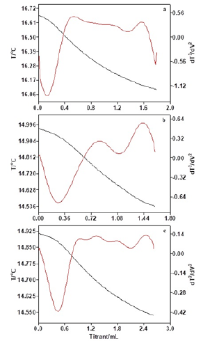
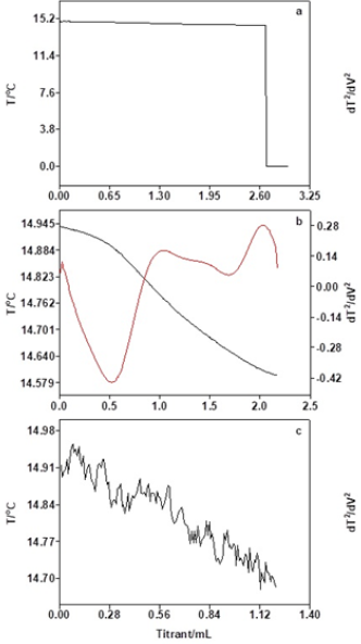
As listed in Table 1, concentrations of the titrant have great influence on determination results of thermometric titration. The concentration of the titrant is high, and the consumption of the titrant volume is small. In contrast, the concentration of the titrant is low, and the consumption of the titrant volume is large. The thermometric titration curves obtained from titrant with different concentrations are shown in Figure 1, from which we can see clearly that there are obvious temperature changes in thermometric titration curves, where the second-derivative plot is presented along with the solution temperature plot. Due to the lower acid number in jet fuel, which is less than 0.015mg KOH/g, and a greater deviation caused by the higher concentration of the titrant, the 0.01mol/L of titrant is used in all subsequent experiments in order to obtain more precise acid number.

Table 1: The determination results of different concentrations of titrants.


Figure 1: Thermometric titration curves of different concentrations of titrants (a: 0.05mol/L, b: 0.02mol/L, c: 0.01mol/L).


Effect of delivery rates on thermometric titration

Table 2: The determination results of different delivery rates.


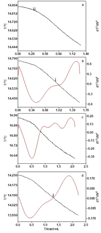
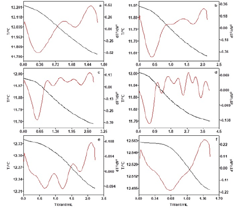
As Table 2 and Figure 2 show, when the delivery rate of the titrant is 0.5mL/min, the thermometric titration curve is smooth without an obvious inflection point. When the delivery rate is between 1.0mL/min and 0.2mL/min, there are obvious temperature changes in thermometric titration curves, where the second-derivative plot with sharp peak is presented along with the solution temperature plot, and the results are accurate and repeatable. Therefore, the delivery rate of titrant plays an important effect on the determination results for thermometric titration, and according to the smallest relative standard deviation (RSD) obtained in the result of titrant consumption, the 1.0mL/min of delivery rate is optimal.

Figure 2: Thermometric titration curves of different delivery rates of titrants (a: 0.5mL/min, b: 1.0mL/min, c: 1.5 mL/min, d: 2.0mL/min).


Effect of stirring rates on thermometric titration

As Figure 3 exhibits, only when the stirring rate is moderate, there are obvious temperature changes in thermometric titration curves, where the second-derivative plot with sharp peak is presented along with the solution temperature plot, and the results are accurate and repeatable. While, when the stirring rate is small, most of paraformaldehyde powders are deposited at the bottom of the titration beaker, which could not be catalyzed by titrant to process endothermic reaction, so that the temperature of the titration system remained basically unchanged. And when the stirring rate is high, the temperature sensor is unable to detect the temperature of the titration system since eddy currents are produced in the titration solution due to the excessive speed of the rotor stirring. Therefore, the moderate stirring rate is optimal.

Figure 3: Thermometric titration curves of different delivery rates of titrants (a: 0.5mL/min, b: 1.0mL/min, c: 1.5 mL/min, d: 2.0mL/min).


Effect of oil mass on thermometric titration

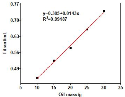
As Table 3 and Figure 4 show, when the oil mass is 5.0g, the temperature change of thermometric titration curve is obvious and the end-point of titration is easily judged. However, due to the small oil mass and the lower acidity, the titration system is relatively vulnerable to other factors, and the repeatability of titration results is not stable. When the oil mass is between 10.0g and 30.0g, the thermometric titration curves are smooth with obvious titration end-point, and the second-derivative plots are presented along with the solution temperature plot, which have sharp peak and obvious titration end-point. As Table 3 exhibits, the results of acid number are basically the same with reasonable RSD value. As Figure 5 shows, the coefficient of correlation (R2) for the linear calibration curve for different oil mass is found to be 0.9949. The results show that better accuracy and repeatability of tested acid number can be obtained, when the concentrations of titrant, delivery rates, stirring rates, and oil mass are 0.01mol/L, 1.0l/min, moderate and 10-30g, respectively.

Table 3: The determination results of different oil mass.


Figure 4: Thermometric titration curves of different oil mass (a: 5g, b: 10g, c: 15g, d: 20g, e: 25g, f: 40g).


Thermometric titration in standard acid samples

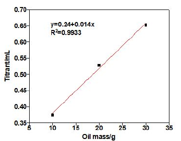
In order to testify the accuracy of the method of catalytic thermometric titrimetry, different mass of benzoic standard acid samples was investigated, and the results are listed in Table 4. The coefficient of correlation (R2) for the linear calibration curve was found to be 0.9924, which was shown in Figure 6. We can see clearly that the acid numbers of three standard acid samples were very similar to real acid number (0.10mg KOH/g) with low RSD. These observations confirm that thermometric titrimetry is an accurate and highly reproducible method, which was easy to determine acid number of colored or additive-containing petroleum oils in titrations.

Table 4: The determination results of different mass of standard acid samples.


Comparison of thermometric and potentiometry titration in jet fuel NO.3

Table 5: Results from the acid number of oil samples by thermometric, potentiometry and visual titrimetry.


Figure 5: Linear regression equation for different oil mass.


Figure 6: Linear regression equation for benzoic acid samples.


The titration of jet fuel NO.3 is investigated by catalytic thermometric titrimetry, employing paraformaldehyde as end-point indicator. By comparison, potentiometric titration is employed to analyze the same oil sample. The acid number of jet fuel NO.3 obtained from thermometric, potentiometry and visual titrimetry titration are shown in Table 5, from which we can see clearly that the acid numbers of jet fuel NO.3 obtained from catalytic thermometric titrimetry are basically consistent with potentiometry and visual titrimetry with less error.

Conclusion

The catalytic thermometric titration is a fast, easy to use, accurate, and highly reproducible method to determine lower acidity in jet fuels, using paraformaldehyde as the end-point indicator. Paraformaldehyde used as the end-point indicator can exhibit strongly endothermic base catalyzed reaction in thermometric titration, which has good agreement with standard acid samples. It can accurately determine the acid number of jet fuels as low as 0.015mg KOH/g or even lower at optimized test conditions. The procedure is fast, easy to use, accurate, and highly reproducible to measure lower acidity in jet fuel. It is very suitable for the routine process and quality control of many types of oils.

References

  1. Shinde H, Bewoor A (2018) Capacitive sensor for engine oil deterioration measurement. AIP Conference Proceedings 1943(1): 020099.
  2. Cui D, Wang Q, Wang P, Chi MS, Pan S, et al. (2018) A comparison of the compositions of acidic and basic fractions in <300 °C fractions from six Chinese shale oils. Fuel 211: 251-260.
  3. He, QJ (2013) Analysis of acid number in crude oil and petroleum product and acid distribution. Guangzhou Chemical Industry 5: 17-20.
  4. Lima RAC, Almeida LF, Lyra WS, Siqueira LA, Gaião EN, et al. (2016) Digital movie based on automatic titrations. Talanta 147: 226-232.
  5. Kahlert H, Meyer G, Albrecht A (2016) Colour maps of acid-base titrations with colour indicators: how to choose the appropriate indicator and how to estimate the systematic titration errors. Chem Texts 2: 7.
  6. Stuart BS, Ferrell JR (2016) Determination of carbonyl groups in pyrolysis bio-oils using potentiometric titration: review and comparison of methods. Energy Fuels 30(2): 1071-1077.
  7. Yu K, Kumar N, Aho A, Roine J, Heinmaa I, et al. (2016) Determination of acid sites in porous aluminosilicate solid catalysts for aqueous phase reactions using potentiometric titration method. Journal of Catalysis 335: 117-124.
  8. Phansi P, Danchana K, Cerdà V (2017) Kinetic thermometric methods in analytical chemistry. TrAC Trends in Analytical Chemistry 97: 316-325.
  9. Hu JQ, Zhang JJ, Yang SZ, Xin YL, Guo L, et al. (2017) Thermometric titration for rapid determination of trace water in jet fuel. Journal of Specroscopy 21: 1-5.
  10. Hu JQ, Xin YL, Yang SZ, Zhang JJ, Guo L (2017) The determination of lower acidity in several coloured oils by catalyzed thermometric titration. Petroleum Chemistry 57: 1099-1104.
  11. Azevedo MCC, Cavaleiro AMV (2012) The acid base titration of a very weak acid: boric acid. Journal of Chemical Education 89(6): 767-770.
  12. Bastos FA, Khan S, Simões EH, Teixeira CA, Tubino M (2013) Thermometric quantitative selective analysis of sodium methoxide in methanol industrial solutions. Journal of the Brazilian Chemical Society 24(8): 1380-1384.
  13. Relli D, Vincent MT (2018) Thermometric titration study of acetaminophen and sodium hypochlorite. Ohio Dominican University, Ohio.
  14. Barin JS, Tischer B, Oliveira AS, Wagner R, Costa AB, et al. (2015) Infrared thermal imaging: a tool for simple, simultaneous, and high throughput enthalpimetric analysis. Analytical Chemsitry 87(24): 12065-12070.
  15. Gumel AM, Annuar MSM, Heidelberg T, Chisti Y (2011) Thermo-kinetics of lipase-catalyzed synthesis of 6-O-glucosyldecanoate. Bioresource Technology 102(19): 8727-8732.
  16. Tao L, Sun H, Gong Y (2012) Determination of total acidity and aluminumion concentration in wastewater of aluminum profile surface treatment by thermometric titration. Metallurgical Analysis 32: 46-50.
  17. Jiang T, Zhan HY, Wang LJ (2011) Experiments and applications of automatic temperature titrator in the control and analysis of alumina production process. Light Metals 46: 22-25.
  18. Thomas KS (2003) Analysis of FFA in edible oils by catalyzed end point thermometric tirimetry (CETT). Journal of the American Oil Chemists Society 80: 21-24.
  19. Carneiro JD, Feres MA, Godinho ES (2002) Determination of the acidity of oils using paraformaldehyde as a thermometric end-point indicator. Journal of the Brazilian Chemical Society 13(5): 692-694.
  20. Cerdà V, González A, Danchana K (2017) From thermometric to spectrophotometric kinetic-catalytic methods of analysis: A Review. Talanta 167: 733-746.
  21. Jharimune S, Sathe AA, Riou RM (2018) Thermochemical measurements of cation exchange in CdSe nanocrystals using isothermal titration calorimetry. Nano Letters 18(11): 6795-6803.
  22. Rivera MRD, Matos JS, Socorro F (2008) Study of the stirring effect in ASTM 2277-205 isothermal titration calorimetric. Journal of Thermal Analysis and Calorimetry 92: 79-82.
  23. Demoz A, Mikula RJ (2011) A thermometric indicator method as a replacement for the filter paper spot test in the titration of clays using methylene blue. Engineering Geology 117(1-2): 12-16.
  24. Luk HN, Peng HY, Han YW, Lin CK, Lin WD, et al. (2018) Detection of acetaminophen using cyclic voltammetry on MnFe2O4/polyaniline. Journal of the Chinese Chemical Society 66(1): 1-6.
  25. Gu QJ, He ZH, Pan QX, Lin ZY, Sun J, et al. (2019) The steric hindrance controlled [2] pseudorotaxanes constructed by V-type stilbene dyes⊂CB [7]. Supramolecular Chemistry 31(2): 1-
  26. Hidayat A, Sutrisno B (2018) Free fatty acids esterification on palm oil sludge using zirconia-supported indonesia natural zeolite as heterogeneous catalyst. Oriental Journal of Chemistry 34(5): 2464.
  27. Čobanov I, Šarac B, Medoš Z, Vraneš M, Gadžurić S, et al. (2018) Effect of cationic structure of surface active ionic liquids on their micellization: A thermodynamic study. Journal of Molecular Liquids 271: 437-442.

For more articles in International Journal of Petrochemical Science
Please click on below link: https://crimsonpublishers.com/pps/

A Close Look at the Application of the Yin-Yang- Based Acupoint Pairs_Crimson Publishers

A Close Look at the Application of the Yin-Yang- Based Acupoint Pairs by Tong Zheng Hong in Advancements in Bioequivalence & Bioavailabi...