Crimson Publishers High Impact Journals

Tuesday, December 7, 2021

Young Rats and Negative Effects of Low-Alcohol and Energy Drinks_Crimson Publishers

 Young Rats and Negative Effects of Low-Alcohol and Energy Drinks by Trubitsyna IE in Gastroenterology Medicine & Research_Gastroenterology Medicine & Research


Abstract

Recently, low-alcohol and energy drinks have become widespread among young people, which are considered “harmless drinks “among them. However, young people get used to these drinks and cannot do without them, the dose of consumption increases. Therefore, it requires study to establish the role of the studied beverages in organizational and mental disorders. The article convincingly proves the causal relationship between alcohol and damage to internal organs and mental state in rats.

Keywords: Low-alcohol and energy drinks; Liver; Kidneys; Lungs; Heart; Psyche

Introduction

The behavior of any living creature is aimed at satisfying a particular need-biological, social. Need can be defined as an objective need for something, for this it must become subjective, normally perceived by the nervous system of the animal, and encourage him to act on its satisfaction, motivate both the animal and man to certain actions. In animals, the main biological motivations are food, drinking, sexual and self-preservation. The emergence of several motivations that require a choice, determine the highest priority. In animals, this is solved by an innate hierarchy of needs-the main one is self-preservation, which always prevails over the rest-when life is threatened, sexual, drinking or eating decreases. When drinking alcoholic beverages, the motivation for self-preservation is suppressed instantly and the craving for alcohol comes out in the first place, animals struggle for being at the drinker with an alcoholic beverage [1,2].

Purpose

To study the behavior of young rats in the use of low-alcohol (LA) and energy drinks (E).

Material and Methods

Studies were conducted on “young” white rats of the Wistar line, weighing 100-120g-15- 16 years by human standards. Three groups were identified: control (C)- animals had free access to water and food. Experimental groups (O) – O1 animals had free access to water and food. Additionally, a drinking bow 1.) with E “Red Bull”. 2.) was added, O2 - drinking bowl with LA “Black Russian” was added. Each group has 5 rats. Autopsy of animals after 14 and 45 days. The damage index (PI) is the number of injuries by the number of animals. In addition to animals, University teachers observed the behavior of young people (students) who used E and LA.

Results and Discussion

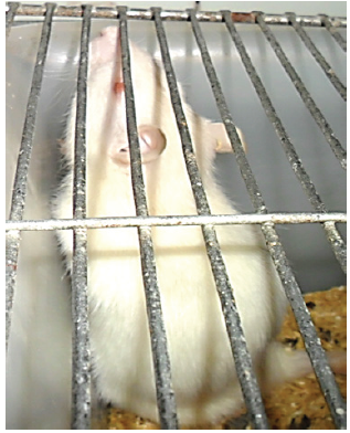
When observing the behavior of experimental animals, it was established that after 60minutes, the rats began to drink water, after 4-5 hours, the entire volume of the proposed drink was drunk-350ml. Control rats, during this time, drank 60ml of water. All the animals of O1 preferred e to water. In the future, they ate food 50% less. The water did not drink. Through 7 days persists preference E, but on 20% increased consumption feed and this raising persisted until the end of experiment. From the first day the behavior of animals changed, increased contact between animals, grooming. So, the first day there was activity and curiosity, after 72 hours the activity decreased, the rats became drowsy and apathetic. Continued to drink E drinks, feed consumption increased by 30%. Communication of rats was limited to communication of individuals within a cage, “grouping” was created, and external stimuli did not distract them from each other. Being in a vertical stand took 20-30% of the time of observation of animals (8 hours), normally 10-15%. An increase in the number of vertical uprights indicates an internal alarm. On day 10 reaction only when adding a new portion of the drink. The absence of E caused concern, the animals were near the drinking trough and waiting for a new portion (Figure 1).

Figure 1:Rats waiting for a new portion.


LA-the effect of these cocktails is similar to the action of energy drinks, but the effect is faster and more pronounced. Through 20-30 minutes they already not departed from drinkers with LA. Dynamics behavior animals: in the first 60-90 minutes arousal, then drowsiness, expressed intoxicating effect, through 5-6 hours increased communication between animals, declined reaction on external stimuli, rats await only drink. Water consumption decreased by 10%, food by 40-50%. Animals are waiting every day they drank 350ml of the drink and demanded more. LA’s absence caused anxiety and aggression. Aggression manifests itself even relative to employees, which daily fed animals, to which animals are accustomed. From the first hours They closed on communication within the “community” of animals, limited to the cell. Being in a vertical rack takes 30-40% of the time, normally 10-15%. This demonstrates the presence of internal anxiety. On day 10, the external reaction only when adding a new portion of the drink. Low feed consumption is maintained. The lack of water is alarming; the animals are next to the drinker and waiting for a new portion. In this group there can be a fight. Rats wait for supplements, which drink, if you add them to the drinker. One rat from 4-’ s unsupportable to drink water and there is fodder, and through 7 days it is died, Figure 3. The attraction to LA and E did not depend on the sex of the experimental animals [3, 4]. The dependence on the age of rats is traced. After autopsy, it was found that young rats have lesions characteristic of old rats- pneumonia, dystrophy and hemorrhages in the myocardium, uneven blood filling of the myocardium uneven blood filling of the myocardium there are areas of weak blood filling alternating with foci of venous-capillary fullness. After that, the behavior of the animals changed while in the cage, a group of animals “closed in their circle”, they almost did not respond to external irritation-the approach of employees to the cage or noise effects: voice, tapping on the cage. Communication between animals was active. They adopted a vertical stance, touching each other with their front paws, which was different from the usual behavior (they sleep during the day). There was an active reaction to the drinking bowl. On the morning on additive a new helpings drinks, in another time on removing this drinker, especially ranging with 3 -’s days use water.

Figure 2:Rats are waiting for a soft drink low alcohol.


Figure 3:White rats, control weight bodies 200 propulsion A. after E mass bodies becomes 350g; after reception LA-120g.


Figure 4:A. Liver closeup, subcapsular hemorrhage, B. Lungs, edema, hemorrhage, C. Pancreatic edema, enhanced vascular pattern. 14 days after using LAD.


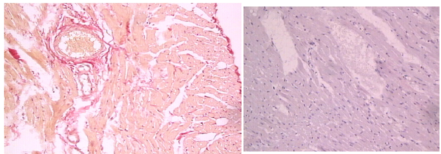
Autopsy on day 14: -in the liver, subcapsular hemorrhage in each animal. Autopsy on day 45: “YOUNG” - starting from day 14, damages in the liver, lungs. pancreatic (Figure 3). Pneumonia in three rats, lungs - abscesses in two rats. Young rats quickly get used to it, increases tolerance to alcoholic beverages. In rats there is a habit and craving for an alcoholic beverage, the intoxicating effect occurs faster than at the beginning of drinking 45% alcohol. There are diseases (pneumonia, myocardial damage) characteristic of older animals. Alcoholic myocardial damage (Figure 4 & 5). All these changes occur faster and more pronounced in those animals that have previously drunk E, it is alarming that by LA and E there is a habit and a need to increase the dose of these drinks. Hierarchy of needs is changing, the instinct of self-preservation is practically no drinking or food craving, especially after LA. The survey of students is anonymous, the data were not disclosed. Students drink these drinks to increase the tone and ability to assimilate new educational material, but teachers note the opposite effect. A small temporary rise in tone (5-6 hours) was replaced by drowsiness and loss of strength. If a student consumed several cans of e, then memorization and assimilation of new skills decreased

Figure 5:Rats. Microphotographs of the myocardium after LA (h&e x160).


Conclusion

Young animals lose sense of self-restraint relative to LA and E. They drink all, that give and require still, in the absence of drinks emerges aggression to other animals and to service personnel. In young animals, the motivation for self-preservation is reduced. In the cell, groups of animals are created, showing interest in each other only within the group. In young animals, damage to the heart, lungs, kidneys, liver was noted, occurring only in old rats. Rejuvenation of chronic diseases. Reception of the studied drinks facilitates the transition to stronger alcoholic beverages (vodka), control animals do not drink vodka of any concentration. Observation of animals allows you to dispense drinks. We know the beginning of the use and could assess the behavior of animals for a long time period. Young people who used e did not have the effect that they expected, but there was an addiction to E. We believe that we can translate the data obtained from animals to humans.

References

  1. Trubitsyna IE, Fedotova TF, Mikhalev IV, Efremov LI, Kleschev VN, et al. (2018) Alcohol-associated internal injuries in rats. Clin Exp Gastroenterol 6(154): 90-93.
  2. Trubitsyna I, Fedotova T, Varvanina G (2019) Metabolic changes in violation in the system of neuro-endocrine regulation after consumption of energy and small-alcoholic beverages. “2nd World Congress on Gastroenterology & Hepatology”, London, UK, p.12.
  3. Acheson A, Mahler SV, Chi H, De Wit H (2006) Differential effects of nicotine on alcohol consumption in men and women. Psychopharmacology (Berl) 186(1): 54-63.
  4. Barrett SP, Tichauer M, Leyton M, Pihl RO (2006) Nicotine increases alcohol self-administration in non-dependent male smokers. Drug Alcohol Depend 81(2): 197-204.


For more articles in Gastroenterology Medicine & Research
Please click on below link: https://crimsonpublishers.com/gmr/

Monday, December 6, 2021

Molecular Detection of Epstein Barr Virus, Human Papilloma Virus Types 16,18 in Breast Cancer Patients in Khartoum State Sudan_Crimson Publishers

 Molecular Detection of Epstein Barr Virus, Human Papilloma Virus Types 16,18 in Breast Cancer Patients in Khartoum State Sudan by Khalid A Enan in Novel Approaches in Cancer Study_Journal of Cancer Research

Abstract

Background: Breast cancer is the most common among female, constituting about 18% of all female cancers, with 1.7 million new cases reported in the world each year. Recently some studies reported that approximately 18% of cancer cases can be linked to infectious agents including viruses particularly Human Papilloma Virus (HPV) and Epstein Barr virus (EBV).

Objective: Molecular detection of EBV and HPV types 16 and 18 in breast cancer patients in Khartoum State, Sudan.

Methods: Paraffin embedded blocks of tumor specimen from 70 Sudanese patients with breast cancer were collected from Omdurman teaching Hospital, Sudan, during the period from March to June 2018. PCR was used to investigate the presence of EBV and HPV type 16, 18 viruses in these specimens.

Results: The results show that eight out of 70 patients were positive for EBV virus (11.4%) Of these positive patients, 3(4.2%) were 30-50, 3(4.2%) were 51-80 and 2(2.8%) were 81-100 years old, respectively. Seven out of the 70 patients were positive for HPV type 18(10%). Of these positive patients, 5 (7.1%) were 30-50years old and 2 (2.9%) were 50-80 years old. None of the patients were found positive for HPV type 16. No significant differences were found between age group as regards infection by EBV or HPV type 18 viruses.

Conclusion: The incidence of EBV and HPV types 18 in breast cancer patients in Khartoum State was documented through the molecular detection of these two virus’s DNA. Detection of EBV and HPV type18 using PCR was established. Generally, these findings are useful for future studies since there is little information available about of EBV and HPV types 16, 18 in Sudan.

Keywords: EBV and HPV16,18; Breast Cancer; Khartoum State; Sudan

Introduction

Breast cancer is a type of cancer originating from breast tissue, most commonly from the inner lining of milk ducts or the lobules that supply the ducts with milk [1]. The size, stage, rate of growth, and other characteristics of the tumor determine the kinds of treatment. Treatment may include surgery, drugs (hormonal therapy and chemotherapy), radiation and/ or immunotherapy [2]. Many factors including radiation, chemicals and viruses, have been found to induce human cancer [3]. Viral factors are the most important class of the infectious agents associated with human cancers [4]. It was estimated that 17-20 % of the worldwide incidence of cancers was attributable to a viral etiology [5]. Infections with oncogenic viruses have been investigated as possible risk factors for a breast cancer aetiology including mousemammary tumor virus (MMTV), Epstein-Barr virus (EBV) and human papilloma virus (HPV) especially types 16, 18, and 33; however, their presice role is not clear. EBV was the first human virus to be directly implicated in carcinogenesis. Epstein Barr virus; a common infection affecting over 90% of the world’s population is one of the viruses that have some unclear and controversial points over its ability to trigger the development of certain tumors [6] such as Burkett’s lymphoma, nasopharyngeal carcinoma, Hodgkin’s disease, gastric carcinoma and post-transplant lymphoproliferative disease [7].

The small untranslated RNAs EBER-1 and -2 are accumulated at high levels during all forms of latency and regulate apoptosis through different mechanisms which play a critical role in efficiency of EBV-induced growth transformation of primary B cells.EBER-1 interacts with the interferon-inducible protein kinase R (PKR), and inhibits its activation by double stranded RNAs, protecting infected cells from IFN-induced apoptosis [8]. However, Wu et al. [9] also reported that EBVencoded RNA 2 (EBER2) but not EBER1 playing a critical role in EBV-induced B-Cell growth transformation. In Sudan, breast cancer is characterized by a geographically focal nature, early onset and aggressive course of the disease [10]. BRCA1, BRCA2 and p53 mutations are infrequent in Sudanese breast cancer patients. Epigenetic changes are suggested as alternative mechanisms to account for the minor contribution in the genetic alterations in three tumor suppressor genes, BRCA1, BRCA2, and p53, in both sporadic and familial breast cancer cases in Sudan [11].

Materials and Methods

Patient criteria and specimen collection

Paraffin embedded blocks of tumor specimens from 70 Sudanese patients with breast cancer were collected from Omdurman Teaching Hospital Sudan during the period from March to June 2018. The collected specimens were stored at room temperature until the test was performed.

Specimen deparaffinization: This was carried out as recommended in the protocol using the DNA extraction kit (Acrogene, USA) The specimens were deparaffinized according to the protocol of the manufacturerDNA extraction procedure was carried out according to manufacturer’s protocol (Acrogene, USA).

DNA extraction

The procedure was carried out according to manufacturer’s protocol (Acrogene ,USA).

Polymerase Chain Reaction (PCR) for detection of EBV: The PCR was performed using primers that are specific for the EBV (gB) conserved regions. The primers used consisted of forward primer SL1, 5-GGACCTCAAAGAAGAGGGGG-3 and the reverse primer SL3, 5-GCTCCTGGTCTTCCGCCTCC-3. The reaction was performed in 25μL volume of PCR PreMix master mix (Intron Biotechnology, Korea). The volume included 5μL master mix, μL forward primer (10pg), 1μL reverse primer (10pg), 5μL extracted DNA and 12μL distilled water. The DNA was amplified in a thermo cycling condition using PCR machine (Techno, Japan) as follow: initial denaturation at 95ºC for 10min, followed by 35 cycles of denaturation at 95ºC for 45 sec, annealing at 60ºC for 45sec and extension at 72ºC for 60sec, with final extension at 72ºC for 10min. The expected size of UTR gene amplicon was 80bp [12].

Polymerase Chain Reaction (PCR) for detection of HPV types 16 and 18: Samples were amplified separately for HPV 16 and 18 with a thermocycler (Techno, Japan), and primers specific for each viral type derived from previously published sequences (12,40). The sequence of primers for HPV 16 DNA were F, 5'TTTlGGGTTACA CAT'TACAAG3' (residues 7864 to 7885), and R, 5'TGTC TGCTJT'J'7'ATACTAACCG3' (residues 57 to 78), which generated a 119-bp product from the URR region. The sequence of primers for HPV 18 DNA were, F 5'GACACCTTAATGAAAAACGACGA3'(residues 460 to 482), and R,5'CGTCGTTGGAGTCGTTCCTG3' (residues 543 to 562), which amplified a 103-bp fragment from open reading frame E6. The reaction was performed in 25μL volume of PCR PreMix master mix (Intron Biotechnology, Korea). The volume included 5μL master mix, 1μL forward primer (10pg), 1μL reverse primer (10pg), 5μL extracted DNA and 12μL distilled water. DNA was amplified as follow: 35 cycles of denaturation at 94ºC for 45sec, annealing at 54ºC for HPV 16 or 58ºC for HPV 18 for 1min 45sec and extension at 74ºC for 30sec [13].

Agarose gel electrophoresis

10μL of amplified product was analyzed by gel electrophoresis in 2% agarose stained with 0.15% ethidium bromide and visualized by using UV gel documentation system (INGeNiuse Germany).

Statistical analysis

Collected data were analyzed using statistical package for social science (SPSS version 12.0). A p value of ≤ 0.05 was considered significant.

Result

Detection of EBV using PCR in breast cancer patients

Eight out of 70 patients were positive for EBV virus (11.4%). Of these positive patients, 3(4.2%) were 30-50, 3(4.2%) were 51-80 and 2(2.8%) were 81-100 years old respectively. Seven out of the 70 patients were positive for HPV type 18(10%). Of these positive patients, 3(4.2%) were (30-50), 3(4.2%) were (51-80) and 2(2.8%) were (81-100) years old, respectively as shown in (Table 1) but with no significant differences.

Detection of HPV types 16, 18 using PCR in breast cancer patients

Table 1: Detection of EBV, HPV types 16, 18 in breast cancer patients.

*means significant difference Note: No of patients in age group 30-50 is 43, in age group 50-80 is 20 and in 81-100 is 3.


Seven out of 70 patients were positive for HPV type 18(10%). Of these positive patients, 5 (7.1%) were 30-50 years old and 2 (2.9%) were 50-80 years old) as shown in (Table 1). All the samples were negative for HPV type 16.

Discussion

Breast cancer is the most common malignancy among females and comprises about 18% of all cancers affecting them. About 1.7 million new cases are reported in the world each year. Based on the most global recent data, approximately 12.3% of women are diagnosed with breast cancer at a point of time during their life. Little is known about the viral causes of breast cancer and their epidemiology in Africa. Even much less is known about the epidemiology of viruses associated with breast cancer in Sudan in particular and in Africa in general. Our current study in Sudan is one of the few studies to report directly measured rates of EBV and HPV types 16, 18 associated with breast cancer patients in Sudan. Some studies have reported on the findings that EBV, HPV types 16, 18 play a role in the development of breast cancer including studies from Sudan [14-18], where this were reported in Sudan [16,19]. On the other hand, several studies failed to detect HPV positivity in breast carcinoma [20] even by studying HPV subtypes 6b, 11, 13, 16, 18, 30, 31, 32, 33, 45, and 51 in 95 women with breast cancer without detecting any of the subtypes [21] researched the HPV subtypes 6, 11, 16, and 18 in 13 IDC, 15 papillomas, and 15 papillary carcinomas cases, and there was no evidence of HPV infection [22] did a similar study by using six different primers, including a total of 40 subtypes 16, 18, 31, 33, and 45 in 81 Swiss women cases with breast cancer with no positivity of HPV detected [17].

When studying the prevalence of subtypes of high (16, 18, 33, and 45) and low risks (6, 11) in 50 breast cancers in women in France by PCR with the general primer GP5+/GP6+, where none of these subtypes were detected in both cases [23]. This may be due to the lack of standardized technique to detect the presence of HPV, since there are different types of primers for different HPV subtypes. False negatives and false positives may occur when PCR overestimates the association between HPV and breast cancer because it cannot indicate which types of cells the virus has infected. Contamination while handling the sample may be partially responsible for the high frequencies of HPV positivity that were reported in several papers. Other risk factors might affect the outcome of the results. For example, studies have shown that women under the age of 25 have a higher prevalence of HPV positivity detection with linear decreasing rate as age increases [24]. Storage of specimens may affect the results, since some researchers found that positive specimens became negative after being frozen for 3 months [25]. Demographic features and genetic backgrounds may also contribute to the geographic difference and HPV infection in breast cancer. The present study focused on the molecular detection of EBV and high-risk HPV types 16 and 18 in breast cancer patients in the Khartoum State. Elnoubi [19] was the first to detect HPV type 16,18 in breast cancer patients in Sudan. The author detected HPV genotype 16, in 21(31%), and HPV genotype 18, in 10(15%) of the tested patients. On the other hand, Adam detected EBV genome in 49 (53.3%) and 10 (11%) patients by LMP-1 and EBNA-4 PCR, respectively. Based on the results of the present study, findings of Elnoubi [19] and Adam AA [16] that suggest that, EBV, HPV type 18 infection may be common in Sudan. The present study in addition to the above-mentioned studies could serve as a baseline for future plans aiming at introducing the vaccine against EBV and HPV in the Sudan. Finally; these findings should highlight the need for the establishment in Sudan of rapid, sensitive, and specific diagnostic techniques (such as ones used here) to better understand the role played by various viruses in the aetiology of breast cancer development in Sudan.

References

  1. Florescu A, Amir E, Bouganim N, Clemons M (2011) Immune therapy for breast cancer in 2010-hype or hope. Current Oncology 18(1): e9-e18.
  2. De villiers EM (2003) Relationship between steroid hormone contraceptives and HPV, cervical interepithelial neoplasia and cervical carcinoma. Int J Cancer 103(6): 705-708.
  3. Mao C, Hughes JP, Kiviat N, Kuypers J, Lee SK, et al. (2003) Clinical findings among young women with genital human papillomavirus infection. Am J Obstet Gynecol 188(3): 677-684.
  4. Clifford GM, Smith S, Aguado T, Franceschi S (2003) Comparison of HPV type distribution in high-grade cervical lesions and cervical cancer: A meta-analysis. Br J Cancer 89(1): 101-105.
  5. Vokes EE, Liebowitz DN, Weichselbaum RR (1997) Nasopharyngeal carcinoma. Lancet 350(9084): 1087-1091.
  6. Thornhill Cher (2008) Epstein Barr virus implicated in bladder cancer progression. Int J Urol 15(5): 429-434.
  7. Nanbo A, Inoue K, Adachi-Takasawa K, Takada K (2002) Epstein-Barr virus RNA confers resistance to interferon alpha- induced apoptosis in Burkitt’s lymphoma. Embo J 21(5): 954-965.
  8. Abe T, Nobuo S, Mitsuhiro T, Toru H, Ataru S, et al. (2008) Infiltration of Epstein Barr virus harboring lymphocytes occur in a large subset of bladder cancers. Int J Urol 15(5): 429-434.
  9. Wu Y, Maruo S, Yajima M, Kanda T, Takada K (2007) Epstein-Barr Virus (EBV)-Encoded RNA 2 (EBER2) but not EBER1 plays a critical role in EBV-induced B-cell growth transformation. Journal of Virology 81(20): 11236-11245.
  10. Masri MA, Abdel Seed NM, Fahal AH, Romano M, Baralle F, et al. (2002) Minor role for BRCA2 (exon11) and p53 (Exon 5-9) among Sudanese breast cancer patients. Breast Cancer Research and Breast Cancer Res Treat 71(2): 145-147.
  11. Burchill SA, Bradbury MF, Pittman K, Southgate J, Smith B, et al. (2005) Detection of epithelial cancer cells in peripheral blood by reverse transcriptase-polymerase chain reaction. Br J Cancer 71(2): 278-281.
  12. Margall N, Matias-Guiu X, Chillon M, Coll P, Alejo M, et al. (1993) Detection of human papillomavirus 16 and 18 DNA in epithelial lesions of the lower genital tract by in situ hybridization and polymerase chain reaction: cervical scrapes are not substitutes for biopsies. J Clin Microbiol 31(4): 924-930.
  13. Shunji M, Hiromi S, Yuji T, Hoichi K, Hiroshi W, et al. (1997) Absence of human papillomavirus-16 and -18 DNA and Epstein–Barr virus DNA in esophageal squamous cell carcinoma. Jpn J Clin Oncol 27(1): 1-5.
  14. Bensaber HS, Bicout DJ, Medjamia M, Bensnouci AM, Comez A, et al. (2017) Molecular detection of Epstein Barr virus in women with breast cancer in the west Algeria. Journal of Cancer Therapy 8(3): 16.
  15. Fessahaye G, Elhassan AM, Elamin EM, Adam AAM, Ghebremedhin A, et al. (2017) Association of Epstein - Barr virus and breast cancer in Eritrea. Infectious Agents and Cancer 12: 62.
  16. Yahia ZA, Adam AA, Elgizouli M, Hussein A, Masri MA, et al. (2014) Epstein Barr virus: A prime candidate of breast cancer aetiology in Sudanese patients. Infectious Agents and Cancer 9(1): 9.
  17. Delgado-García S, Martínez-Escoriza JC, Alba A, Martín-Bayón TA, Ballester-Galiana H, et al. (2017) Presence of human papillomavirus DNA in breast cancer: A Spanish case-control study. BMC Cancer17(1): 320.
  18. Choi J, Kim C, Lee HS, Choi YJ, Kim HY, et al. (2016) Detection of human papillomavirus in Korean breast cancer patients by real-time polymerase chain reaction and meta-analysis of human papillomavirus and breast cancer. Journal of Pathology and Translational Medicine 50(6): 442-450.
  19. Elnoubi, Osman Abdalla Eltayeb (2016) Molecular diagnosis of HPV isolated from breast cancer patients in Radiation and Isotopes Center Khartoum (RICK) -Sudan, Shendi University, Sudan.
  20. Wrede D, Luqmani YA, Coombes RC, Vousden KH (1992) Absence of HPV 16 and 18 DNA in breast cancer. British Journal of Cancer 65(6): 891-894.
  21. Bratthauer GL, Tavassoli FA, O'Leary TJ (1992) Etiology of breast carcinoma: no apparent role for papillomavirus types 6/11/16/18. Pathology Research and Practice 188(3): 384-386.
  22. Lindel K, Forster A, Altermatt HJ, Greiner R, Gruber G (2007) Breast cancer and human papillomavirus (HPV) infection: No evidence of a viral etiology in a group of Swiss women. Breast 16(2): 172-177.
  23. Girianelli VR, Thuler LCS, Silva GAE (2010) Prevalence of HPV infection among women covered by the family health program in the Baixada Fluminense, Rio de Janeiro, Brazil. Revista Brasileira de Ginecologia e Obstetricia 32(1): 39-46.
  24. Chang P, Wang T, Yao Q, Lv Y, Zhang J, et al. (2012) Absence of human papillomavirus in patients with breast cancer in north-west China. Medical Oncology 29(2): 521-525.

For more articles in Journal of Cancer Research
Please click on below link: https://crimsonpublishers.com/nacs/


Friday, December 3, 2021

Food Proteins as a Tool in Human Longevity: A Mini-Review_Crimson Publishers

 Food Proteins as a Tool in Human Longevity: A Mini-Review by Olga Luisa Tavano in Novel Techniques in Nutrition and Food Science_Food science journals

Abstract

Demographic transition and interest in human longevity increasingly guide studies on the best quality of the human diet, a factor directly related to the lifespan. As the world's population is aging, with more people over 65, dietary recommendations specific to this context should be discussed and adopted. Proteins are related to this theme both for their fundamental role as nutrients, but also as nutraceutical potentials that act not only for the maintenance of health, but for the prolongation of life. Thus, there is also a growing interest in research on the effects of ingestion of peptides with different bioactivities.

Keywords: Longevity; Older people; Elderly; Lifespan; Protein intake; Bioactive peptides

Introduction

Human longevity is a highly complex phenomenon influenced by a variety of social, cultural, environmental, genetic and epigenetic factors that isolated or associated influence the duration of human life. Due to the demographic transition, the percentage of people over 65 has increased and the idea of living beyond 120 is not a distant utopia, as discussed by Finkel [1]. To reach an extremely advanced age, genetics is gaining relevance considering that dysregulation of transcription and chromatin networks is probably a strategic target. In this sense, so-called epigenetic diets can be an important contribution to healthy aging and longevity and human proteins deserve a prominent role [2]. Many evidence-based studies recognize nutrition as a key factor in life extension [3-5]. Proteins are important dietary factors associated with maintaining life, promoting health both for its nutrient performance and for its nutraceutical potential.

Nutritional aspects

Life expectancy and metabolic health are strongly influenced by dietary interventions [6], whereas in this context the control of the quantity and quality of protein intake is of great impact [7]. For example, evidence‐based studies indicate that protein supplementation may help prevent sarcopenia, an age-related decrease in muscle mass and performance [8]. Richter et al. [9] discuss the need to adopt differentiated reference values when it comes to protein intake for older people, and they indicate the need to adopt a reference value of 1.0g protein/kg body weight per day for adults >65 years, above 0.8g protein/kg bw/day currently recommended for adults of any age. This differentiated protein intake would be recommended to prevent sarcopenia [10] and to promote better disability trajectories in the elderly. Mendonça et al. [11] present data that suggest further progress in this discussion, opening the need for perhaps considering the division into more age groups to establish recommendations specific to each need. When they determined a relationship between protein intake and disability for adults older than 85 years, they found that higher protein intake (such as 1.0g/kg body weight/day or higher) was associated with lower protein intake and development of disability in older adults Houston et al. [12], when studying the relationship of protein intake by adults between 70 and 79 years and their loss of lean mass, found that those who consumed an average of 1.1g/kg/day lost 40% less lean mass than those averaged 0.8 g/kg bw/day. The PROT-AGE Study Group highlight two important recommendations for healthy older adults (> 65): “To maintain and regain muscle, older people should consume an average daily intake in the range of 1.0 to 1.2 g/kg bw/d”, and still: “25 to 30g protein per meal, containing about 2.5 to 2.8g leucine” [13]. But while some studies cite increased leucine intake as positive, caution is needed, as others associate high dietary BCAA as a cardiometabolic risk [14]. Not only can the intake of some specific amino acids be considered, but the ratio between them seems to be important to lifespan.

Romano et al. [15] pointed out that an adequate intake of essential and non-essential amino acids is crucial for preserving cellular integrity and healthy whole-body metabolism in mice. They observed that different proportions of essential and non-essential, limiting essential or exceeding non-essential, may increase or decrease lifespan, and high or unbalanced non-essential amino acids intake provided by food proteins can be detrimental to life expectancy. It is important to highlight that the issue of protein consumption should be better discussed, since the influence of food matrix interaction has been briefly explored and may bring new considerations about the benefits of the protein sources themselves [7]. Some authors, for example, already approach plant-based proteins in this context, highlighting the advantages of these proteins in the context of their matrix [16].

Nutraceutical aspects

Although the classic nutritional aspect of the proteins has been present in the majority of studies that provided evidence about relationship protein and healthy lifespan, the nutraceutical contribution of proteins is important too. Intact proteins and free amino acids may have impact functions on our metabolism, but peptides have shown greater potential for bioactivity. Compared to intact proteins, peptides have the advantage that they can be absorbed into the human gut and penetrate cells where their function could be performed. Bioactive peptides have been demonstrating their potential for positive interference in human metabolism, both in a curative and preventive axis of different metabolic aspects that may be directly or indirectly linked to life expectancy. In general, bioactive peptides are short food protein sequences composed mainly of 2 to 20 amino acid residues and it is these protein-derived molecules that can have metabolic and “epigenetic effect [17,18]. “

The delivery of peptides may be in different ways, including release during natural digestion of foods or oral administration of peptides synthesized or obtained by hydrolysis of different protein sources. They can be absorbed through the small-intestinal epithelium by passive diffusion mechanisms, carrier-mediated transport via PEPT1, endocytosis or paracellular mechanisms [19]. The application of bioactive peptides as health promoters has been highlighted, among other advantages, they are considered natural agents with low toxicity. Different bifunctionalities have been demonstrated in in vitro and in vivo studies. Many of them closely related to the promotion of human longevity. Following are some of these potential peptide bioactivities:

a) Antihypertensive peptides- The antihypertensive activity of peptides is one of the most cited in the literature, and these peptides have appropriate sequences to inhibit angiotensin-I converting enzyme (ACE-I), an enzyme of the renin angiotensin system. This inhibition promotes an important regulation of the cardiovascular function and blood pressure by both competitive and non-competitive modes. Peptides with few amino acids, and with N-terminal containing leucine, isoleucine and valine, and/or C-terminal with aromatic amino acids have been pointed as the most active [20].

b) Antioxidant peptides- peptides may have antioxidant activity. Although the exact mechanism for this function is unclear, it is suggested that its action is due to different mechanisms such as: inhibition of singlet oxygen-quenching potential, harmful lipid oxidation, free radical scavenging, and/or chelation of metal ions. Antioxidants can neutralize the deleterious effect of the excess of free radicals which can produce negative effects on human health, such as damage in proteins and cell membranes or DNA mutations. Characteristics like hydrophobic amino acids in their N-terminal regions and/or the presence of Cys, Met, His, Phe, Trp and Tyr may favor the antioxidant properties [21,22].

c) DPP-IV inhibitors- Many peptides present as potential as inhibitors of the dipeptidyl peptidase IV, an enzyme involved in the regulation of serum glucose, this is, in the control of type 2 diabetes in humans. In general, hydrophobic amino acids are present in DPP-IV inhibitory peptides, what increase the interaction with the active site of DPP-IV [23].

d) Anti-cancer peptides- Some peptides have the ability to induce cytoplasmic membrane disruption, both through micellization and pore opening, inducing apoptosis. The cationic characteristic has been found among the most potent [24].

e) Neuroprotective peptides- Some peptides can help maintain a healthy brain by contributing to the maintenance of cognitive functions, which would be an important factor in preventing the cognitive impairment or dementia, debilitating age-related disorders. Considering that brain neurogenesis decreases during ageing, peptides that induced proliferation and increased survival and maturation of neural progenitor cells into neurons could prevent age-related cognitive decline [25].

f) Anti-inflammatory properties- Peptides have been studied as possible anti-inflammatory agents. Considering that chronic inflammation may be related to causes of life-shortening illnesses, such as obesity, type-2 diabetes or cardiovascular diseases, but chronic use of anti-inflammatory drugs may lead to side effects such as functional renal impairment in elderly subjects [26]. The application of these peptides can be of great importance. Many anti-inflammatory peptides contain positively charged amino acids, such as Arg and Lys, especially at their C- or N- terminus, and prominent hydrophobicity, which is a major factor associated with their anti-inflammatory potential [27].

g) Antithrombotic peptides- Peptides can present anticoagulant activity. A classic example is the hirudin an anticoagulant peptide produced in the salivary glands of leech, able to bind on thrombin and avoiding the cleavage of fibrinogen and the formation of the fibrin clot [28]. If well administered, they can help in the treatment of thrombotic diseases [29].

h) Multifunctional properties- The same peptide may have the ability to exert more than one benefit to human health. For example, with respect to the prevention of cardiovascular disease, the peptides may be hypocholesterolemia and antidiabetic, or further accumulate functions such as lumazine, which is hypocholesterolemia, antioxidant and anti-inflammatory [30].

Conclusion

Specific recommendations for age subgroups over 65 may be useful for the prevention of lifelong disabilities of older people. Research evidence indicates that an intake of at least 1g protein/kg body weight/day is significantly beneficial for healthy longevity. It is useful to guide protein intake considering its balance between essential and nonessential amino acids and even the potential for release and absorption of bifunctional peptides during digestion. Bifunctional peptides may play an important role in preventing diseases such as cancer, cardiovascular disease, and which both threaten the duration of life and may impair its quality.

References

  1. Finkel T (2019) The enlightenment of age. Nature 573: 193-194.
  2. Morris BJ, Willcox BJ, Donlonb TA (2019) Genetic and epigenetic regulation of human aging and longevity. Biochim Biophys Acta Mol Basis Dis 1865(7): 1718–1744.
  3. Abbot A (2018) Reduced-calorie diet shows signs of slowing ageing in people. Nature 555(7698): 570-571.
  4. Costa D, Scognamiglio M, Fiorito C, Benincasa G, Napoli C (2019) Genetic background, epigenetic factors and dietary interventions which influence human longevity. Biogerontology 20(5): 605-626.
  5. Chrysohoou C, Stefanadis C (2019) Longevity and diet. Myth or pragmatism? Maturitas 76(4): 303-307.
  6. Kitada M, Ogura Y, Monno I, Koya D (2019) The impact of dietary protein intake on longevity and metabolic health. EBioMedicine 43: 632-640.
  7. Burd NA, McKenna CF, Salvador AF, Paulussen KJM, Moore DR (2019) Dietary protein quantity, quality, and exercise are key to healthy living: a muscle-centric perspective across the lifespan. Front Nutr 6: 83.
  8. Beasley JM, Shikany JM, Thomson CA (2013) The role of dietary protein intake in the prevention of sarcopenia of aging. Nutr Clin Pract 28(6): 684-690.
  9. Richter M, Baerlocher K, Bauer JM, Elmadf I, Heseker H, et al. (2019) Revised reference values for the intake of protein. Ann Nutr Metab 74(3): 242-250.
  10. Phillips SM (2017) Determining the protein needs of “older” persons one meal at a time. Am J Clin Nutr 105(2): 291-292.
  11. Mendonça N, Granic A, Hill TR, Siervo M Mathers JC, et al. (2019) Protein intake and disability trajectories in very old adults: The Newcastle 85+ study. J Am Geriatr Soc 67(1): 50-56.
  12. Houston DK, Nicklas BJ, Ding J, Harris TB, Tylavsky FA, et al. (2008) Dietary protein intake is associated with lean mass change in older, community-dwelling adults: The health, ageing, and body composition (Health ABC) study. Am J Clin Nutr 87(1): 150-155.
  13. Bauer J, Jentoft AJC, Morley JE, Phillips S, Sieber C, et al. (2013) Evidence-based recommendations for optimal dietary protein intake in older people: A position paper from the PROT-AGE study group. J Am Med Dir Assoc 14(8): 542-559.
  14. White PJ, Newgard CB (2019) Branched-chain amino acids in disease. Science 363(6427): 582-583.
  15. Romano C, Corsetti G, Flati V, Pasini E, Picca A, et al. (2019) Influence of diets with varying essential/nonessential amino acid ratios on mouse lifespan. Nutrients 11(6): E1367.
  16. Lonnie M, Hooker E, Brunstrom JM, Corfe BM, Green MA, et al. (2018) Protein for life: review of optimal protein intake, sustainable dietary sources and the effect on appetite in ageing adults. Nutrients 10(3): E360.
  17. Bhandari D, Rafiq S, Gat Y, Gat P, Waghmare R, et al. (2019) A review on bioactive peptides: physiological functions, bioavailability and safety. Int J Pept Res Ther 3: 1-12.
  18. Chalamaiaha M, Ulug SK, Hong H, Wu J (2019) Regulatory requirements of bioactive peptides (protein hydrolysates) from food proteins. J Func Foods 58: 123-129.
  19. Williams WMM, Stevens BR, Moughan PJ (2014) Are intact peptides absorbed from the healthy gut in the adult human? Nutr Res Rev 27(2): 308-329.
  20. Premkumar J, Malini M, Joshy VA (2019) A critical review on food protein derived antihypertensive peptides. Drug Inv Today 12(3): 474-479.
  21. Tavano OL, Murcia AB, Secundo F, Lafuente RF (2018) Biotechnological applications of proteases in food technology. Compr Rev Food Sci Food Saf 17(2): 412-436.
  22. Nwachukwu ID, Aluko RE (2019) Structural and functional properties of food protein‐derived antioxidant peptides. J Food Biochem 43(1): 1-13.
  23. Nongonierma AB, FitzGerald RJ (2017) Features of dipeptidyl peptidase IV (DPP-IV) inhibitory peptides from dietary proteins. J Food Biochem 43(1): 1-11.
  24. Aaghaz S, Gohe V, Kama A (2019) Peptides as potential anticancer agents. Curr Top Med Chem 19(17): 1491-1511.
  25. Katayama S, Nakamura S (2019) Emerging roles of bioactive peptides on brain health promotion. Int J Food Sci Technol 54(60): 1949-1955.
  26. Henry D, Page J, Whyte I, Nanra R, Hall C (1997) Consumption of non-steroidal anti-inflammatory drugs and the development of functional renal impairment in elderly subjects. Results of a case-control study. Br J Clin Pharmacol 44(1): 85-90.
  27. Guha S, Majumder K (2019) Structural-features of food-derived bioactive peptides with anti-inflammatory activity: A brief review. J Food Biochem 43(1): 1-14.
  28. Krstenansky JL, Owen TJ, Yates MT, Mao SJT (1987) Anticoagulant peptides. Nature of the interaction of the C-terminal region of hirudin with a noncatalytic binding site on thrombin. J Med Chem 30(9): 1688-1691.
  29. Cheng S, Tu M, Liu H, Zhao G, Du M (2019) Food-derived antithrombotic peptides: Preparation, identification, and interactions with thrombin. Crit Rev Food Sci Nutr 59(sup1): S81-S95.
  30. Lammi C, Aiello G, Boschin G, Arnoldi A (2019) Multifunctional peptides for the prevention of cardiovascular disease: A new concept in the area of bioactive food-derived peptides. J Func Foods 55: 135-145.

https://crimsonpublishers.com/ntnf/fulltext/NTNF.000590.php


For more articles in Food science journals
Please click on below link: https://crimsonpublishers.com/ntnf/

Thursday, December 2, 2021

Towards Green Cements: The Metakaolin Route_Crimson Publishers

 Towards Green Cements: The Metakaolin Route by Henry F Meier in Aspects in Mining & Mineral Science_Open access journals in Mining & Mineral Science


Opinion

One of the main challenges to be faced by the industry in this 21st century is the drastic reduction of its gaseous emissions, whether with the presence of particulate matter or substances that are aggressive to the environment. Both are problems found in the cement industry, which is responsible for up to 7% of carbon dioxide emissions produced by industrial human activity, which leads to the greenhouse effect [1]. This is due to two reasons: the first is that the raw material used for the production of clinker in rotary cement kilns is the limestone that undergoes a high temperature decarbonation reaction, with the transformation of carbonates into oxides and CO2 emission in the gaseous form, releasing fossilized carbon that had been purged from the natural carbon cycle in the atmosphere; the second is that the thermal energy used to supply the energy demand for the reactions comes from the use of solid fossil fuels such as petroleum coke. The CO2 average rate production is of the order of 800 kg of CO2/ton of cement, due to the calcination of limestone. Of this amount, 50% comes from decarbonation and 40% from burning fossil fuels to supply energy demand. Efforts have been made to reduce emission rates, such as the use of biofuels, increase of clays in the raw material, and absorption of CO2 emissions. An alternative that has been evaluated is the use of pozzolanic clays in a reactive state as supplementary material [2], a technology that has been developed but which is not yet widely applied in the cement industry. The calcination of clays promotes the dehydroxylation of the molecules by the action of heat, forming only water vapor as a gaseous emission and an amorphous material called metakaolin. Metakaolin is highly reactive with water and forms a higher concentration of porous, low density spheres, with high absorption capacity and large surface area, suitable for clinker replacement. Metakaolin is known as low carbon cement, or “green cement”, and can be used as a substitute or as an additive to conventional cement since it has better pozzolanic properties than Portland cement, such as [3]: increased resistance to compression and flexion, chemical attack and durability; the decrease in alkali-silica permeability and reactivity; facilitating the application of concrete, improves finish and appearance. In addition to the precursor kaolin, metakaolin can be obtained from recycled paper sludge, thus responding to more than one environmental appeal.

The calcination process of clays can be carried out in different ways, but the most promising is the “flash” calcination, in which the particulate solid kaolin is exposed to a gas stream at high temperature (500-900 °C) and in an adequate proportion for a few seconds, being cooled quickly afterwards [4,5]. Another way of production is through slow calcination in rotary kilns or in fixed beds (“soak calcination”), which requires residence times in the order of tens of minutes to hours due to low heating rates. San Nicolas et al. [5] noted that the metakaolin produced by the “flash” process, with a temperature between 600 and 700 °C, contained a greater amount of spherical particles compared to that produced in a rotary kiln, which made it easier to apply, in addition to finding no significant difference regarding the pozzolanic activity of the products of each process. Claverie et al. [6] also found that the spherical particles were composed of gases and aluminosilicates with varied crystallinity. This heterogeneity was credited to the temperature gradient and the cyclonic movement of the flow in the calciner. By controlling the particles residence time in the calciner, different degrees of reactivity and dehydroxylation can be obtained. However, when exposed to temperatures greater than 900 °C and/or maintained for an excessive period in the calciner, metakaolin changes to other physical forms such as amorphous silica, mullite and glassy/crystalline silica. These latter compounds are no longer interesting as supplementary material, since they lose pozzolanic activity. Since kaolin calcination requires greater control of the mass ratio between hot gases and kaolin, temperature and residence time of the particles, its large-scale production becomes an engineering challenge. If this process is not well controlled, metakaolin can be transformed into undesirable materials. Still, the optimum temperature and calcination period are not yet known [7]. Investigations of the thermofluidodynamics of this reaction system, therefore, are fundamental for the deep understanding of calcination and, consequently, to advance in the development of this technology. However, greater attention has been paid to mathematical models for calcination of limestone, a fundamental raw material for cement, than for kaolin calcination. Possibly the first attempt in this direction was made by Salvador and Davies [8,9]. Teklay et al. [9] proposed a mathematical model for the chemical and physical conversions that occur in the kaolinitic clay particle, and subsequently evaluated the “flash” calcination in a pilot-scale reactor [9].

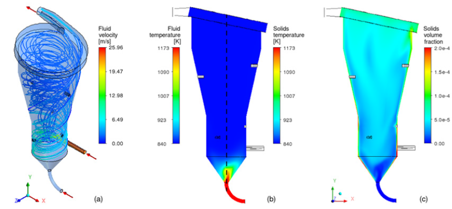
Numerous experimental studies of dehydroxylation in a rapid reaction system (“flash”) with clays from different regions of Brazil have been carried out, showing that the process is technically and economically viable. Given that experimental tests have already been carried out successfully on a pilot scale, the transfer of this knowledge to industrial application remains, which requires a methodology adjusted for the complex calcination of kaolin in large scale. The proper investigations of reactors with large geometric proportions can be done using the Computational Fluid Dynamics (CFD) technique. In this sense, simulations are performed with a reactive multiphase model, capable of predicting in detail the pressure, velocity, temperature and chemical species concentration under reactive and turbulent conditions with heat and mass transfer. (Figure 1) presents simulation results of the rotary flow of kaolin and hot gases in an innovative configuration of calciner, in which a good thermal exchange is observed between the phases with the upward transport. Combining CFD simulations with optimization techniques, it is possible to define the geometric details of the reactor in order to guarantee an adequate configuration to leave the scale of the pilot plant where kg/h are produced, for the industrial prototype scale with ton/h production of metakaolin. Metakaolin appears as an attractive and viable alternative for cement production, as it is capable of reducing up to 50% of CO2 emissions without drastic changes in the industrial park [10,11]. Numerical simulations can be used to evaluate the scale-up of the metakaolin production process, bringing it to high-tech status, hence facilitating the mitigation of CO2 emissions by replacing limestone calcination with the production of low carbon cement.

Figure 1: Results from numerical simulation of kaolin in a calciner (a) Fluid stream lines, (b) Kaolin temperature and (c) Volume fraction.


Acknowledgement

The authors gratefully acknowledge the financial support from FAPESC (code 2018TR1532) for this research.

References

  1. Worrell E, Price L, Martin N, Hendriks C, Meida LO (2001) Carbon dioxide emissions from the global cement industry. Annual Review of Environment and Resources 26: 303-329.
  2. Bridson D, Daavies T, Harrison D (1985) Properties of flash-calcined kaolinite. Clays and Clay Minerals 33(3): 258-260.
  3. Teklay A, Yin C, Rosendahl L, Bojer M (2014) Calcination of kaolinite clay particles for cement production: A modeling study. Cement and Concrete Research 61-62: 11-19.
  4. Salvador S, Pons O (2000) A semi-mobile flash dryer/calciner unit to manufacture pozzolana from raw clay soils -application to soil stabilization. Construction and Building Materials 14: 109-117.
  5. San Nicolas R, Cyr M, Escadeillas G (2013) Characteristics and applications of ash metakaolins. Applied Clay Science 83(84): 253-262.
  6. Claverie M, Martin F, Tardy JP, Cyr M, De Parseval P, et al. (2015) Structural and chemical changes in kaolinite caused by flash calcination: Formation of spherical particles. Applied Clay Science 114: 247-255.
  7. Rashad AM (2013) Metakaolin as cementitious material: History, scours, production and composition-A comprehensive overview. Construction and Building Materials 41: 303-318.
  8. Salvador S, Davies TW (1994) Modeling of combined heating and dehydroxylation of kaolinite particles during flash calcination production of metakaolinite. Process Adv Mater 9: 128-135.
  9. Teklay A, Yin C, Rosendahl L, Kohler LL (2015) Experimental and modeling study of flash calcination of kaolinite rich clay particles in a gas suspension calciner. Applied Clay Science 103: 10-19.
  10. Salvador S (1995) Pozzolanic properties of flash-calcined kaolinite: A comparative study with soak-calcined products. Cement and Concrete Research 25(1): 102-112.
  11. Siddique R, Klaus J (2009) Influence of metakaolin on the properties of mortar and concrete: A review. Applied Clay Science 43(3-4): 392-400.

For more articles in open access journals in Mining & Mineral Science
Please click on below link: https://crimsonpublishers.com/amms/

Wednesday, December 1, 2021

Contour Augmentation after Guided Bone Regeneration with Porous Titanium Granules: A Clinical, Histologic and Histomorphometric Evaluation_Crimson Publishers

 Contour Augmentation after Guided Bone Regeneration with Porous Titanium Granules: A Clinical, Histologic and Histomorphometric Evaluation by Hadi Gholami in Modern Research in Dentistry_Open Access Dentistry and oral journals Impact factor


Abstract

Objectives: To determine clinical, histological, and histomorphometrial results following ridge preservation via contour augmentation with high resistance biomaterials.

Material and Methods: A 54-year-old female was referred by her general dentist due to her crowded and periodontally hopeless maxillary right canine and premolar teeth. Two dental implants were surgically placed in palatal positions. The exposed implant surfaces were covered with a slowly-resorbable, synthetic, nanocrystalline hydroxyapatite bone substitute and overbuilding was done with porous titanium granules (PTGs) mixed with the patient’s blood. A long-lasting collagen membrane was fixed over the graft sites. Patient was recalled six months later for the second-phase surgery. At that time, a core biopsy from the augmentation site was taken.

Result: The patient’s cone beam computed tomography scan showed that hard tissue width and height increased from 8.4 and 10.6mm to 9.2 and 12.3mm, respectively. The histological sections revealed that new bone was bridging between the PTGs and neighboring particles. The new bone matrix consisted of 12.41% mineralized bone matrix and 2.82% osteoid. Concerning the osteoconductivity of PTGs, 27.0% new mineralized bone, 10.2% osteoid, and 62.8% soft tissue were found covering the titanium particles.

Conclusion: Overbuilding the ridge via contour augmentation with non-resorbable titanium granules could serve as a valid approach based on optimal clinical and biological results.

Clinical Relevance: Immediate implant placement and use of PTGs and collagen membrane all at the same time can improve clinical periodontal parameters and implant stability in the short-term.

Keywords: Guided Bone Regeneration; Contour Augmentation; Ridge preservation; Titanium Granules

Introduction

Tooth extraction causes inevitable changes in the supporting structures, which may lead to complications for dental implant placement [1]. Since the 1960s, many studies have been conducted to assess the loss of hard and soft tissues after tooth extraction, and innumerable efforts have been made to prevent or minimize bone loss [2]. Different methods have been suggested to preserve and reconstruct bone volume and prevent alveolar ridge resorption following traumatic events such as tooth extraction. The guided bone regeneration (GBR) technique has presented promising results in repairing bone defects. Autogenous bone grafts are considered the gold standard for GBR, due to their osteoconductive, osteoinductive, and osteogenic properties [3,4]. Despite their high efficacy for bone reconstruction, the need for a second surgical site, unpredictable remodeling rate and bone loss have been regarded as their main disadvantages.

Thus, more recent studies have aimed to find a suitable alternative for autogenous bone grafts. Bone substitutes have found their niche within the field of dentistry and have shown promising results [5-7]. Hydroxyapatite-based materials are commonly used for this purpose. Rothamel et al. [8] evaluated the efficacy of nanocrystalline hydroxyapatite paste; however, he claimed that it is not efficient for ridge preservation due to its unpredictable resorption pattern. Since nonresorbable materials can withstand external loads and are resistant to deformation, they can be used with high success rate in bone defect reconstruction, especially for contour augmentation [5, 7,9-11]. The biocompatibility of titanium has been proven in the recent years, and its use in implant dentistry and orthopedics is widely growing. Titanium is highly resistant to corrosion in body fluids. Furthermore, it is considered a potentially appropriate bone substitute material due to its nonresorbable properties [10,11].

Titanium particles can stimulate the activation of complement system and platelets and can consequently increase the level of platelet-derived growth factor (PDGF). PDGF has been shown to promote bone growth, and this capability along with large surface area is advantageous for bone reconstruction [12]. Porous titanium granules (PTG) (Natix™, Tigran Technologies AB, Malmo, Sweden) possess these properties and contain 700-1000μm diameter granules. The porous nature of these granules enables bone infiltration between the particles. PTGs consist of irregular, highly porous, nonresorbable granules of commercially pure titanium; they were first introduced for treatment of peri-implant defects. When implanted, the granules are able to interlock with each other, providing a suitable environment for intra- and inter-granule osteogenesis. A stable porous structure is formed as such, which provides an optimal environment for bone ingrowth. Moreover, PTGs have been successfully used in different circumstances within the specialty of implant dentistry. They have also been suggested for management of periodontal furcation involvement of teeth [13-15]. On the basis of the mean total amount of regenerated bone, Tavakoli et al. [16] reported that the use of Natix™ bone substitute with a membrane can promote bone regeneration in the process of healing of extraction sockets in dogs [16]. Considering the valuable characteristics properties of titanium granules as nonresorbable materials, they may be suitable for contour augmentation with different GBR procedures. Therefore, the purpose of this case report was to determine post-extraction dimensional changes following ridge preservation via contour augmentation with PTGs and a collagen membrane for horizontal GBR. As a second objective, the histological composition of the grafted extraction socket was evaluated after 8 months of follow-up.

Material and Methods

Patient

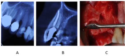
A 54-year-old female was referred to our clinic complaining of crowded and periodontally hopeless maxillary right canine and premolar teeth. Due to excessive loss of tooth structure and severe bone loss detected circumferentially, the suggested treatment plan included extraction of teeth (right canine and premolars) and immediate implantation with GBR. Initial cone beam computed tomography data showed that there was no buccal bone plate over the existing teeth (Figure 1).

Figure 1: (A) Coronal plane of CBCT scan: Bone defect present around maxillary right canine and first and second premolars. (B) Sagittal plane of CBCT scan: Absence of buccal bone plate over the maxillary right canine. (C) Clinical view of bone defect around maxillary right canine and first and second premolars after surgical flap elevation and tooth extraction.

Presurgical treatment

The patient’s medical and dental history showed that she had no contraindication for dental implant surgery. The cone beam computed tomography scans, periapical radiographs and clinical photographs were obtained, dental cast was poured, and a clinical periodontal examination was carried out, which included assessment of bleeding on probing (BOP), pocket depth, and plaque index. The patient underwent scaling and root planning and oral hygiene and plaque control instructions were reinforced.

Surgical treatment (stage 1)

Local anesthesia was administered at the surgical site (buccally and palatally) using 2% lidocaine with 1:100,000 epinephrine. A sulcular incision was made extending from tooth #2 to tooth #6 with a releasing incision at the mesial of tooth #6. A full thickness mucoperiosteal flap was elevated to expose the labial and palatal bone plate. Interdental papillae were preserved and reflected buccally. The hopeless teeth (canine and premolars) were extracted via a conservative approach using piezo surgery and periotomes. The tooth extraction sockets were then curetted to remove all soft tissue remnants. The bone defects and surgical sites were thoroughly irrigated with sterile saline solution. Two dental implants (4.2x11.5mm SIC max for anterior socket and 4.0x11.5mm SIC ace for posterior socket; SIC, Switzerland) were installed in palatal positions. Due to inadequate buccal bone, 4-8 threads of inserted implants remained exposed. The exposed implant surfaces were covered with a slowly-resorbable, synthetic, nanocrystalline hydroxyapatite bone substitute (NanoBoneÒ, Artoss, Germany) and overbuilding was done beyond the resorbed or lost buccal plate with PTGs (Natix™; Tigran Technologies AB, Malmo, Sweden) mixed with the patient’s blood (obtained from a vein). A long-lasting collagen membrane (SIC membrane; SIC, Germany) was fixed with two tacks (FRIOS bone fixation system, FRIOS, Germany) at the most apical parts in the buccal side and subsequently at the palatal side to firmly cover the entire grafted area. The membrane covered not only the defect site, but also a 3mm margin of sound bone at the mesial, distal, apical and palatal.

The flaps were closed with tension-free sutures (3-0 silk) (Figure 2). Postoperatively, the patient was instructed to take 500mg amoxicillin three times a day for a total of 10 days. Also, 0.2% chlorhexidine gluconate mouth rinse (DarouPakhsh Holding Co., Iran) was prescribed for plaque control, twice daily. The patient’s initial postoperative visit was 10 days after surgery; the silk sutures were removed. The patient received professional cleaning on a daily basis alternately between a periodontist and general dentist for the first two weeks. Mechanical oral hygiene practice, consisting of tooth brushing and inter-dental brushing, was initiated by the patient on the 28th post-operative day. Post-operative care included oral hygiene reinforcement and professional plaque removal done on demand.

Figure 2: (A) Placement of two implants at the sites of maxillary right canine and second premolar teeth. (B) Covering the buccal bone defect with PTGs. (C) Placement of collagen membrane over PTGs. (D) Returning the flap and suturing.


Surgical treatment (stage 2)

The patient was recalled six months later for the second-phase surgery and uncovering of implants. A 2.7mm x 6.0mm trephine bur was used to take a core biopsy from the augmentation site under copious saline irrigation. The biopsy core was immersed in 10% formalin solution. The patient was followed-up weekly until completion of soft tissue healing and then monthly until healing abutment placement (Figure 3). The trephine biopsy core was sent to the Robert K. Schenk Laboratory of Oral Histology (School of Dental Medicine, University of Bern, Switzerland) for histologic and histometric evaluation.

Figure 3: (A) Flap elevation in the second-stage surgery. (B) Uncovering the implants. (C) Obtaining a core biopsy. (D) Returning the flap and suturing. (E) Soft tissue healing six months after surgery. (F) Opening of healing abutment to place the temporary abutment.


Sample preparation and descriptive histology

The specimen was dehydrated in ethanol series and embedded in methyl methacrylate. Undecalcified sections measuring ~500 microns in thickness were cut using a low speed diamond saw under coolant (Varicut® VC-50, Leco, Munich, Germany). After mounting the sections on acrylic glass slabs, they were ground and polished to a final thickness of about 100µm (Knuth-Rotor-3, Struers, Rodovre/Copenhagen, Denmark). Most sections were stained with toluidine blue and basic fuchsine and the two most central ground sections were used for qualitative and quantitative analyses. A few sections were used for polarized light microscopy. Digital photography was performed using a ProgRes® C5 digital camera (Jenoptik Laser, Optik Systeme GmbH, Jena, Germany) connected to a Zeiss Axioplan microscope (Carl Zeiss, Göttingen, Germany). Image processing and morphometric analyses were performed directly under the microscope. One experienced examiner (D.B.) performed descriptive histological analysis.

Histomorphometry

The area fractions (%) of the newly formed mineralized bone, osteoid, filler material 1 (PTG), filler material 2 (NanoBone) and soft tissue (ST) occupying the biopsy volume were determined by point counting directly under the microscope, using a square grid (distance between 6x6 test lines: 0.067mm) at x250 magnification. The percentage of PTG surface covered with the newly formed bone (osteoconduction) was evaluated by counting the intersections using the same grid and magnification as mentioned above.

Result

Clinical results

Figure 4: (A) Assessment of bone width and height on preoperative CBCT scan of patient; (B) Assessment of bone width and height on postoperative CBCT scan of patient.
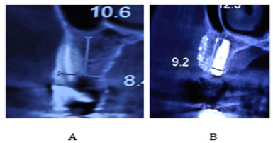

Our patient was a 55-year-old female with no systemic disease, who was referred to our clinic due to failed endodontic treatment of the maxillary right canine and premolar teeth. After extraction of the afore-mentioned teeth due to their hopeless prognosis, two implants were surgically placed at the sites of the maxillary right canine and second premolar. After implant surgery, hard and soft tissue assessment was meticulously done until prosthetic loading of implants six months after the second-stage surgery every two months. During the follow-up sessions, parallel periapical radiographs were obtained, scaling and root planning was performed, and the oral hygiene instructions were given and reinforced. Periodontal parameters including pocket depth, BOP and pus formation were also assessed. Clinical assessment showed no signs of inflammation around the dental implants. Probing depth in the buccal, palatal and interproximal areas of implants was 6 mm and BOP was present. This value was 3mm without BOP or pus after treatment. The patient’s cone beam computed tomography scan showed that width and height of bone increased from 8.4mm and 10.6mm before treatment to 9.2mm and 12.3mm after treatment, respectively (Figure 4). No signs of graft loss or gingival recession were noted around dental implants.

Histology

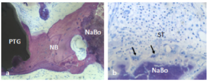
The histological sections revealed the presence of PTGs, new bone, soft tissue and NanoBoneÒ (Figure 5-7). The PTGs were variable in shape, size and density. Most of the new bone was lamellar bone (Figure 6a); however, osteoid lined by osteoblasts was occasionally observed (Figure 6b). The intervening soft tissue was non-fibrous and rich in small and medium-sized blood vessels (Figure 6a,c). New bone was observed in direct contact with PTGs (Figure 6c) and in the pores of PTGs (Figure 6d). Furthermore, new bone was bridging between PTGs and neighboring NanoBoneÒ particles (Figure 7a). The NanoBoneÒ particles were lined by new bone (Figure 7a) and few multinucleated, osteoclast-like, giant cells (Figure 7b). Such multinucleated giant cells were not observed on the PTG surface. The soft tissue peripheral to the NanoBoneÒ particles was cell-rich (Figure 7b).

Figure 5: Histological section revealed the presence of PTG: porous titanium granules, NB: new bone, ST: soft tissue, and NaBo: remnant particles of nano hydroxyapatite synthetic bone substitutes.


Figure 6: ST: soft tissue and NB: New bone in direct contact with PTGs: porous titanium granules.
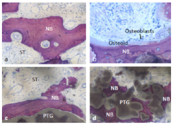

Figure 7: NB: new bone was bridging between PTGs: porous titanium granules and neighboring NaBo: synthetic nano hydroxyl apatite bone substitutes (a). Multinucleated, osteoclast-like, giant cells (arrows) and cell-rich soft tissue were observed peripheral to Nano Bone particles (b).

Histomorphometry

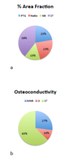
The area fractions of new bone, soft tissue, PTGs, and NanoBoneÒ were 15.2%, 39.3%, 22.4%, and 23.1%, respectively (Figure 8a). The new bone matrix consisted of 12.41% mineralized bone matrix and 2.82% osteoid. Concerning the osteoconductivity of PTGs, 27.0% new mineralized bone, 10.2% osteoid, and 62.8% soft tissue were found to cover the titanium particles (Figure 8b).

Figure 8: The area fractions of NB: new bone, ST: soft tissue, PTGs: porous titanium granules, and NaBo: NanoBone (a). The new bone matrix consisted of MNB: mineralized bone matrix, ST: soft tissue, and O: osteoid (b).

Discussion

In this case study, PTGs were used to cover the bone defect in the buccal surface of implants in the first phase of surgery. PTGs were first introduced by Bystedt et al. [17] as an osteoconductive material for bone grafting in sinus floor augmentation surgery [17]. Today, PTGs are used for treatment of bone defects [18], inflammatory peri-implant defects [19] and healing of tooth extraction sockets [20]. In our study, some clinical periodontal parameters such as pocket depth, BOP and pus formation were assessed at baseline and every two months after the second-stage surgery. Wohlfahrt et al. [21] reported that clinical attachment level and gingival recession were not significantly different from the baseline values 12 months after treatment of grade II furcal defects of mandibular molars with PTG, but pocket depth and gingival index significantly decreased after treatment and BOP remained unchanged [14]. An animal study by Wohlfahrt et al. [21] showed that probing depth increased six weeks after treatment of grade II furcal defects with PTGs compared to baseline, but gingival index decreased after treatment. Horizontal probing depth was lower in group treated with PTGs compared to the deproteinized bovine bone mineral and sham groups, although the difference between the groups was not significant [21].

In our study, pocket depth was 6mm before treatment and BOP was present in the areas with bone loss around teeth. After implant placement and bone grafting, pocket depth improved to 3mm and BOP was no longer present. For bone grafting and dental implant placement, graft material may be applied before or simultaneously with placement of dental implants at the site of bone loss. Applying bone graft material prior to implant placement requires an additional surgical procedure, and the associated risks and costs must be taken into account. In our study, bone grafting and dental implant placement were done simultaneously. Table 1 [22-34] lists some studies that used this technique with different bone graft materials. In all the studies listed in Table 1 [22-34], this technique yielded favorable radiographic and clinical results. This technique eliminates the need for an additional surgical procedure; however, there is a risk of implant movement and subsequent movement of graft material. Therefore, this technique requires high precision, and immediate loading of implants is not encouraged and should be avoided.

Moreover, stable bone graft materials must be used. In our study, implant loading was performed six months after the second-stage surgery and a resorbable membrane was placed over PTGs for further stabilization. In most of the studies listed in Table 1 [22-34], GBR technique was used for bone grafting. The results of a review study by Aghaloo et al. [35] showed that the implant survival rate in GBR technique for augmentation of edentulous maxillary ridge was between 96.1-100%, which was slightly higher than that of other technique [36]. Also, collagen membrane was used in the majority of the studies cited in Table 1 and also in our study. Collagen membrane has excellent biocompatibility [37] and more flexibility than ePTFE and titanium mesh [38] and does not need additional surgery for its removal. It appears that use of membrane along with regenerative material enhances the short-term results of bone augmentation since Delgado Ruiz et al. [39] showed that six weeks after bone defect coverage using PTGs along with collagen membrane, the percentage of cortical defect closure was significantly higher than that of the PTG group without the collagen membrane. In the PTG group without collagen membrane, evidence of tissue inflammation was noted [40]. Roos et al. [41] stated that use of resorbable membrane along with bone substitute had no significant difference from the use of bone substitute alone for treatment of peri-implantitis defects with regard to the improvement of clinical and radiographic parameters in long-term (5 years) [42]. The above-mentioned findings indicate that as long as the membrane has not been resorbed, it protects bone graft materials against movement and degradation and can therefore enhance the short-term results [43,44].

Conclusion

Tooth extraction followed by implant placement and use of PTGs and collagen membrane all at the same time can improve clinical periodontal parameters and implant stability in the shortterm. Long-term randomized clinical trials with large sample size are required in this respect. Moreover, future studies are recommended to compare different bone graft materials for simultaneous placement with implant insertion.

References

  1. Murakami Y, Honda Y, Anada T, Shimauchi H, Suzuki O (2010) Comparative study on bone regeneration by synthetic octacalcium phosphate with various granule sizes. Acta Biomater 6(4): 1542-1548.
  2. Rickert D, Slater JJ, Meijer HJ, Vissink A, Raghoebar GM (2012) Maxillary sinus lift with solely autogenous bone compared to a combination of autogenous bone and growth factors or (solely) bone substitutes. A systematic review. Int J Oral Maxillofacial Surg 41(2): 160-167.
  3. Neovius E, Engstrand T (2010) Craniofacial reconstruction with bone and biomaterials: review over the last 11 years. J Plast Reconstr Aesthet Surg: JPRAS 63(10): 1615-1623.
  4. Rubin JP, Yaremchuk MJ (1997) Complications and toxicities of implantable biomaterials used in facial reconstructive and aesthetic surgery: a comprehensive review of the literature. Plast Reconstr Surg 100(5): 1336-1353.
  5. Gottlow J, Nyman S, Lindhe J, Karring T, Wennstrom J (1986) New attachment formation in the human periodontium by guided tissue regeneration. Case reports. J Clin Periodontology 13(6): 604-616.
  6. Papapanou PN, Tonetti MS (2000) Diagnosis and epidemiology of periodontal osseous lesions. Periodontol 2000 22: 8-21.
  7. Trombelli L, Mayfield LJ, Needleman I, Moles D, Scabbia A (2002) A systematic review of graft materials and biological agents for periodontal intraosseous defects. J Clinical Periodontology 29 Suppl 3: 117-135.
  8. Rothamel D, Schwarz F, Herten M, Engelhardt E, Donath K, et al. (2008) Dimensional ridge alterations following socket preservation using a nanocrystalline hydroxyapatite paste: a histomorphometrical study in dogs. Int J Oral Maxillofac Surg 37(8): 741-747.
  9. Lynch SE, Williams RC, Polson AM, Howell TH, Reddy MS, et al. (1989) A combination of platelet-derived and insulin-like growth factors enhances periodontal regeneration. J Clin Periodontol 16(8): 545-548.
  10. Reynolds MA, Reidy ME, Mays GL, Gunsolley JC (2003) The efficacy of bone replacement grafts in the treatment of periodontal osseous defects. A systematic review. Ann Periodontology 8(1): 227-265.
  11. Shirmohammadi A, Chitsazi MT, Lafzi A (2009) A clinical comparison of autogenous bone graft with and without autogenous periodontal ligament graft in the treatment of periodontal intrabony defects. Clin Oral Investig 13(3): 279-286.
  12. Gholami GA, Kadkhodazadeh M, Ardakani M, Tehranchi M, Aghaloo M, et al. (2010) Histologic and histomorphometric evaluation of bone substitutes in experimental defects. Res J Biol Sci 5(7): 465-469.
  13. Gholami G, Kadkhodazadeh M (2012) Porous titanium granules for management of a severe intra bony defect. Journal of Clinical Periodontology 39:134.
  14. Wohlfahrt JC, Lyngstadaas SP, Heijl L, Aass AM (2012) Porous titanium granules in the treatment of mandibular Class II furcation defects: a consecutive case series. J Periodontol 83(1): 61-69.
  15. Wohlfahrt JC, Lyngstadaas SP, Ronold HJ, Saxegaard E, Ellingsen JE, et al. (2012) Porous titanium granules in the surgical treatment of peri-implant osseous defects: a randomized clinical trial. International J Oral Maxillofac Implants 27(2): 401-410.
  16. Tavakoli M, Moghareabed A, Farsam T, Abbas FM, Badrian H, et al. (2012) Evaluation of dental socket healing after using of porous titanium granules: Histologic and histomorphometric assessment in dogs. Den Res J 9(5): 600-606.
  17. Bystedt H, Rasmusson L (2009) Porous titanium granules used as osteoconductive material for sinus floor augmentation: a clinical pilot study. Clin Implant Dent Relat Res 11(2): 101-105.
  18. Wohlfahrt JC, Monjo M, Ronold HJ, Aass AM, Ellingsen JE, et al. (2010) Porous titanium granules promote bone healing and growth in rabbit tibia peri-implant osseous defects. Clin Oral Implants Res 21(2): 165-173.
  19. Mijiritsky E, Yatzkaier G, Mazor Z, Lorean A, Levin L (2013) The use of porous titanium granules for treatment of peri-implantitis lesions: preliminary clinical and radiographic results in humans. Br Dent J 214(5): E13.
  20. Verket A, Lyngstadaas SP, Ronold HJ, Wohlfahrt JC (2014) Osseointegration of dental implants in extraction sockets preserved with porous titanium granules - an experimental study. Clin Oral Implants Res 25(2): e100-108.
  21. Wohlfahrt JC, Aass AM, Ronold HJ, Heijl L, Haugen HJ, et al. (2012) Microcomputed tomographic and histologic analysis of animal experimental degree II furcation defects treated with porous titanium granules or deproteinized bovine bone. J Periodontol 83(2): 211-221.
  22. Demarosi F, Varoni E, Rimondini L, Carrassi A, Leghissa GC (2016) Immediate implant placement after removal of maxillary impacted canine teeth: A technical note. Int J Oral Maxillofac Implants 31(1): 191-194.
  23. Stricker A, Fleiner J, Stubinger S, Fleiner H, Buser D, et al. (2016) Ridge preservation after ridge expansion with simultaneous guided bone regeneration: a preclinical study. Clin Oral Implants Res 27(11): e116-e124.
  24. Kolerman R, Nissan J, Rahmanov A, Zenziper E, Slutzkey S, et al. (2016) Radiological and biological assessment of immediately restored anterior maxillary implants combined with GBR and free connective tissue graft. Clin Implant Dent Related Res 18(6): 1142-1152.
  25. Jung RE, Benic GI, Scherrer D, Hammerle CH (2015) Cone beam computed tomography evaluation of regenerated buccal bone 5 years after simultaneous implant placement and guided bone regeneration procedures--a randomized, controlled clinical trial. Clin Oral Implants Res 26(1): 28-34.
  26. Konstantinidis I, Kumar T, Kher U, Stanitsas PD, Hinrichs JE, et al. (2015) Clinical results of implant placement in resorbed ridges using simultaneous guided bone regeneration: a multicenter case series. Clin Oral Investig 19(2): 553-559.
  27. Covani U, Canullo L, Toti P, Alfonsi F, Barone A et al. (2014) Tissue stability of implants placed in fresh extraction sockets: a 5-year prospective single-cohort study. J Periodontol 85(9): e323-e332.
  28. Noelken R, Neffe BA, Kunkel M, Wagner W (2014) Maintenance of marginal bone support and soft tissue esthetics at immediately provisionalized OsseoSpeed implants placed into extraction sites: 2-year results. Clin Oral Implants Res 25(2): 214-220.
  29. Cucchi A, Ghensi P (2014) Vertical guided bone regeneration using titanium-reinforced d-PTFE membrane and prehydrated corticocancellous bone graft. Open Dent J 8: 194-200.
  30. Jung RE, Fenner N, Hammerle CH, Zitzmann NU (2013) Long-term outcome of implants placed with guided bone regeneration (GBR) using resorbable and non-resorbable membranes after 12-14 years. Clin Oral Implants Res 24(10): 1065-1073.
  31. Favero G, Botticelli D, Favero G, Garcia B, Mainetti T, et al. (2013) Alveolar bony crest preservation at implants installed immediately after tooth extraction: an experimental study in the dog. Clin Oral Implants Res 24(1): 7-12.
  32. Caneva M, Botticelli D, Morelli F, Cesaretti G, Beolchini M, et al. (2012) Alveolar process preservation at implants installed immediately into extraction sockets using deproteinized bovine bone mineral-an experimental study in dogs. Clin Oral Implants Res 23(7): 789-796.
  33. Shibly O, Kutkut A, Albandar JM (2012) One-year re-entry results of guided bone regeneration around immediately placed implants with immediate or conventional loading: a case series. J Int Acad Periodontol 14(3): 62-68.
  34. Benic GI, Jung RE, Siegenthaler DW, Hammerle CH (2009) Clinical and radiographic comparison of implants in regenerated or native bone: 5-year results. Clin Oral Implants Res 20(5): 507-513.
  35. Aghaloo TL, Misch C, Lin GH, Iacono VJ, Wang HL, et al. (2016) Bone augmentation of the edentulous maxilla for implant placement: A systematic review. Int J Oral Maxillofac Implants 31 Suppl: s19-s30.
  36. Janner SF, Bosshardt DD, Cochran DL, Chappuis V, Huynh Ba G, et al. (2017) The influence of collagen membrane and autogenous bone chips on bone augmentation in the anterior maxilla: a preclinical study. Clin Oral Implants Res 28(11): 1368-1380.
  37. Wessing B, Urban I, Montero E, Zechner W, Hof M, et al. (2017) A multicenter randomized controlled clinical trial using a new resorbable non-cross-linked collagen membrane for guided bone regeneration at dehisced single implant sites: interim results of a bone augmentation procedure. Clin Oral Implants Res 28(11): e218-e226.
  38. Benic GI, Bernasconi M, Jung RE, Hammerle CH (2017) Clinical and radiographic intra-subject comparison of implants placed with or without guided bone regeneration: 15-year results. J Clin Periodontol 44(3): 315-325.
  39. Delgado Ruiz RA, Calvo Guirado JL, Abboud M, Fernandez RMP, Mate-Sanchez JE, Negri B, et al. (2014) Porous titanium granules in critical size defects of rabbit tibia with or without membranes. Int J Oral Sci 6(2): 105-110.
  40. Benic GI, Ge Y, Gallucci GO, Jung RE, Schneider D, et al. (2017) Guided bone regeneration and abutment connection augment the buccal soft tissue contour: 3-year results of a prospective comparative clinical study. Clin Oral Implants Res 28(2): 219-225.
  41. Roos-Jansaker AM, Persson GR, Lindahl C, Renvert S (2014) Surgical treatment of peri-implantitis using a bone substitute with or without a resorbable membrane: a 5-year follow-up. J Clin Periodontol 41(11): 1108-1114.
  42. Gultekin BA, Cansiz E, Borahan MO (2017) Clinical and 3-dimensional radiographic evaluation of autogenous iliac block bone grafting and guided bone regeneration in patients with atrophic maxilla. J Oral Maxillofac Surg 75(4): 709-722.
  43. Chattopadhyay S, Raines RT (2014) Review collagen-based biomaterials for wound healing. Biopolymers 101(8): 821-833.
  44. Caffesse RG, Nasjleti CE, Morrison EC, Sanchez R (1994) Guided tissue regeneration: comparison of bioabsorbable and non-bioabsorbable membranes. Histologic and histometric study in dogs. J Periodontol 65(6): 583-591.

For more articles in Open Access Dentistry and oral journals Impact factor
Please click on below link: https://crimsonpublishers.com/mrd/

A Close Look at the Application of the Yin-Yang- Based Acupoint Pairs_Crimson Publishers

A Close Look at the Application of the Yin-Yang- Based Acupoint Pairs by Tong Zheng Hong in Advancements in Bioequivalence & Bioavailabi...