Age Related Changes in the Development of Large Intestine in Red Sokoto Goats by Bello A in Open Access Research in Anatomy_Anatomy Research International journals
Abstract
This study was aimed at investigating the age-related changes in the post neonatal development of the large intestine in Red Sokoto Goat, using standard morphological techniques. In this study, five age groups were used (i.e. 0-6 months, 6 months-1 year, 1-2 years, 2-3 years and above 3 years, which were grouped in this study as Group A-Group E) and were all obtained from Wamakko market at Wamakko Local Government, Sokoto State. The Ages were estimated using the knowledge of Eruption and Wearing of the milk and Permanent Teeth of the Rostral dentition. The different segments of the Large intestine; Caecum, Colon and Rectum were found to be present in every age group and there was no change in position. There was colour change with visible increase in size with increase in developmental age. Although there was a gradual increase observed in length, width and thickness of the various segments of the Large intestine, the weight and volume however were noticed to increase rapidly with advancement in development. The last group (i.e., above 3 years of age) showed the highest values, suggesting that as the animal advances in age, it adapts to the new feeding pattern as the nature of feed changes as the animal gets older, which also explains the colour change in the organ with advancement in development.
Keywords: Age; Neonatal; Development; Large intestine; Red Sokoto Goat; Rostral dentition
Introduction
Goats are the principal domesticated small ruminants in terms of total numbers and production of food and fiber products [1], with an estimated population of 800 million worldwide. Goats are herbivorous animals which belong to an integral part of a traditional crop livestock production [2]. They often fit well into biological and economic niches and can be incorporated into existing grazing operations with sheep and cattle, and they can also be used to control weed to help make use of pasture’s diversity. In the tropics such as Nigeria, goat population is estimated to be 34.45 million. Goats have special characteristic features that make it easy for them to thrive in any environment. Goats also produce a considerable amount of manure, which is of special importance in those areas where cattle are of lesser importance [3]. Three main goats are recognized in Nigeria: the Sahel goat, the Red Sokoto goat and the West African dwarf goat [4].
The Red Sokoto goat however is most commonly found in Sokoto area of Nigeria and part of the Niger republic. They are the most widespread and well-known type of goats in Nigeria. The Red Sokoto goat was the source of “Morocco leather” known in Europe from the medieval period onwards. It acquired its name because it was transported across the Sahara by caravans controlled by Moroccan merchants. The Sokoto Red is still known for its suitability for fine leather. The digestive system is the only system, which satisfies the energy need of the body through absorption of nutrients and thus it makes the powerful relation with nature by digesting various types of feed which are digestible to specific animals. Goats being ruminant animals possess a complex stomach (i.e. stomach with four compartments) the small and large intestines [2].
The Large intestine is the termination of the ileum to the anus. The large intestine is comprised of the caecum, colon and rectum. The function of large intestine is the absorption of considerable quantity of water, vitamins and electrolytes and production of mucus to allow easy passage of faeces. The capacity of the large intestine in goats ranges from 11/4to 11/2 gallons. So far, relatively little work has been done to describe the Gastro-intestinal changes associated with normal ageing and in many instances normal data on which to base clinical comparison are not available.
During postnatal development, the structure of the gastrointestinal tract is affected by several factors including diet, age, genetic determinants, and hormones secreted in the intestine and in the other organs [5]. The mammal growth period between birth and maturity seems to be particularly interesting in the context of the development of intestinal mucosa, a tissue that is associated with exchange and absorption processes. At the same time, the form and function of the alimentary tract develop to meet the increased metabolic demands during the growth period. Many studies on goat’s digestive system has been done; [6-9] on the Histo-architecture of the large intestine, [8-11] on the Comparative histological study of the rectum in different species of animals. These studies have shown slight differences in the general morphology and histology of the large intestine in different species of animals. However, there is paucity of information on the age-related changes of the development of the large intestine in the Red Sokoto goat.
The large intestine plays a great role in the absorption of water and electrolytes from the ingesta and also functions in the production of mucus for easy passage of faeces, hence makes this organ very important and necessary for the process of digestion to be completed. Therefore, there is a great need to study the agerelated changes of the large intestine for better understanding of the animal’s digestive capability, so as to enhance goat production in Nigeria. The data obtained from this study will help to bridge the existing gap on the morphology and histology of the large intestine of Red Sokoto goat in different age groups. The aim of the study was to study the age related changes in the development of the large intestine in the Red Sokoto goat, while the Specific objectives was to determine the gross observation (Morphology) of the different segments of the large intestine in relation to their age groups and to determine the biometric parameters of the large intestine in relation to their age groups.
Material and Method
The study was conducted in Sokoto metropolis, the capital of Sokoto State of Nigeria. Geographically, the state is located at latitude 12015N and 050E, and is 308m above the sea level. Sokoto state occupies an area of short grass savannah vegetation in the south and thorn in the north. It shares boundaries with Zamfara state to the east, Niger republic to the north and Kebbi state to the west and southwest.
animals of either sex was used in this study. The animal’s digestive tract was purchased from Wammako market at Wammako Local Government and transported by road to the gross Anatomy Laboratory of the Department of Veterinary Anatomy, Usmanu Danfodiyo University, Sokoto. The animals were aged using the rostral dentition method of ageing, the digestive tract was dissected out from the abdominal cavity then the whole large intestine caecum, colon and rectum was removed from the entire digestive system.
Gross examination
The components of the samples were observed thoroughly based on shape, size, and colour of each organ in situ and separately internally and externally based on the help of eyes. The large intestine is the final part of the gastrointestinal tract that consists of the caecum, colon and rectum. The caecum is the first segment of the large intestine. It is much larger in diameter than the small intestine. It is almost horizontally positioned, on the right side of the abdominal cavity. The colon is a capacious tube that roughly surrounds the loops of small intestine as an arch. The colon has three major segments: the ascending, transverse and descending colon. The transverse is constricted and located between the ascending and descending colon. The descending colon is directed backwards, and the rectum connects to the anus.
Biometric
The samples were assigned into five post-natal groups according to their age variation (0-6 months, 6 months-1 year, 2 years-3 years and above 3 years). The biometrical measurements like length, weight, width, thickness and volume were all measured as follows:
Length: The length of the entire large intestine was measured as well as the length of the various segments of the large intestine using a calibrated measuring tape (Butterfly) and recorded in centimeters (cm).
Weight: The weight of the entire large intestine as well as the various segments of the large intestine was measured using a weighing balance (Mettler®) and recorded in kilograms (kg).
Width: The width which is the distance between two lateral aspects of the intestines was measured using a meter rule and recorded in centimeters (cm).
Thickness: The thickness of the intestines was also measured and recorded in millimeters (mm) using a vernir caliper (Starrett®).
Ageing in ruminants
According to Jeffrey (1996) ruminant age determination was carried out using knowledge of eruption and wearing of milk teeth and permanent teeth, as well as other anatomical features such as increase in interdental space and change in shape of teeth, angulation of the teeth, degree of wearing of the teeth. Goats, as with other ruminant animals, lack upper incisors. Instead, a harddental pad on the frontal part of the upper jaw serves in place of teeth. In kid goats, the first pair of milk teeth incisors occurs at birth to 1 week of age. The second pair of milk teeth incisors erupts at one to 2 weeks of age, the third pair at 2 to 3 weeks of age, and the fourth pair of milk incisors appears at 3 to 4 weeks of age.
Permanent incisor teeth as a guide to age estimation in small ruminants. The sequence for the eruption of permanent incisors is 1 to 2 years of age for the first pair of incisors, 2 to 3 years for the second pair, 3 to 4 years for the third, and above 4years for the fourth pair of incisors. A full mouth of permanent teeth is in place by the time the goat reaches 4 years of age. Data obtained were processed and presented in mean + standard deviation (mean ± S.D) using Microsoft excels software 2010 (Figure 1).
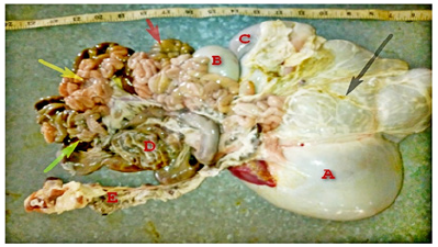
Figure 1: Photograph of the entire digestive tract of Red Sokoto Goat (2-3 years) showing A-Rumen; B-Reticulum; C-Omasum; D-Colon; E-Rectum; Omentum (Black arrow); Duodenum (Red arrow); Jejenum (Yellow arrow); Ileum( Green arrow).
Result and Discussion
A total of 10 samples were used for the study and grouped into 5 of the groups are as follows: 0-6 months, 6months-1 year, 2-3 years and above 3 years. The large intestine was found to be located towards the caudal end of the abdominal cavity in all ages. It consisted of three parts i.e. the caecum, colon and rectum. It begins with the caecum and ends in the rectum; it is a continuation of the small intestine.
Caecum
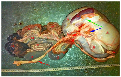
The caecum was found to be the 1st part of the large intestine, which is somewhat dark in colour, voluminous and shaped like a big comma. It begins at a junction called the ileocaecal junction in this specie but varies in other species such as the equine specie; the large intestine begins at the illeo-caeco-colic junction in the equine species (Figure 2).
Figure 2: Photograph of the entire digestive tract of Red Sokoto Goat (3 year and above) showing A-Portion of the diaphragm; B-omasum; C-Rumen; D-Spleen; E-Centripetal and Centrifugal coiling of the Colon; F- Duodenum; G- Body of the Caecum; H-Ileum; I-Part of the Duodenum; J-Apex of the caecum; K-Jejunum; L-Rumen; M-Mesentery; Spleen (Green arrow); Point of entry of the oesophagus into the Rumen (Blue arrow); Rectum (Red arrow).
Colon
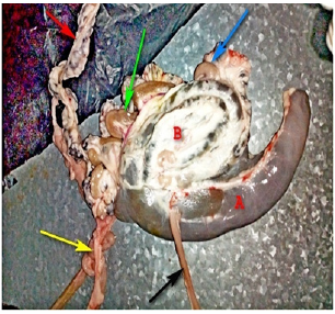
The colon was the next part of the large intestine located just immediately after the caecum and connected to the caecum by an indistinctive junction known as the caeco-colic junction and constituted of the ascending, descending and transverse colon and also the centripetal and centrifugal coiling. The ascending colon was found to be the first part of the colon, attached to the caecum and continued by the centripetal coiling. The centripetal and centrifugal coiling was observed to be coiled closely around each other, hence the name. The centrifugal and centripetal coiling are somewhat darker in colour than the entire length of the large intestine and held together in place by fatty tissues lining the external walls of these organs. The transverse colon is short and is a continuation of the centrifugal coiling of the colon. It lies transversely in between the centrifugal coiling and the descending colon. The descending colon however is found to be located between the transverse colon and the rectum and projects downwards towards the rectum (Figures 3-5).
Figure 3: Picture of the small and large intestines showing A-Centripetal and Centrifugal coiling of the Colon; B-Body of the Caecum; Rectum (Blue arrow); Ileum (Yellow arrow); Jejunum (Green arrow); Duodenum (Red arrow).
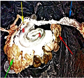
Figure 4: Photograph of the Large intestine in Red Sokoto Goat s (above 3 years) showing A-Body of the Caecum; B-Apex of the Caecum; Rectum (Green arrow); Mesentery (Yellow arrow); Mesenteric lymphnode (Red arrow); Ileocaecal junction (Black arrow).

Figure 5: Image of the Large intestine of Red Sokoto Goat (2-3 years) showing A-Caecum; B-Colon; Mesenteric lymphnode (Blue arrow); Transverse colon (Green arrow); Rectum (Red arrow); Urethra (Yellow arrow); Ileum at the ileocaecal junction (Black arrow).
Rectum
Figure 6: Photograph of the entire digestive tract of Red Sokoto Goat (0-6 months) showing A-Caecum (apex); B-Colon; C-Mesentery; Ileocaecal junction (Yellow arrow); Transverse colon (Black arrow); Rectum (Red arrow).
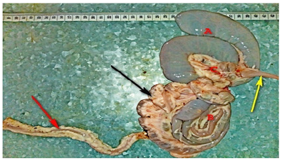
The rectum was the last part of the large intestine that terminates in the anus. A short segment of the large intestine, nature of faeces found in this region differed from that found in the entire length of the large intestine. The wall of the rectum was found to be slightly thicker than the entire length of the large intestine and the walls were found to be lined by a large number of fatty tissues (Figure 6).
Caecum
Table 1: Age estimation in goats.

The length, thickness and volume of caecum at various postnatal ages of Red Sokoto Goat. The Results show that the weight, length, width, thickness and volume of the various ages of the caecum where increasing in values with advancement of developmental ages chronologically as shown below in (Table 1). Although there is no significant difference in value of the weight and volume in the first two groups i.e., A and B (Table 1).
Ascending colon
The length, thickness and volume of ascending colon at various postnatal ages of Red Sokoto goat shown that the colon was divided into 5 segments as observed grossly of the first part of the colon. Morphometric observations show that there is a position increase in weight, length and volume of the organ with the advancement in post-natal developmental ages as shown below in (Table 2).
Table 2: Mean ± SD of the length, width, thickness and volume of caecum in various post-natal ages.

Key: Group A: (0-6 months), Group B: (6 months-1 year), Group C: (1-2 years), Group D: (2-3 years), Group E: (above 3 years).
Centripetal colon
Table 3: Mean ± SD of the length, width, thickness and volume of the Ascending colon in various post-natal ages.

Key: Group A: (0-6 months), Group B: (6 months-1 year), Group C: (1-2 years), Group D: (2-3 years), Group E: (above 3 years).
The length, thickness and volume of centripetal colon at various postnatal ages of Red Sokoto goat. Observations show in (Table 3) below, the increase from (group A to group E) in values with advancement of developmental ages.
Centripetal colon
Table 4: Mean ± SD of the length, width, thickness and volume of the centripetal colon in various post-natal ages.

Key: Group A: (0-6 months), Group B: (6 months-1 year), Group C: (1-2 years), Group D: (2-3 years), Group E: (above 3 years).
The length, thickness and volume of centripetal colon at various postnatal ages of Red Sokoto goat. The results have shown that the weight, length, width, thickness and volume of the segment of the centrifugal colon were increasing from (group A to group E) in values with advancement of developmental ages as shown in (Table 4).
Transverse colon
The length, thickness and volume of transverse colon at various postnatal ages of Red Sokoto goat. Observations have shown that there is increase in the biometric values from (group A to group E) with advancement of developmental changes (Table 5).
Table 5: Mean ± SD of the length, width, thickness and volume of the Centrifugal Colon in various post-natal ages.

Key: Group A: (0-6 months), Group B: (6 months-1 year), Group C: (1-2 years), Group D: (2-3 years), Group E: (above 3 years).
Descending colon
The Length, Thickness and Volume of descending colon at Various Postnatal Ages of Red Sokoto Goat. Observations have shown an increase in the biometric values of the descending colon from (group A to group E) with advancement of developmental ages (Table 6).
Table 6: Mean ± SD of the length, width, thickness and volume of the Transverse Colon in various post-natal ages.
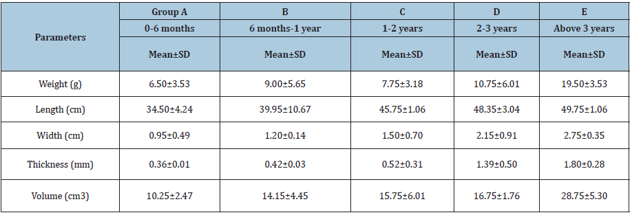
Key: Group A: (0-6 months), Group B: (6 months-1 year), Group C: (1-2 years), Group D: (2-3 years), Group E: (above 3 years).
Rectum
The length, thickness and volume of rectum at various postnatal ages of Red Sokoto goat. Results have shown that the weight, length, width, thickness and volume of the rectum were increasing from (group A to group E) in values with advancement of developmental ages as shown in (Table 7) below.
Table 7: Mean ± SD of the length, width, thickness and volume of the Descending colon in various post-natal ages.
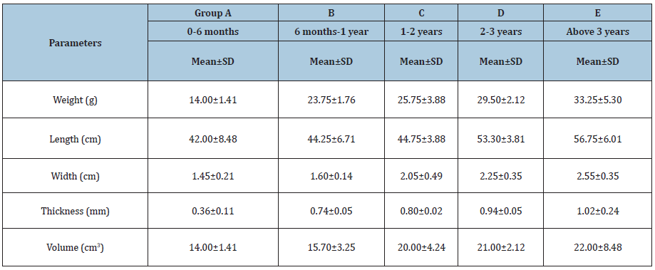
Key: Group A: (0-6 months), Group B: (6 months-1 year), Group C: (1-2 years), Group D: (2-3 years), Group E: (above 3 years).
Discussion
This research showed that with advancement in post-natal developmental ages, there was corresponding increase in the morphometric parameters. In accordance with the findings of Kadam on the study of the histo-architecture of large intestine in goat. The large intestine was found to be located towards the caudal end of the abdominal cavity and consists of three parts: the caecum, colon and rectum (Table 8).
Table 8: Mean ± SD of the length, width, thickness and volume of the Rectum in various post-natal ages.

Key: Group A: (0-6 months), Group B: (6 months-1 year), Group C: (1-2 years), Group D: (2-3 years), Group E: (above 3 years).
In this research, it was observed that the caecum is the first part of the large intestine, which connected with the small intestine at a junction called the ileocaecal junction. A substantial histological and anatomical study has been reported on the large intestine of ruminants [8,10,12]. Also, by on the Morphometric and histological studies of the caecum in mongrel dogs, which are in line with this research. Although the findings of [13-15] are in contradiction to these findings. The ileocaecal junction however also forms the basis of comparism in other species such as the equine species that have the illeo-ceco-colic junction instead. The caecum is the proximal blind end pouch of the large intestine, voluminous and shaped like a very big coma, it is almost horizontally positioned on the right side of the abdominal cavity and the size of this organ was observed to have increased with advancement in the post-natal developmental ages (Table 9) [16-21].
Table 9: Table showing the relationship of organ index of the large intestine in relation to various post-natal developmental ages.

Note: Large intestine organ index = weight of caecum/weight of large intestine x 100 Colon organ index = volume of colon/ volume of large intestine × 100 Rectum organ index = weight of rectum/weight of large intestine x 100
The colon was located immediately after the caecum forming a junction called the caeco-colic junction. The colon is divided into the ascending colon, descending colon, transverse colon, centripetal and centrifugal coiling. The ascending colon was found to be attached to the caecum as a proximal loop, forming the most proximal part of the colon and continued by the centripetal and centrifugal coiling. The centrifugal and centrifugal coiling however coiled closely around each other, somewhat dark in colour and lined by a large number of fatty tissues. The centrifugal coiling however was continued by a short transverse colon which was found to lie transversely between the centrifugal coiling and the descending colon. The descending colon which was found to project downwards towards the rectum was the last part of the colon (Table 10).
Table 10: Table showing the relationship of volumetric index of the large intestine in relation to various post-natal developmental ages.

Note: Large intestine volumetric index = volume of large intestine/volume of GIT × 100 Caecum volumetric index = volume of caecum/ volume of large intestine × 100 Colon volumetric index = volume of colon/ volume of large intestine × 100 Rectum volumetric index = volume of rectum/volume of large intestine × 100
The rectum was observed to be the terminal part of the large intestine, which is in line with the findings of Getty [17] and Majeed [21], it is situated in the pelvic cavity. It is short, thicker than the entire length of the large intestine and the external walls were covered by a large number of fatty tissues. It was also observed that the nature of faeces in this part of the intestine was harder and pelleted in this species of animals unlike the pasty faeces which was observed in the caecum and colon. The caecum and the ascending colon were both found to lie on the greater Omentum. The above findings on the morphology of the large intestine were observed in all ages that were used in this study [22].
In this study, the biometry of the large intestine was found to be progressively increasing with the advancement in post-natal development. The observed increase is in direct proportion to the advancement in age due to a variety of factors that contribute in the growth of an animal. One of such factors includes change in the nature and size of feed consumed by the animals as their ages advance [23].
The weight and volume of the caecum, ascending colon and rectum were found to increase substantially and rapidly with advancement of post-natal developmental ages, these findings are in line with the findings of on the gross and ultra-structural studies on the large intestine in Uttar fowl, who observed that the length and weight of the caecum and colorectum and their diameter and thickness at proximal, middle and distal portions in all the age groups increased with advancing age. From the observed length, the increment was in accordance with the findings of on the morphometric study of small and large intestine of Mus musculus during post-natal development which showed that the length and surface area of the various segments of the large intestine gradually increased with age [24].
The length as observed in this study increased gradually with increase in post-natal developmental ages in the caecum colon and rectum of the large intestine. The thickness and width were also found to increase gradually with advancement of post-natal developmental ages. The increase was not as substantial and rapid as that observed in the weight and volume of the same segments of the large intestine.
Conclusion
Grossly, the various segments of the large intestine which have been established from this present study as the caecum, colon and rectum are found to be present in every post-natal age group worked on in this study. The position of these segments also were not noticed to have changed in all age groups, however the colour of this organ may differ with advancement in post-natal developmental ages probably due to other factors such as the change in nature of the contents as the animal advances in age [25].
The biometrical parameters were also established in every age group used in this study. It was also established in this study that the biometric parameters such as weight and volume increased rapidly and substantially in relation to the advancement in postnatal developmental ages. There were also changes observed in the length, thickness and width of the various segments of the large intestine. However, these changes were not as rapid and substantial as that noticed in the weight and volume in relation to the advancement in post-natal developmental ages.
Recommendation
Based on the above results and findings, it is recommended that more work on the histological development of the large intestine in Red Sokoto goat using both light and electron microscopy should be advocated in order to have a deep sense of conclusion.
References
- Nawathe DR, Sohael AS, Umo I (1985) Health management of a dairy herd on the Jos Plateau (Nigeria). Bull Anim Hlth Prod Africa 33: 199-205.
- Seyoum S (1992) Economics of small ruminant meat production Ans consumption in Sub Saharan Africa, Small ruminant research and development in Africa. ILRAD, Nairobi, Kenya, pp. 25-46.
- Winrock International (1983) Sheep and goats in developing countries: Their present and potential role. Winrock int morrilton, Arkansas, USA.
- Ngere LO, Adu IF, Okubanjo IO (1984) The Indigenous goats of Animal genetic resources information 3: 1-9.
- Walthall K, Cappon GD, Hurtt ME, Zoetis T (2005) Post-natal development of the gastrointestinal system: A species comparison. Birth Defects Res B Dev Reprod Toxicol 74(2): 132-156.
- Dellman HD (1993) Textbook of veterinary histology. (4th edn), Delta Society, USA.
- Estacio AC, Maala CP (1996) Abstracts of completed studies. UPLB College, Philippines.
- Ghosh RK (1995) Primary veterinary anatomy. (1st edn), Current international publications, India, pp. 133-135.
- Singh UB, Sulochana S (1978) A Laboratory manual of histological and histochemical technique. (1st edn), Kothari Medical Publishing House, India, pp. 35-37.
- Ozung PO, Nsa EE, Ebegbulem VN, Ubua JA (1964) The potentials of small ruminant production in cross river rain forest zone of Nigeria: A review. Continental Journal of Animal and Veterinary Research 3(1): 33-37.
- Ramkrishna VG (1998) The systemic histology. (1st edn), Jaypee Brothers’ Medical Publishers Pvt Ltd, India.
- Dellman HD, Brown EM (1987) Textbook of veterinary histology. (3rd edn). Philadelphia, USA, pp. 209-253.
- Miller ME, Howard E, Christensen G (1993) Millers anatomy of the Dog. (3rd edn), Philadelphia, USA, pp. 444-445.
- Kumar A (2004) Veterinary surgical techniques. (4th edn), Vikas publishing house Pvt Ltd, India, pp. 298-300.
- Budras KD, Patrick H, McCarthy PH, Fricke W, Richter R (2007) Anatomy of the dog:(5th revised edn), schlutersche, Germany, pp. 56-57.
- Cerbo DAR, Manfredi MT, Zanzani S, Stradiotto K (2010) Gastrointestinal infection in goat farm in Lombardy (Northern Italy): Analysis on community and spatial distribution of parasites. Small Rumin Res 88(2-3): 102-112.
- Sisson S, Grossman JD, Getty R (1975) Sisson and Grossman’s anatomy of the domestic animals. (5th edn), MacMillan Company of India Ltd, India, pp. 739-740.
- Goligher J (1884) Surgery of the anus, rectum and colon. Bailliere Tindall, London, UK, pp. 1-47.
- Levi AC, Borghi F, Garavoglia M (1991) Development of the anal canal muscles. Dis Colon Rectum 34(3): 262-266.
- Maala CP, Cummings JF (1985) Anatomia Histologia Embryologia 14: 116-141.
- Majeed MF, Al-Asadi FS, Nassir AN, Rahi EH (2009) The morphological and histological study of the caecum in broiler chicken. Bas J Vet Res 8(1): 19-25.
- Mukherjee KL (1990) Medical laboratory technologies. (3rd edn), Tata Mac Graw Hill Publishing Co Ltd, India, pp. 1111-1119.
- Nobles VP (1984) The development of human anal canal. Dis Colon Rectum 34(3): 262-266.
- Sisson S, Grossman’s, Daniel J, Robert G (1975) The anatomy of the domestic animals.
- Skandalakis JE, Gray SW, Ricketts R (1994) The colon and rectum. Embryology for surgeons. The embryological basis for the treatment of congenital anomalies. Baltimore: Williams and Wilkins 242-281.


No comments:
Post a Comment