A Study on the Biometrical Changes of the Small Intestine of Red Sokoto Goat (Capra aegagrus hireus) by Bello A in Open Access Research in Anatomy_International Journal of Anatomy and Research
Abstract
This study is aimed at investigating the age-related changes in postnatal development of small intestine of red Sokoto goat. In this study, a total of thirty (30) red Sokoto goat digestive tract samples were used and grouped into five (5) age categories (group A to E). The goat ages were estimated using dentition eruption and wearing. The small intestines were identified and separated from the other part of digestive tract. The gross identification revealed that the small intestine was composed of three (3) segments that named; duodenum, jejunum and ileum with anatomical demarcations between them. The biometric study of weight, length, width, thickness, and volume was found to be increasing with advancement in postnatal ages. The mean weight value of the duodenum were found to range between 16.75 to 42.75 and the mean value of length, width, thickness and volume were observed to be within the range of 71.00 to 199.50, 1.05, to 2.01, 0.28 to 0.66 and 8.50 to 35.00 from group A to group E respectively. Based on the result obtained, it was concluded that the mean weight of the digestive tract, small intestine and various segment of the small intestine (jejunum and ileum) tends to increase with age that is from group A to group E as in the duodenum. However, there is variation in the mean weight, length, width, thickness and volume of various intestinal segments that is duodenum, jejunum and ileum with the jejunum had larger value compare to the duodenum and ileum.
Keywords: Anatomy; Age related changes; Biometric study; Capra aegagrus hireus; Postnatal development
Introduction
Goats are one of the oldest domesticated species and have been used for milk, meat, hair and skins in most countries [1]. They are considered as small livestock animals, compared to bigger animals such as cattle, buffalo, horses and camel but larger than micro livestock stock such as poultry, rabbits and guinea pig. Each recognized breed of goat has specific weight ranges. Goats are of economic value or useful of humans as a renewable provider of milk manure and fiber and then as meat and hide [2]. Some provide goats to impoverished people in poor countries, because goats are easier and cheaper to manage than cattle and have multiple uses [2].
The importance of small intestine in Utilization and Absorption of Food substance create the need to study the developmental changes of different small intestinal segment for better understanding of the anatomy, physiology, and pathology of the organs of small intestine. The information obtained from this study will help to bridge the existing gap on the normal morphology morphometry and histological of the small intestinal segment of goat at different age categories.
Material and Method
Study area
The study was conducted in Sokoto metropolis, the capital of Sokoto State of Nigeria. Geographically the state is situated on Latitude 120 15N and 05oE and is 308m above the sea level. Sokoto state occupies an area of short grass savannah vegetation in the south and thorn in the north. It shares boundaries with Zamfara State to the east, Niger Republic to the North and Kebbi State to the west and southwest.
The State was ranked second in the nation livestock population with an estimated number of 3 million cattle, 3.85million sheep, 4 million goats, 0.8 million camels, 2 million chickens and 1 million poultry [3]. These animal species are one of the major sources of proteins to the inhabitants of the state and over 75% of them are reared or are raised in traditional free-range system living on close association with human settlements [3].
Experimental design
Goat small intestines were collected from Slaughtered Goats at the Sokoto Metropolitan abattoir and transported to the Anatomy Laboratory, Department of Veterinary Anatomy, Faculty of Veterinary Medicine, Usmanu Danfodiyo University Sokoto. The goats were sexed using external genitalia. The goat ages were estimated using the dentition eruption and wearing.
This is as follows
0 - 6 months - Eruption of all the decidua’s teeth.
6 months - I year - Wearing of all the deciduate teeth.
1 year-1 ½ year - I1
1 ½ year- 2 year - I2
2 year- 3 year - I3
Above 3 year - I4
Where I1 = Central incisor
I2 = Second Incisor
I3 = Third Incisor
I4 = Canine or Corner incisor
Note that the I1, I2, I3 and I4 of the lower jaw.
Based on this the small intestinal sample were categorized into five groups as follows:
1st - 0 to 6 months
2nd - 6monthsto 1 year
3rd - 1 year to 2 year
4th - 2 year to 3 year
5th - Above 3 years
Biometrical landmarks
A. Weight of the entire tabular organs: The weight of the entire tabular organs was weighed using weighing scale in kilogram.
B. Weight of small intestine: The small intestines were weight using electrical weighing balance and recorded in grams.
C. Weight of Duodenum: the duodenums were weighed using digital scale and recorded in grams.
D. Weight of Jejunum: the jejunums were weighed using digital scale and recorded in grams.
E. Weight in Ileum: The Ileums were weighed using digital scale and recorded in grams.
F. Weight of pancreas: The pancreases were weighed using digital scale and recorded in gram.
G. Length of small intestine were measured using measuring tape from the pyloric region of the duodenum to the ileocaecal junction of the intestine and recorded in centimeter.
H. Length of Duodenum: The lengths of the duodenum were measured using measuring tape and recorded in centimeter.
. Length of jejunum: The lengths of jejunum were measured using measuring tape and recorded in centimeter.
J. Length of Ileum: The lengths of Ileum were measured using measuring tape and recorded in centimeter.
K. Length of Pancreas: The length of pancreas was measured and recorded in centimeter with of small intestine were measure using metric.
L. Width of Duodenum: The widths of duodenums were measured using metric rule and recorded in centimeter.
M. Width of Jejunum: The widths of ileums were measured using metric rules and recorded in centimeter.
N. Width of ileum: The widths of ileum were measured using metric rules and recorded in centimeter.
O. Width of pancreas: The widths of pancreas were measured using metric rule and recorded in centimeter.
P. Volume of small intestine: The volumes of small intestine were measured by the volume of water displaced as described by Archimedes principle and recorded in cubic centimeter.
Q. Volume of Duodenum: The volumes of Duodenum were measured by the volume of water displaced as described by Archimedes principle and recorded in cubic centimeter.
R. Volume of Jejunum; The volume of Jejunum was measured by the volume of water displaced as described by Archimedes principle and recorded in cubic centimeter.
S. Volume of Ileum: The volumes of the ileums were measured by the volume of water displaced as described by Archimedes principle and recorded in cubic centimeter.
T. Volume of pancreas: The volumes of pancreas were measured by the volume of water displaced as described by Archimedes principles and recorded in cubic centimeter.
U. Thickness of Duodenum: Thicknesses of Duodenums along the luminal diameter were measured using the digital caliper and recorded in Millimeter.
V. Thickness of the jejunum: The thicknesses of a Jejunum along the luminal diameter were measured using the digital caliper and recorded in millimeter.
W. Thickness of ileum: The thicknesses of an ileum along the luminal diameter were measured using the digital caliper and recorded in millimeter. Thickness of pancreas: The thicknesses of a pancreas were measured using the digital caliper and recorded in millimeter.
Data obtained were presented in mean standard deviation. Using Microsoft excel 2012.
Result and Discussion
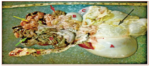
The small intestine of Red Sokoto goat was shown to be situated in the right ventral caudal area of abdominal cavity; it begins at the pylorus and ended at the caecum in ileocaecal junction. Observations shown that it consists of three segments that named Duodenum, Jejunum and Ileum (Figure 1). It connects with the large intestine in the lower most part of the alimentary canal which begins with the caecum and ends with the rectum. The entire organ shown to be a musculomembranous structure and elastic throughout it course with slight differences between the segments (Figure 1).
figure 1: Photograph of the digestive tract of red Sokoto goat, it shows A: Rumen; B: Omasum; C: Abomasum; D: Colon; E: Rectum; (black arrow): Omentum; (red arrow); Duodenum; (yellow arrow): Jejunum; (green arrow): Ileum.

The biometric study of red Sokoto goat shows that the small intestine had different in the mean, weight value at different age categories in which there is an increase in the value with age. Also, the mean value of the duodenum, jejunum and ileum in weight, length, width thickness and volume increase with age that is at different various postnatal age categories.
The mean ± SD weight value of the duodenum were found to range between 16.75SD to 42.75SD and the mean value of length, width, thickness and volume were to be 71.00SD to 199.50SD, 1.05SD, to 2.01SD, 0.28SD to 0.66SD and 8.50SD to 35.00SD from group A to group E respectively (Table 1). The result revealed that the mean weight of the digestive tract, small intestine and various segment of the small intestine (duodenum, jejunum and ileum) tends to increase with age that is from group A to group E as in the duodenum. However, there is variation in the mean weight, length, width, thickness and volume of various intestinal segments that is duodenum, jejunum and ileum with the jejunum had larger value compare to the duodenum and ileum as shown in the (Table 1, 2 & 3).
Table 1: Mean ± SD value of the duodenum in relation to various postnatal ages.

The mean value of pancreas in various postnatal ages increase with age, that is the weight, length, width thickness and volume mean, and value elevated as shown in (Table 4, 5 & 6). The organs index and volumetric index of duodenum, jejunum and ileum and pancreas were revealed to increase with age. The table below shows the result of biometrical readings comprising of weight, length, width, thickness and volume of small intestine segment together with the pancreas. The remarkable significant variation of weight, length, width, thickness and volume of each part of small intestine in the red Sokoto goat at different postnatal ages categories may be due to increase feed consume high contents of fiber in their diet which need more time and large area for digestion and absorption.
Table 2: Mean ± SD value of the jejunum in relation to the various postnatal ages.

Table 3: Mean ± SD value of the ileum in relation to the various postnatal ages.

Table 4: Mean ± SD values of the pancreas in relation to the various postnatal ages.

Table 5: Table showing small intestine organ index.

Table 6: Volumetric index.

The observed biometric study of small intestine of Red Sokoto Goat was found to be progressively increasing with advancement in postnatal ages. These results are in accordance with the findings of [4]. Similar measurement was recorded by [5] in bovines, ovines and caprine, [6] in pampas deer [7] in Giraffe. However, there is variation in the mean weight, length, width, thickness and volume of various intestinal segments that is duodenum, jejunum and ileum with the jejunum had larger value compared to the duodenum and ileum. These results were compatible with the result obtained by Luay & Najlaa [8] in adult indigenous Gazelle and Sabuj et al. [9] in white New Zealand Rabbit. But however, variations exist in the value of duodenum and ileum in which the duodenum has lower value than ileum in the present study which is incompatible with the results of Luay & Najlaa [8] and Sabuj et al. [9] in which the duodenum has large value compared to the ileum. These variations may be due different in species use in carrying out the researches [10-15].
Conclusion
It was concluded that the small intestine in Red Sokoto goat is situated in the right ventral caudal area of abdominal cavity which start from the pylorus and ended at the caecum in ileocaecal junction [16-21]. It is composed of three segments (duodenum, jejunum and ileum) the significant increase of length, weight, width and volume was in the jejunum compared to duodenum and ileum that have large biometric value in their thickness than the jejunum at different postnatal ages [22-33].
Recommendations
Based on the above results it was recommended that further studies should be conducted in different domestic species and breeds, in Nigeria for the purpose of teaching and research all over the world.
References
- Coffey L, Margo H, Wells A (2011) Goats sustainable production overview.
- Mahmoud AA (2010) Present status of the world goat populations and their productivity. Lohmann Information 45(2): 42-52.
- Buhari BK (2008) Sokoto state investment promotion committee trade investment Nigeria 6(2): 35.
- Yildirim A, Ulutas Z, Olak N, Sirin E, Aksoy Y (2014) A study open gastrointestinal tract characteristic of ram lams of the same weight from six Turkish sheep breeds. South Africa Journal of Animal Science 44: 90-96.
- Klaus DB, Robert EH (2003) Bovine anatomy. An illustrated text 7: 68-77.
- Pérez W, Clauss M, Ungerfeld R (2008) Observation on the macroscopic anatomy of the intestinal tract and its mesenteric fold in the pampas deer. Anat Histol Embryol 37(4): 317-321.
- Pérez W, Lima M, Clauss M (2009) Gross anatomy of the intestinal in the giraffe (Giraffa camelopardalis). Anat Histol Embryol 38(6): 432-435.
- Hamza LO, Siwan NA (2017) Morphological features of the small intestine in the adult indigenous gazelle (Gazelle Subgutturosa). International Journal of Science and Nature 8(2): 223-229.
- Nath SK, Das S, Kar J, Afrin K, Dash AK, et al. (2016) Topographical and biometrical anatomy of the digestive tract of white New Zealand rabbit (Oryctolagus cuniculus). J Adv Vet Anim Res 3(2): 145-151.
- Althanaian TA, Alkhodair KM, Albokhadaim IF, Ramdam RO, Ali AM (2012) Gross anatomical studies on duodenum of one humped camel (Camelus dromedaries). International Journal of Zoological Research 8(2): 90-97.
- Andleeb R, Rajesh R, Massarat K, Baba MA, Dar FA, et al. (2016) Histomorphological study of the small intestine in Gaddi Goat. Indian Journal of Veterinary Anatomy 28(2): 10-13.
- Barnwal AK, Yadava RCP (1975) Studies on the histological structures of small intestine of Indian buffaloes (Bubalus bubalis). Indian J. Anim. HIth 14(1): 19-23.
- Copenhaver WM, Kelly DE, Wood RL (1978) The digestive system. In: Bailey’s textbook of Histology. 17th (edn), Williams and Wilkins Company, Baltimore, USA.
- Deinz K, Kum S (2016) A Histological and histochemial study of the small intestine of the dromedary camel (Camelus dromedarius). Journal of Camel Practice and Research 23(1): 111-116.
- Dyce KM, Sack WO, Wensing CJ (2002) Textbook of veterinary anatomy. 4th (edn), Sounders Publishers, Washington, USA, pp. 129-136.
- Ergun E, Ergun L, Asti RN, Kurum A (2003) Light and scanning electron microscopic morphology of Paneth cells in the sheep small intestine. Revue Med Vet 154(5): 351-355.
- Eurell JA, Frappier BL (2006) Dellmann’s textbook of veterinary histology. 6th (edn), Blackwell publishing, State Avenue, USA, pp. 194-199.
- http://Faostat.fao.org/default.aspx
- Frandson RD, Wilke WL, Fails AD (2011) Anatomy and physiology of farm animals. 7th (edn), Wiley Blackwell Publication, Hoboken, New Jersey, USA, p. 350.
- Hasanzadeh S, Monazzah S (2011) Gross morphology, histomorphology and histomorphology of jejunum in the adult river buffalo. Iranian J Vet Res 12: 99-106.
- Kumar P, Kumar P, Singh G, Poonia A (2013) Histological architecture and histochemistry of duodenum of the sheep (ovis aries). Indian J vet Anat 25: 30-32.
- Gahlot KP, Kumar P, Singh G (2017) Histoarchitecture and histochemistry of duodenum and jejunum of the goat (Capra hircus). Haryana Vet 56(1): 37-40.
- Kumar P, Kumar P, Singh G, Poonia A, Parkash J (2014) Histological architecture and histochemistry of jejunum of sheep. Haryana Vet 53(1): 55-57.
- Lalitha PS (1990) Vacuolated cells in the crypts of lieberkuhn of small intestine of Indian Buffalo (Bubalus buhalis). Indian J Vet Anat 2: 31-32.
- Mason IL (1988) A world dictionary of Livestock breeds, type and varieties. 3rd (edn), CAB International, Wallingford, UK.
- Perez W, Vazqueze N (2012) Gross anatomy of the gastrointestinal tract in the Brown Brocket deer (Mazama guoaubira). J Morphol Sci 29(3): 148-150.
- Perez W, Erdogan S, Ungerfeld R (2014) Anatomical study of the gastrointestinal tract in free-living axis deer (Axis axis). Anat Histol Embryol 44(1): 43-49.
- Hassan SA, Moussa EA (2015) Light and scanning electron microscopy of the small Intestine of goat (Capra hircus). Journal of cell and Animal Biology 9(1): 1-8.
- Sheahan DJ, Jarvis HR (1976) Comparative histochemical of gastrointestinal mucosubstances. Am J Anat 146(2): 103-131.
- McGeady TA, Quinn PJ, Patrick ESF, Ryan MT (2006) Veterinary embryology. 1st (edn), Development and rotation of the intestines in domestic animals. Wiley Blackwell, Hoboken, New Jersey, USA, p. 217.
- Talukar M (1999) Gross anatomical histomorphological and histochemical studies on the stomach and intestine of cross breed adult pig. Assam Agricultural University Khanapara, Guwahati, India.
- Taylor RE, Field TG (1999) Growth and development scientific farm animal production: An introduction to animal science. 6th (edn), Prentice Hall, New Jersey, USA, pp. 321-324.
- Trautmann A, Fiebiger J (1957) The digestive system. In: Fundamentals of the histology of domestic animals. Habel RE, Biberstein EL (Eds.), 2nd (edn), Constock Publishing Associates, New York, USA, pp. 199-216.


No comments:
Post a Comment