Abstract
The mains aim of our research group in epistemology of
imagination-a theoretical proposal by the first author of this paper, is
shift the veterinary
education in imagen-ology, grounded in the representation of anatomical
structures to be interpreted by students, toward cognitive actions by
means
of which students can constructs their own representations. In short,
move from representations for student’s interpretation, toward cognitive
representative actions in which the students construct the radiologic
plates. Our proposal for the construction of representative actions
entails the
imagination of actions grounded in geometric think, the representative
actions must be grounded in imaginary actions, then, we change the
concept of
imaging in radiology, for imagen-ology in imagination.
Keywords: Veterinary; Education; Epistemology; Imagination;
Imagen ology; Radiology; Geometric think; Geometric imagination; Mental
configurations
Introduction
The role of geometric think is the construction of mental
configurations of geometric images of anatomical structures
of the animal body serving for the spatial location, as well as for
the establish their relationships with the different regions and
cavities of animal’s body. Hence, the aim goal of education is to
encourage the process for which the student can imagine the things
that he cannot see. In anatomy, by means of the dissection, the
student can see structures like muscles, bones and the organs of
the body, but in a living animal, he cannot see it. Then, the role of
geometric imagination in imagen-ology is the construction of three dimensional
mental configurations due that animal anatomy is
three-dimensional, and the radiologic plates are two-dimensional.
We need mental constructions of three-dimensional representaction
of the radiological plates as near as possible to our three dimensional
configuration achieved throughout imagine-action.
Epistemology of imagination in imagen-ology by
radiology
In radiology, the proposal of the epistemology of imagination is
the construction of three-dimensional (3D) mental images by mean
of two-dimensional (2D) image of the radiologic plates, passing
from the interpretation of radiologic plates, to the construction of
them by the students. We need then give to students the technical
and theoretical foundations about the construction of radiologic
plates, in which is recorded the shadow of the anatomical structure
of the body. From epistemological point of view, this process takes
us to remind the allegory of Plato´s Cave in which the people watch
shadows of men and animals projected in a wall, used by Plato to
compare the effect of education and the lack of it in our nature.
The epistemology of imagen-ology in Veterinary, understood
as the relationship between student as subject and animal as
object. The X-ray that go across the structures of animal with less
impediment, is recorded in radiologic plate as radiolucent or black
areas, while the areas that are not reached by X-ray show up as
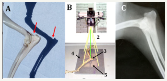
opaque or white areas [1,2]. So, what we observe in a radiologic
plate is the shadow of the object structures reached by the X-ray,
in analogy with the shadows by effect of the sunlight, as too in the
allegory of Plato´s Cave, as can be observed in panel A of the Figure
1.
Figure 1:Shadows projected by sunlight (visible light) and X-rays (outside the spectrum of visible light).
For the creation of radiographs, it is necessary to play with
the X-ray beam so that it penetrates in a certain direction and
thus obtain a two-dimensional image, based on knowledge of
the anatomy, creating the shadow left by each of the structures.
Because the anatomical structures are three-dimensional, at least
two orthogonal radiographic images corresponding to the X and
Y Cartesian planes are required, which, when combined, allow us
to mentally construct the three-dimensional configuration of the
anatomical structure learned in the dissections. Even, in some
cases, we can play with the direction of the rays and make oblique
shots, which will generate a more complete three-dimensional
mental image and not overlook any structure that overlaps with
others by having only a two-dimensional plane X and Y obtained.
Geometric imagination in radiology by teaching of
medical imagen-ology
In a recent work of our research group [3], reference was
made to the role that geometric thought plays in the imaginary
mental configuration of histological structures, as well as their
relationships, with the Cartesian coordinates X and Y coming into
play, which for three-dimensionality the Z-axis is also considered.
As already mentioned, the images obtained by X-rays are twodimensional
shadows generated when projecting the X-ray beam
to specimen. A three-dimensional mental configuration based on a
system of axes is required, of the X, Y and Z coordinates represented
in Figure 2, to obtain at least two orthogonal radiographs for threedimensionality.
This way, the X axis goes from left to right, the Y axis
from bottom to top and the Z axis from back to front [4]. Applying
this in the abdominal cavity, in a lateral radiograph, Figure 3, the
X axis would be long, from cranial to caudal, the Y axis would be
wide, from ventral to dorsal, and the Z axis would be thickness from
side to side of the cavity. Now, in a ventro-dorsal (VD) radiograph
shown in Figure 4, the X axis is from side to side, the Y is caudal to
cranial and the Z is from dorsal to ventral. Then, the positions are
calculated by measuring the lateral position, height and depth for
our spatial location.
Figure 2:X, Y and Z axis system.
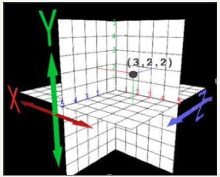
Figure 3:SX, Y and Z axis system applied to L abdominal radiograph.
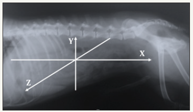
Figure 4:X, Y, Z axis system applied to a VD abdominal radiograph.
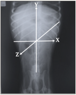
From the epistemology of the imagination and with the
topographical approach, we can say that, within the cavities,
the anatomical location of each structure or organ, as well as its
relationships, is studied, taking this geometric thought to the
areas of topographic anatomy. The student must understand the
relationships, cranial, caudal, dorsal, ventral and lateral. In this way,
having the mental configuration of the anatomical organization
inside the cavities, supported by the geometric thought and
using the three axes as mentioned, can be represented with some
accuracy the place in the space in which each are located organs
and their relationships. This imaginary configuration through
geometrical thinking will allow the student to decide how to place
the patient and what projection would give the X-ray beam to obtain
the required radiographs and move from the interpretation of what
is represented on the plates, to representations created by the own
actions of the students. Transform representations into representactions,
where the imagination turns into imaginary-actions.
Imagine-actions and represent-actions in tridimensional
mental configurations in radiology
The proposal of the epistemology of imagination is that mental
imaginary configurations becomes in real images in imagen-ology,
throughout the imagination of actions that we call imagine-actions.
A practical example of this proposal is the mental configuration of
organ location and their relationship with other organs contained in
the abdominal cavity of the dog-especially gastrointestinal, taking
into account their tridimensional structure grounded in anatomy
previous knowledge, in order to locate, for example, ingested bodies,
common in them. Thus, we know that the cecum and ascending
colon are to the right of the median plane of the abdominal cavity
and the descending colon is to the left, both connected with the
transverse colon that goes from right to left [5-7]. Considering the
nomenclature used to designate the radiographic images, in the LD
projection in the abdominal cavity, despite the contrast medium
applied -barium enema, cecum and ascendant colon cannot be
distinguished of descendent colon, since these overlaps.
So, we don’t know where an injury or a foreign body could be,
if in the cecum, descending colon or ascending colon. Regarding
the transverse colon [3] its travel from one side to another
is not appreciated. For this, taking into account the technical
recommendation of the orthogonal shots (having two shots in
90 c), in the VD projection of the abdomen and with the use of
contrast medium, all its parts could be clearly distinguished: cecum,
ascending colon, transverse colon and descending colon, whose
numbering is equal to panel A. In addition, it would be possible to
accurately determine its anatomical situation within the cavity and,
therefore, where the possible injury could be.
One might think that with the VD projection it would be
sufficient for this study of the large intestine, but this is not the
case, since with this projection the location of the colon and rectum
[5] in the dorsal to ventral direction could not be specified, as can
be seen in Figure 4 is located at a medium height, discarding some
constipation where the descending colon changes its position,
with a displacement towards the ventral or an inflammation of
lympho-nodes, among other anomalies. With the imagine-actions,
it is possible to carry out in an appropriate way all the steps to
follow for taking a radiographic plate, whatever the case, from how
to place the patient (position), the intensity of the X-rays, time of
exposure, X ray beam projection (alignment) and development
process. In this way, all the actions carried out to create the
representation of the reality expressed in the radiographic plates
as it represents-actions [8], will have their origin in the imaginary
action of the student or imagine-actions and would be guided by
the exercise of the geometric thought, within the framework of the
epistemology of the imagination. The names of the structures and
organs mentioned in this work are based on the Nomina Anatomica
Veterinaria [9].
Acknowledgement
Project SIP: 20180338
Project PIAPIME ID 2.11.09.18 FES Cuautitlán UNAM.
Project DGAPA-UNAM PAPIME PE 205717.
References
- Thrall DE (2003) Manual de diagnóstico radiológico veterinario. (4th
edn), Elsevier, Barcelona, Spain.
- Muhlbauer MC, Kneller SK (2013) Radiography of the dog and cat: Guide
to making and interpreting radiographs. (1st edn), Wiley-Blackwell, USA.
- Oliver González MR, García Tovar CG, Soto Zárate CI, Garrido Fariña
G, Rodríguez Salazar LM (2017) Epistemología de la imaginación: el
pensamiento geométrico en la enseñanza de la anatomía y la histología.
Lat Am J Sci Educ 22115.
- Garrosa S (1998) El concepto de dimensión. Los ejes XYZ.
- Pérez SAP (2007) Atlas de anatomía radiográfica: órganos digestivos
abdominales y tránsito gastrointestinal en perro adulto (Canis
familiaris). Tesis profesional. FES-Cuautitlán UNAM.
- Rodríguez SLM (2015) Epistemología de la Imaginación: el trabajo
experimental de Ørsted. Corinter (Ed.), Capítulo, México, p. 5.
- Evans HCE, de Lahunta A (2013) Miller’s Anatomy of the Dog, (4th edn),
Elsevier Saunders, USA.
- Oliver GRS (2017) Epistemología aplicada a las ciencias biológicas
básicas. 3er Congreso de Ciencia, Educación y Tecnología, Facultad de
Estudios Superiores Cuautitlán (FES-C) UNAM.
- Gasse H (2012) Nomina anatomica veterinaria. International Committee
on Veterinary Gross Anatomical Nomenclature. (5th edn), Editorial
Committee, Hannover, Germany.

