Symptomatic Nephroptosis (SN) Causes the Loin Pain and Haematuria Syndrome (LPHS) by Ahmed N Ghanem* in Surgical Medicine Open Access Journal_ Surgical Medicine Open Access Journal

Abstract
The
objective of this letter is to report recent advances on the diagnosis and
therapy of the loin pain hematuria syndrome (LPHS). The patho-aetiological link
of LPHS with symptomatic nephroptosis (SN) is a novel discovery of the 21st
century that was first preliminary reported in 2002 [1]. The link was confirmed
in 2016 in an article that précised the exact pathoaetiology of LPHS; ischemic
nephritis and sympathetic neuropathy of SN. It also advanced a 100% curative
therapy for LPHS namely; the renal sympathetic denervation and nephropexy
(RSD&N) surgery [2]. Symptomatic nephroptosis has been known for centuries
[3] but was disparaged [4] >70 years ago and omitted from all textbooks.
Hence it is a forgotten disease. The loin pain hematuria syndrome was first
reported in 1967 [5] and has remained without any effective therapy till now
The
discovery of SN causing LPHS and the successful curable therapy are based on a
prospective cohort 10 years study [1,2] of 190 patients diagnosed with SN among
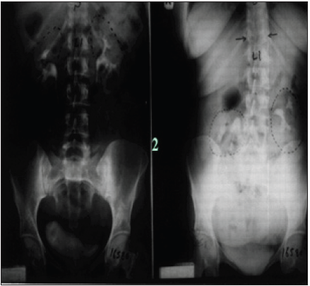
whom 36(18.9%) patients developed LPHS. Two new signs that demonstrate renal
pedicle stretch of SN (Figure 1) causes vessel stenosis and ischemic nephritis
as evidenced by the new IVU7 sign and tube stretch hypothesis (Figure 2 &
3). These signs demonstrate that stretching of renal artery in pedicle at erect
posture causes vessels stenosis, diminished blood flow and induces ischemic
nephritis and sympathetic neuropathy.
This is illustrated by producing the same effect on stretching a rubber
catheter tube causing stenosis (reduced diameter) that diminishes lumen flow.
Lumen flow of the blood vessel or catheter is completely obliterated when
stretch of more than double the length of vessel or tube is accompanied by
rotation twist. Ischemic nephritis affects the renal medulla long before the
cortex, particularly eroding the medullary papillae creating fistulae between renal blood vessels and the
collecting system explaining the venous shunt causing the hematuria. This is
demonstrated only on retrograde pyelography (RGP) (Figure 4 & 5). So, the
diagnosis of SN is only possible on intravenous urography (IVU) with erect film
(IVU-E) on which the IVU7 sign demonstrates renal drop and pedicle stretch
(Figure 1 & 2) and RGP that demonstrates medullary damage (Figure 4 &
5) while all other imaging including CAT and MRI are useless being feasible
only on supine posture
https://crimsonpublishers.com/smoaj/fulltext/SMOAJ.000562.php
Crimson
Publishers: https://crimsonpublishers.com/
For
more articles in Surgical Medicine Open Access Journal,
Please
click on below link: https://crimsonpublishers.com/smoaj/


No comments:
Post a Comment